When facing unexplained pain or swelling, the question of whether a CT scan can reveal blood clots is crucial. This advanced imaging technique plays a vital role in diagnosing conditions that could threaten your health. Understanding how a CT scan works and its effectiveness in detecting clots can empower you to make informed decisions about your health care.
Blood clots can lead to serious complications, such as pulmonary embolism, making timely diagnosis essential. A CT scan, especially when performed with contrast dye, is often the first line of defense in identifying these dangerous obstructions. This non-invasive procedure provides detailed images of your body, which can lead to life-saving interventions.
As you delve into this article, you’ll learn about the CT scan procedure, its benefits, and what to expect. This knowledge not only reduces anxiety but also keeps you proactive in your healthcare journey. Understanding the imaging process is your first step towards clarity and peace of mind.
Understanding CT Scans and Blood Clots
A CT scan, or computed tomography scan, is a powerful diagnostic tool that provides detailed images of the internal structures of the body, helping healthcare professionals identify various medical conditions, including blood clots. Blood clots can occur in veins or arteries and may lead to serious complications if not detected and treated promptly. When a healthcare provider suspects a patient may have a clot-such as in cases of deep vein thrombosis (DVT) or pulmonary embolism-getting a CT scan can be a crucial step in confirming or ruling out the presence of these potentially life-threatening obstructions.
The process of how CT scans diagnose blood clots involves the use of advanced imaging technology that combines X-ray images taken from different angles. These images are processed by a computer to create cross-sectional views of the body, enabling detailed visualization of blood vessels and surrounding tissues. Specifically, a CT pulmonary angiography (CTPA) is often performed to investigate clots in the pulmonary arteries, while a CT venography (CTV) is used for clots in the veins. The use of a contrast dye, which is injected into a vein before the scan, enhances visibility of the blood vessels and helps in accurately detecting the location and size of any clots present.
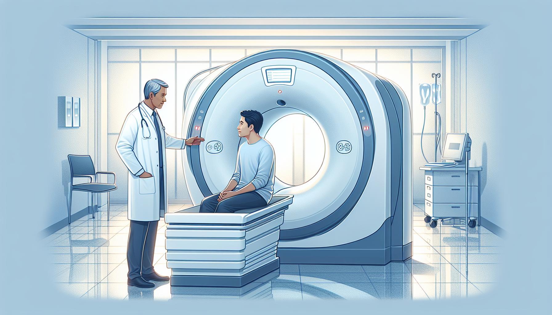
Understanding the preparation and expectations surrounding a CT scan can ease patient anxiety. Typically, patients are advised to avoid eating or drinking for a few hours before the procedure if contrast dye is to be used. During the scan, patients lie down on a table that slides into the CT machine, which resembles a large doughnut. The procedure is quick, usually taking just a few minutes, and it is painless. Once completed, radiologists interpret the results, providing vital information that can lead to an appropriate treatment plan for managing blood clots. Although CT scans are generally safe, they should be performed with appropriate considerations regarding radiation exposure and contrast material allergies, emphasizing the need to discuss any concerns with healthcare professionals prior to the scan.
By illuminating the intricate details of blood vessels, CT scans play an indispensable role in the timely diagnosis of blood clots, ultimately guiding effective treatment and improving patient outcomes. Knowledge about this procedure not only empowers patients but also reinforces the essential partnership between patients and their healthcare providers in addressing serious health issues like blood clots.
How CT Scans Diagnose Blood Clots
CT scans are a cornerstone in the diagnosis of blood clots, enabling healthcare providers to get a clear view of the internal structures of the body. When a doctor suspects the presence of a clot, such as in cases of deep vein thrombosis (DVT) or pulmonary embolism, the need for precise imaging can be critical. The technology behind CT scans combines multiple X-ray images taken from various angles, producing detailed cross-sectional images of the body. This process allows clinicians to visualize blood vessels and surrounding tissues effectively, identifying clots that may not be easily seen through other imaging methods.
Typically, two specific types of CT scans are utilized in diagnosing blood clots: CT pulmonary angiography (CTPA) and CT venography (CTV). CTPA is employed to assess potential clots in the pulmonary arteries, which can be life-threatening if not treated promptly. Conversely, CTV focuses on detecting clots in the venous system, particularly in the legs or abdomen. Both types of scans frequently use a contrast dye, which is injected into a vein, improving the visibility of the blood vessels on the images. This enhancement is essential for accurately determining the size and location of any clots present.
For anyone preparing for a CT scan, it’s helpful to understand what to expect, which can significantly reduce anxiety. Generally, patients are advised to refrain from eating or drinking a few hours before their appointment if a contrast dye is to be used. During the scan, the patient lies down on a table that gently slides into the CT machine, which resembles a large, doughnut-like structure. The actual scanning process is quick, often lasting only a few minutes, and is designed to be painless, although some may experience a warm sensation from the contrast dye. Once the scan is complete, radiologists meticulously analyze the images and share their findings with the healthcare provider, who will discuss the results and potential next steps based on the diagnosis.
Understanding how CT scans operate in diagnosing blood clots empowers patients to take an active role in their healthcare. Knowledge of the procedure can alleviate fears and foster a collaborative relationship between patients and their medical team. Always consult your healthcare provider for personalized advice and additional information regarding diagnoses and treatments related to blood clots.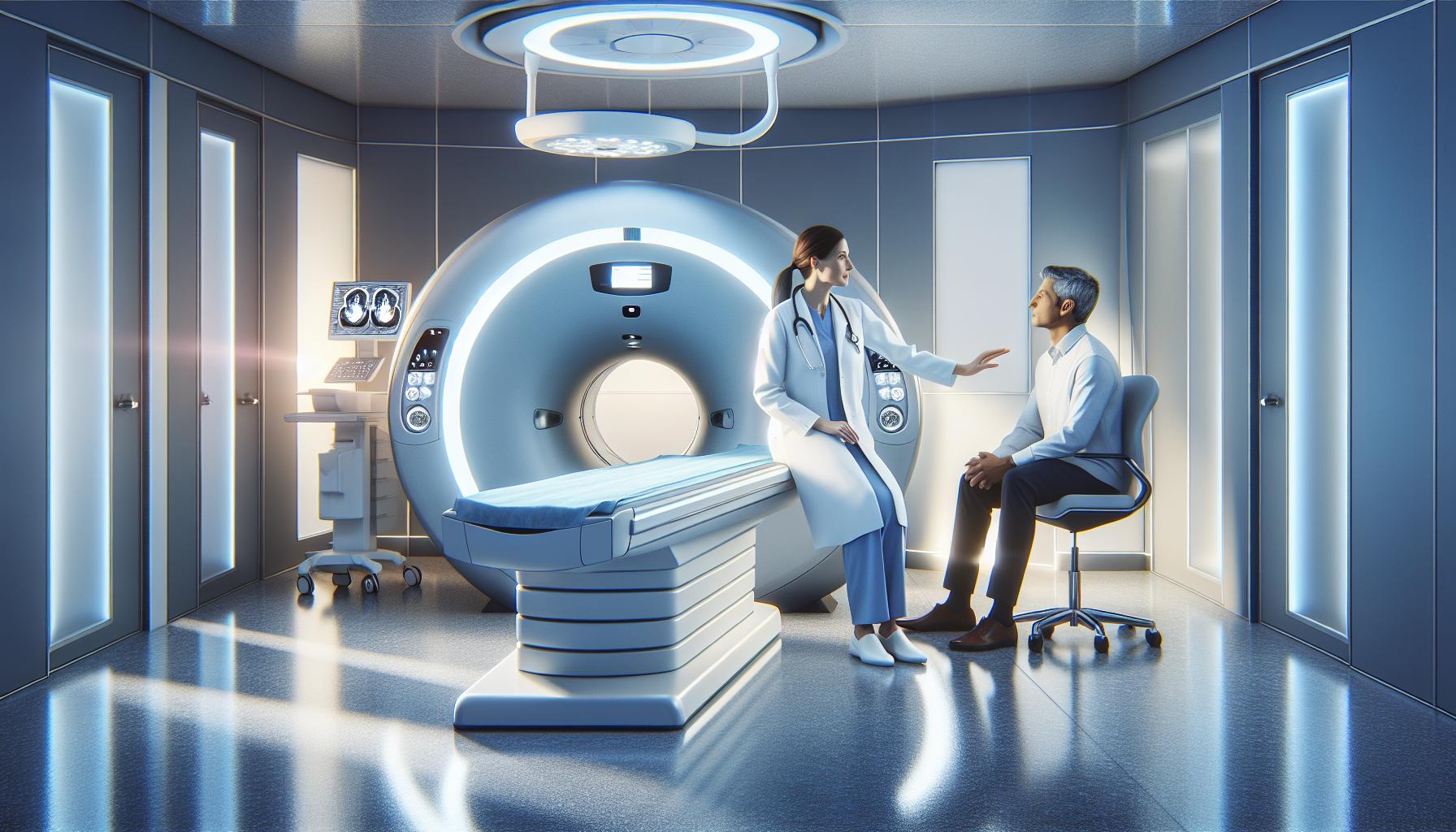
Types of CT Scans Used for Detecting Clots
A CT scan is a powerful tool in the medical field, particularly for diagnosing blood clots, as it provides detailed images that can pinpoint the presence of clots quickly and accurately. Among the various types of CT scans available, two are most commonly employed for detecting blood clots: CT pulmonary angiography (CTPA) and CT venography (CTV). Both methods rely on advanced imaging technology and often utilize a contrast dye to enhance visibility.
CT pulmonary angiography (CTPA) is specifically designed to examine the pulmonary arteries located in the lungs. It’s particularly vital for diagnosing pulmonary embolism, a condition where a clot travels to the lungs and can become life-threatening. In this scan, the contrast dye is injected into a vein, allowing the radiologist to visualize the blood flow and identify any obstructions. This imaging is swift, often taking just a few minutes, and can inform immediate treatment decisions, helping to save lives.
In contrast, CT venography (CTV) is focused on the venous system, primarily in the legs and abdomen, and is effective in identifying conditions such as deep vein thrombosis (DVT). Similar to CTPA, CTV employs contrast dye to outline the veins, making it easier to spot clots that might not be visible through other forms of imaging. For those experiencing symptoms of DVT, such as leg swelling or pain, CTV can provide crucial information to direct the appropriate interventions.
Understanding these different approaches allows patients to feel more informed about their diagnostic journey. If you are facing a CT scan, knowing whether it will be a CTPA or CTV can ease any anxiety. It’s important to have open discussions with your healthcare provider about which type of scan is suitable for your specific situation, ensuring that you receive tailored care aligned with your health needs. Always remember, the goal of these imaging techniques is to provide clarity and facilitate timely, effective treatment.
Preparation Steps Before a CT Scan
Undergoing a CT scan can be a pivotal moment in diagnosing conditions like blood clots, and preparing adequately is crucial to ensure accurate results and a smooth experience. Proper preparation helps minimize anxiety and enhances the effectiveness of the imaging process.
The first step in preparing for a CT scan is to communicate openly with your healthcare provider about any existing medical conditions, allergies, or medications you are taking, especially if they include blood thinners or anticoagulants. Your medical team will provide specific instructions based on your individual health profile and the type of scan being performed, such as CT pulmonary angiography or CT venography.
It is common for patients to be asked to fast for a few hours before the scan, particularly if a contrast dye will be used. This is essential because the dye can cause nausea or other reactions if administered on a full stomach. Drinking plenty of water before the procedure is advisable as staying hydrated can help improve the clarity of the images. If you have concerns regarding the contrast dye or any past reactions, be sure to discuss this with your healthcare team in advance to explore alternative options if necessary.
On the day of the scan, wear loose-fitting clothing without metal fasteners, zippers, or belts, as these items can interfere with the imaging. You may also be asked to change into a hospital gown. Arriving early for your appointment allows time for registration and any last-minute questions you might have, ensuring that you feel calm and informed before entering the scanning room.
Being well-prepared can significantly enhance your CT scan experience, emphasizing the importance of following your doctor’s instructions and maintaining open lines of communication. Your proactive approach not only aids in achieving the best imaging results but also empowers you on your path to understanding and managing your health conditions effectively.
What to Expect During Your CT Scan
Undergoing a CT scan can be a crucial step in diagnosing potential blood clots, and understanding what happens during the procedure can significantly reduce any anxiety you may feel. As you enter the imaging room, it’s likely to be well-lit and filled with advanced technology. The central focus is a large, doughnut-shaped machine known as the CT scanner, which will take detailed images of your body to help your healthcare provider evaluate your condition.
Once you’re comfortably positioned on the scanning table, the radiologic technologist will help you find the right placement, often asking you to lie flat on your back. If you’re receiving contrast dye, it may be administered through an IV in your arm, which can create a warmth or flushing sensation as it enters your bloodstream. This is a normal response and generally resolves quickly. The scanning process itself usually lasts only a few minutes. During this time, you might be asked to hold your breath briefly while the images are taken; this is to prevent motion blur, which could affect the quality of the results.
To ensure a clear picture, you’ll need to remain as still as possible. The machine will rotate around you, capturing multiple cross-sectional images of the area being examined. It’s important to listen to the technologist’s instructions-if you have any urgent concerns, such as extreme discomfort or difficulty breathing, don’t hesitate to let them know. Rest assured that throughout the process, you will be in direct communication with the medical staff, who are present to assist you.
After the scan is complete, you’ll typically be asked to wait a short while while the images are processed. This allows the radiologist to assess the results accurately and determine if any further imaging or evaluation is necessary. Once everything is finished, you can return to your regular activities. Keep in mind that your healthcare provider will discuss the results with you, ensuring you understand the implications regarding blood clots or any other findings. Remember, your comfort and understanding are paramount, so take this opportunity to ask questions and seek clarity about your CT scan experience.
Interpreting CT Scan Results
Interpreting the results of a CT scan can be both a moment of anticipation and anxiety for patients and their families. Understanding what these results mean is crucial, especially when it comes to diagnosing blood clots, which can pose serious health risks. A CT scan is highly effective for revealing the presence of clots, primarily in areas like the lungs (CT pulmonary angiography) or in veins (CT venography). Radiologists analyze the images taken during your scan to determine whether there are any clots present and, if so, the extent of them.
When reviewing your CT scan results, it’s essential to stay informed about the various indicators that radiologists look for. A clot typically appears as a dark area on the images, contrasting with surrounding tissues. In the case of a pulmonary embolism, for example, the radiologist will examine the pulmonary arteries for any obstructions that a clot may cause. To aid in interpretation, the radiologist might consider factors such as the size and location of the clot, the presence of other indicators of a problem, and any previous imaging history.
Understanding your results involves not only looking at the images but also interpreting the accompanying report. This report will summarize findings, including whether a clot is present, and will often describe recommendations for any further tests or treatments necessary. If your results are unclear or you have questions, don’t hesitate to discuss these with your healthcare provider. They can provide clarity on the importance of your results in the context of your overall health and any additional steps that may be required for your care.
It’s crucial to remember that while the CT scan is a powerful diagnostic tool, it is just one piece of the puzzle. Factors like symptoms, medical history, and additional tests will all play a role in forming a complete picture of your health. Always consult with your healthcare provider for personalized guidance based on your specific condition. They can offer reassurance and help you navigate your path forward, ensuring that you feel supported throughout your medical journey.
Alternative Imaging Techniques for Blood Clots
When it comes to diagnosing blood clots, several alternative imaging techniques can complement or serve as alternatives to CT scans, each with its own unique advantages. Understanding these methods helps patients make informed decisions about their imaging options, especially if they have concerns about radiation exposure or other factors associated with CT scanning.
One of the most commonly used alternatives is ultrasound, particularly for detecting deep vein thrombosis (DVT) in the legs. This technique uses high-frequency sound waves to create images of the blood vessels. Ultrasound is non-invasive, does not involve radiation, and is typically the first line of testing when a DVT is suspected. It can effectively visualize the blood flow and encounter clots, allowing for immediate assessments without the complexities of contrast agents, as might be required in CT imaging.
Another option is magnetic resonance imaging (MRI), which can be particularly useful in visualizing locations difficult to assess with CT, such as the pelvis and abdominal veins. MRI utilizes a magnetic field and radio waves to produce detailed images of soft tissues, offering a clear view of potential clots without exposure to radiation. While MRI is less commonly used for clot detection compared to CT or ultrasound, it may be recommended in certain situations, especially when additional information about tissue health is needed or in patients who cannot tolerate contrast dyes used in other imaging modalities.
In some cases, healthcare providers might also consider a venography. This involves injecting a contrast dye directly into a vein and taking X-rays to visualize blood flow and identify clots. While effective, it is more invasive than the other methods and is used less frequently today due to the rise of ultrasound and CT.
It’s essential for patients to discuss thoroughly with their healthcare providers the best imaging choice based on their unique circumstances, symptoms, and medical history. Each imaging technique has its specific context for use, and understanding the advantages and limitations of each can empower patients to participate in their health care decisions actively.
CT Scan Safety: Risks and Considerations
Undergoing a CT scan can be a pivotal step in diagnosing conditions such as blood clots, but many patients understandably have questions about the safety and implications of this imaging procedure. While CT scans are invaluable tools that provide detailed images of internal structures, they do come with certain risks that warrant consideration.
The primary concern associated with CT scans is the exposure to ionizing radiation. Although the doses are relatively low and the procedure is generally considered safe, repeated exposure over time can increase the risk of developing cancer, especially in vulnerable populations such as children or individuals requiring frequent imaging. It’s crucial for patients to have an open dialogue with their healthcare provider about the necessity of the scan and to weigh the benefits against the risks. For instance, in a situation where a CT scan may save a life by diagnosing a severe blood clot accurately, the potential risks may be deemed acceptable.
Another aspect to consider is the use of contrast material, which is often employed to enhance the clarity of images. While most people tolerate contrast agents well, some may experience allergic reactions ranging from mild itching to more severe reactions that necessitate immediate medical attention. Individuals with pre-existing kidney conditions should also discuss their history with their healthcare provider, as contrast dyes can pose risks to kidney function.
To ensure a smooth and safe experience, patients should prepare for their CT scan by following specific instructions provided by their healthcare team. This could involve fasting for several hours prior to the examination, particularly if contrast material will be used. Understanding these preparatory steps can help to mitigate anxiety and ensure the procedure is effective. Additionally, it’s beneficial for patients to ask about the specific type of CT scan and the potential implications, allowing for a more informed consent process.
Ultimately, the decision to proceed with a CT scan should always be made collaboratively between the patient and healthcare provider, taking into account the urgency of the situation and the medical history of the individual. As with any medical procedure, being informed and prepared can help patients feel more at ease while ensuring their health is prioritized.
Insurance Coverage for CT Scans
Navigating the intricacies of insurance coverage for a CT scan can feel daunting, especially when facing a medical situation involving potential blood clots. Knowing what to expect can alleviate some of that anxiety. Generally, the first step is to verify whether your health insurance plan covers CT scans, as policies can vary significantly. Typically, CT scans are covered when deemed medically necessary by your healthcare provider, particularly for diagnosing conditions like blood clots. It’s essential to obtain prior authorization if required by your insurer, which involves your doctor submitting documentation justifying the need for the scan.
When you’re scheduling your appointment, take the time to discuss costs with both your healthcare provider and the imaging facility. The fee for a CT scan can range widely, influenced by factors such as the specific type of scan needed, the facility’s location, and whether any contrast material is used. If you’re concerned about out-of-pocket expenses, check with your health insurance provider regarding your deductible, co-pays, and any limits on coverage for diagnostic imaging. Many facilities offer payment plans or financial assistance for those facing high medical bills.
It’s also worth noting that certain specialized scans, such as those performed for lung embolism, might have specific coding that affects coverage. Having a clear understanding of your policy can empower you to ask informed questions and advocate for the necessary imaging without undue stress. Consider keeping a list of questions handy for your healthcare provider during discussions, such as:
- Is the CT scan considered medically necessary for my condition?
- Will my insurance require prior authorization for this procedure?
- What are the expected costs and out-of-pocket expenses based on my insurance plan?
Being proactive and informed can help ensure that the imaging you need proceeds smoothly, allowing you to focus on your health and well-being without the added burden of financial uncertainty.
Real-Life Impact: Patient Stories and Outcomes
In times of medical uncertainty, many patients find themselves grappling with anxiety about potential diagnoses, especially regarding serious conditions like blood clots. The stories of individuals who underwent CT scans can provide much-needed insight and reassurance about the power of medical imaging in identifying these life-threatening conditions. For instance, consider the experience of Sarah, a 34-year-old who suddenly developed severe leg swelling and pain. Her healthcare provider recommended a CT scan to rule out a deep vein thrombosis (DVT). The scan not only confirmed the presence of a blood clot but also allowed for timely intervention, which significantly reduced her risk of complications. Sarah remarked how vital the CT scan was, emphasizing that what initially felt like a routine procedure turned into a critical lifesaver.
Similarly, James, a 45-year-old grandfather, experienced unexplained shortness of breath and chest pain. After being rushed to the emergency room, the medical team performed a CT pulmonary angiography. The results revealed a pulmonary embolism-another form of blood clot that can be fatal. Thanks to the speed and accuracy of the CT scan, James received immediate treatment and was able to recover fully. His story is a testament to how these imaging techniques not only diagnose but also guide urgent care interventions that can prevent tragic outcomes.
Empowering Awareness and Prompt Action
Stories like Sarah’s and James’s highlight the importance of seeking medical attention when faced with troubling symptoms. Being informed about the signs and risks associated with blood clots can empower patients to act swiftly. If someone experiences leg swelling, pain, or sudden shortness of breath, it’s crucial to consult a healthcare provider promptly. CT scans play a pivotal role in this process; they provide clear, diagnostic imaging that can confirm or rule out the presence of blood clots, facilitating quick and effective treatment.
Moreover, patients are encouraged to have open discussions with their healthcare providers regarding any fears or concerns they may have about the CT scanning process. Understanding what to expect can alleviate anxiety-knowing that these scans could potentially save lives can transform fear into proactive health management. Resources and patient stories further enhance this journey, providing relatable experiences that can serve as guiding lights in moments of uncertainty.
The Lifesaving Role of CT Scans
As shown through these narratives, the utilization of CT scans for blood clots highlights their lifesaving capabilities and underscores their critical role in modern healthcare. These imaging techniques empower not only the medical teams to provide precise diagnoses but also patients to take control of their health journeys with confidence and awareness. Remember, while individual stories can be inspiring, incorporating professional medical advice is essential for personalized diagnosis and treatment pathways.
FAQs About CT Scans and Blood Clots
Many people wonder whether a CT scan can accurately show blood clots, and the answer is a resounding yes. CT scans are highly effective imaging tools specifically designed to identify clots in various body parts, most notably in the lungs (pulmonary embolism) and veins of the legs (deep vein thrombosis). The imaging technology used in CT scans allows for detailed views of blood vessels, which can clearly highlight any abnormalities such as blockages caused by clots.
Common Concerns About CT Scans
Patients often have questions and concerns about undergoing a CT scan. Here are some frequently asked questions:
- What should I do to prepare for a CT scan?
Your healthcare provider will give you specific instructions, which may include fasting for several hours before the scan and avoiding certain medications. Inform them if you have allergies, particularly to contrast dye, as this may be used during the procedure to enhance visibility. - Are there any risks associated with CT scans?
While CT scans expose patients to radiation, the levels are generally low and the benefits of accurately diagnosing conditions like blood clots outweigh the risks. Discuss any concerns about radiation exposure with your doctor, especially if you have had multiple scans in the past. - How long does a CT scan take?
The actual scanning process is relatively quick, usually lasting around 10 to 30 minutes. However, additional time may be required for preparation and recovery, particularly if contrast dye is used. - When can I expect my results?
Results are often available within hours or by the next business day. Your healthcare provider will explain the findings and discuss the next steps if necessary.
Post-Scan Guidance
After the scan, if you received contrast dye, it’s essential to drink plenty of fluids to help flush the dye from your system. Monitor for any unusual reactions, like itching or swelling, and contact your healthcare provider if any symptoms occur.
Understanding the role of CT scans in diagnosing conditions such as blood clots can alleviate anxiety and ensure you arrive fully informed at your appointment. If you have any lingering questions, don’t hesitate to reach out to your healthcare team; they are there to support you every step of the way.
Frequently asked questions
Q: How accurate are CT scans in detecting blood clots?
A: CT scans are highly accurate for detecting blood clots, especially in the lungs (pulmonary embolism) and legs (deep vein thrombosis). The sensitivity can exceed 90%, making them a crucial diagnostic tool. For further details, refer to the section on “How CT Scans Diagnose Blood Clots.”
Q: What types of blood clots can a CT scan identify?
A: CT scans can identify various types of blood clots, including pulmonary embolisms in the lungs and deep vein thrombosis in the legs. Understanding these types can help you grasp the significance of this imaging technique. For more information, see “Types of CT Scans Used for Detecting Clots.”
Q: Are there any risks associated with CT scans for blood clots?
A: While CT scans are generally safe, potential risks include exposure to radiation and allergic reactions to contrast dye. Discuss safety concerns with your physician before the scan. More on this can be found in the section “CT Scan Safety: Risks and Considerations.”
Q: How should I prepare for a CT scan to check for blood clots?
A: Preparation for a CT scan typically includes fasting for a few hours and informing your doctor about any medications or allergies. Following specific preparation guidelines can enhance the quality of the scan; see “Preparation Steps Before a CT Scan” for details.
Q: What can I expect during a CT scan for blood clots?
A: During a CT scan for blood clots, you will lie on a table as it moves through the scanner. You may hear humming noises, and it’s essential to remain still. Understanding this process can ease anxiety; refer to “What to Expect During Your CT Scan.”
Q: How are the results of a CT scan for blood clots interpreted?
A: Results from a CT scan are interpreted by radiologists, who look for signs of clots in blood vessels. Diagnosis typically takes one to two days, and your doctor will discuss the findings. For more details, see “Interpreting CT Scan Results.”
Q: Can CT scans replace other imaging techniques for blood clot detection?
A: CT scans are often preferred due to their speed and accuracy, but they may not entirely replace ultrasound or MRI, depending on the situation and patient needs. Consult the “Alternative Imaging Techniques for Blood Clots” section for a comparative overview.
Q: How can I address concerns about insurance coverage for a CT scan?
A: To address concerns about insurance coverage for a CT scan, contact your insurance provider directly for specific policy details. You can also discuss options with your healthcare provider. For more insights, check out the “Insurance Coverage for CT Scans” section.
In Summary
Understanding whether a CT scan can reveal blood clots is crucial for your health and peace of mind. Remember, the insights gained from this imaging technique can be life-saving, helping to diagnose serious conditions quickly and efficiently. If you have further questions or concerns, don’t hesitate to consult your healthcare provider for personalized guidance.
For more detailed information on medical imaging, explore our related articles on “Understanding CT Scan Procedures” and “Preparing for Your CT Scan: A Step-by-Step Guide.” Stay informed-your health depends on it!
Ready to take the next step? Subscribe to our newsletter for the latest updates on medical advancements and health tips, or schedule a consultation to discuss your imaging needs. Your proactive approach today can lead to a healthier tomorrow!

