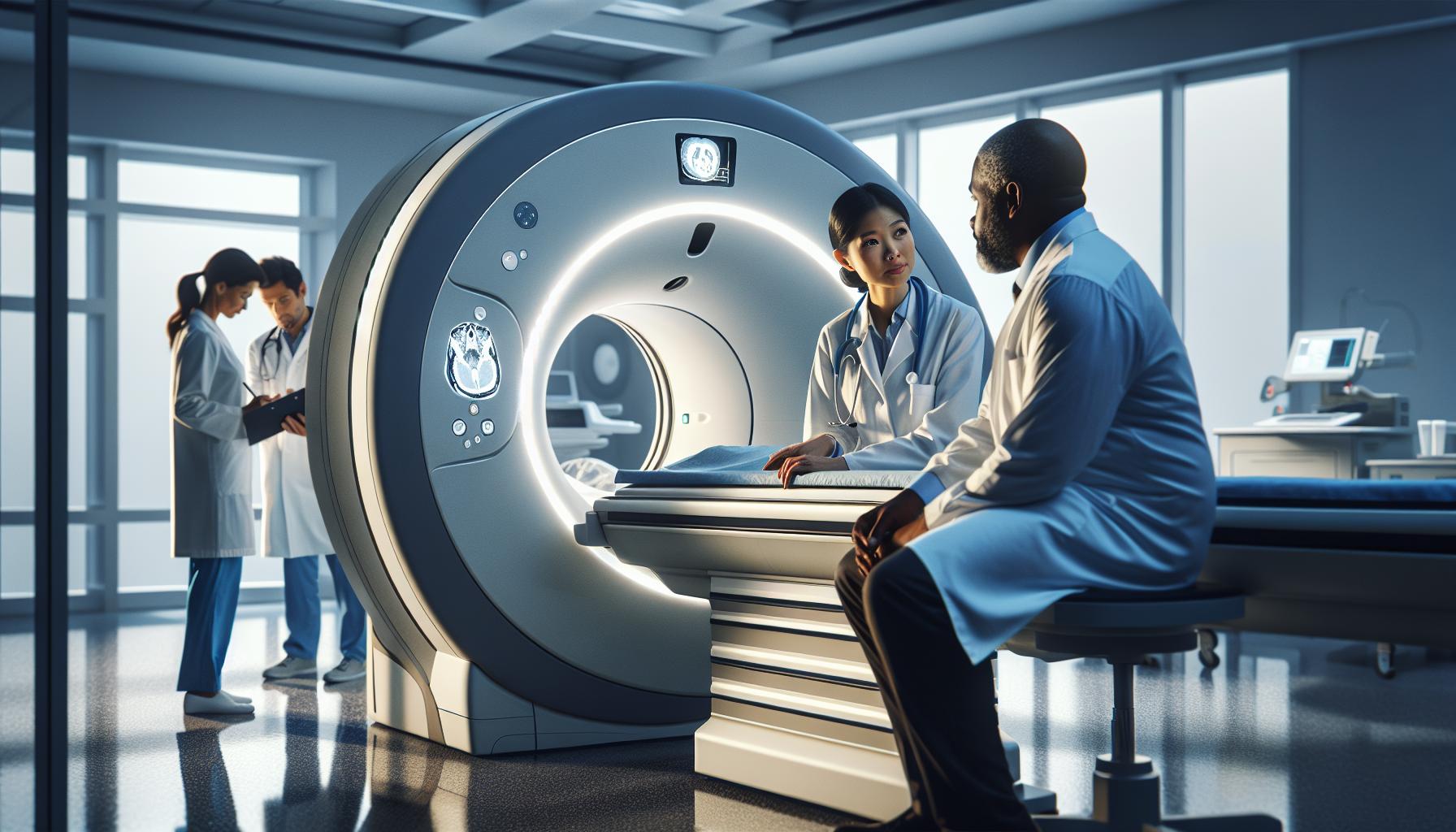
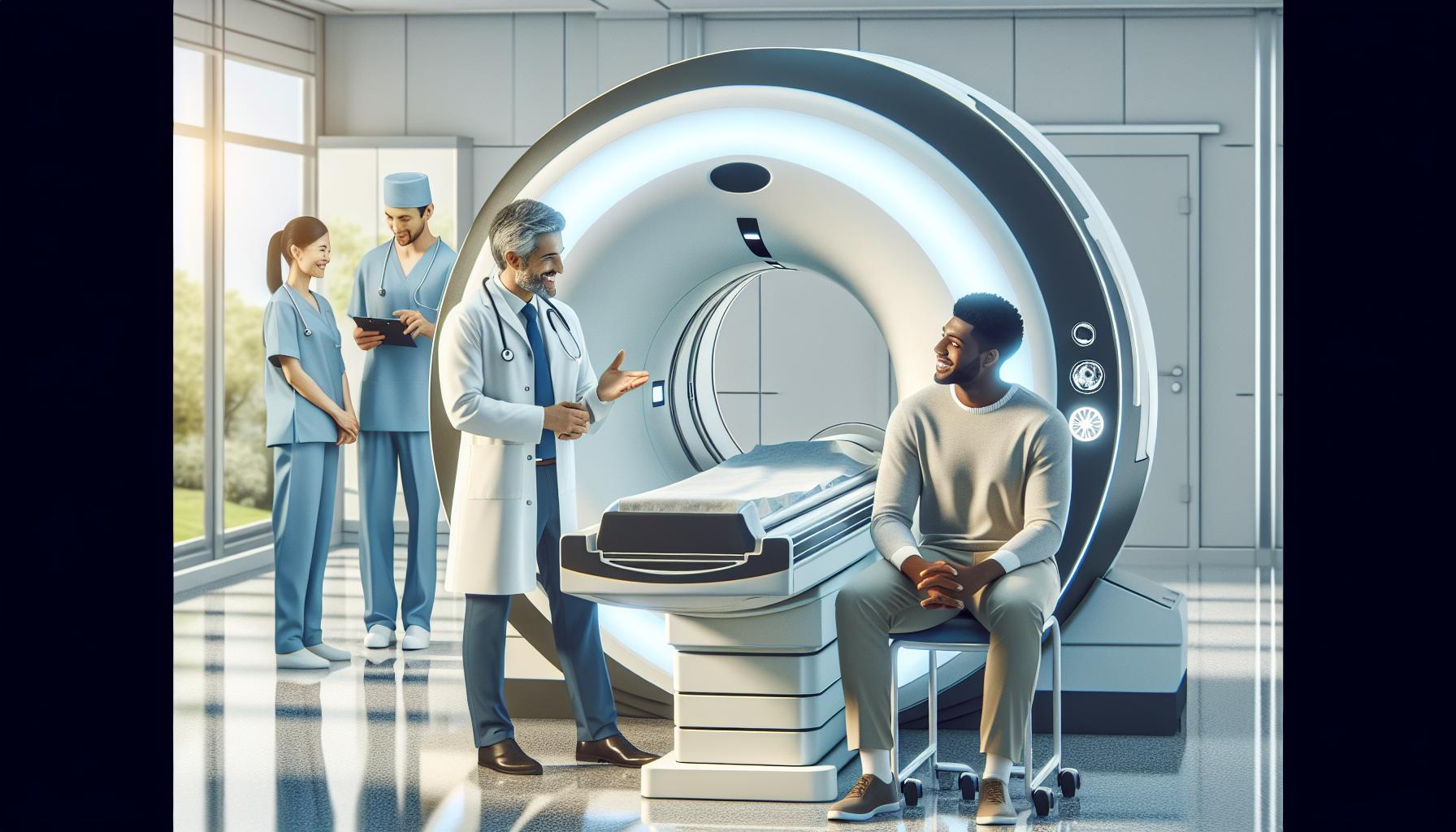
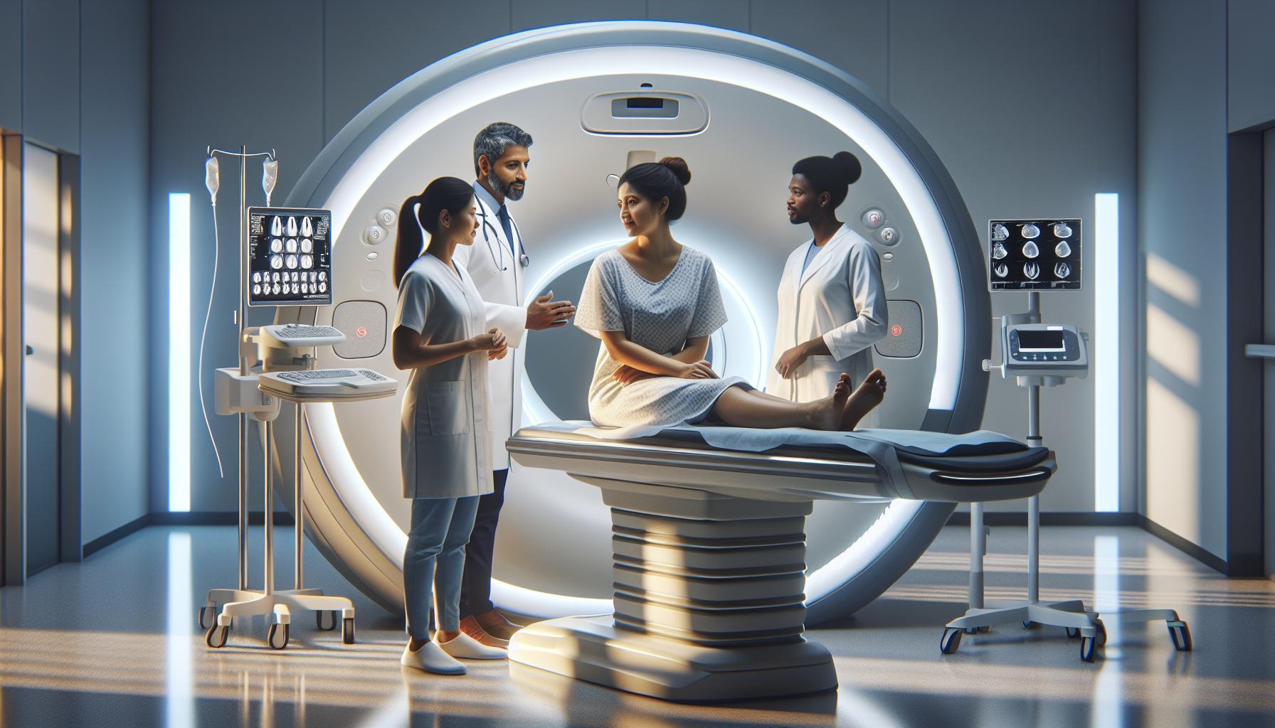
Every year, millions of individuals rely on CT scans for a closer look at their health, but how does this technology actually work? Computed Tomography (CT) combines multiple X-ray images taken from various angles to produce detailed cross-sectional views of the body, offering insights crucial for diagnosis and treatment planning.
Understanding CT scan technology not only highlights its pivotal role in modern medicine but also addresses common concerns around safety, preparation, and the procedure itself. As you dive into the intricacies of how CT machines operate, you’ll discover valuable insights that can alleviate anxiety, clarify expectations, and empower you to engage more fully in your healthcare journey. From the science behind image creation to practical tips for your visit, this article aims to demystify CT scans, providing assurance and clarity along the way.
Understanding the Basics of CT Scan Technology
A CT scan, or computed tomography scan, represents a groundbreaking advancement in medical imaging technology, significantly enhancing how healthcare providers visualize and diagnose health conditions within the body. This sophisticated procedure utilizes a series of X-ray images taken from many different angles around the body and processes them using computer algorithms to create cross-sectional images-often referred to as slices-of bones, blood vessels, and soft tissues. This level of detail not only enables the detection of abnormalities such as tumors, fractures, and internal bleeding but also allows for more precise assessments compared to traditional X-rays.
At the heart of a CT scan machine is a rotating X-ray tube that emits a narrow beam of radiation. As the tube rotates around the patient, sensors capture the X-rays that pass through the body, producing signals that a computer then transforms into images. The result is a comprehensive view of internal structures, presented in a series of two-dimensional cross-sections, which can be reconstructed into three-dimensional images for further analysis. This technology has fundamentally changed diagnostic practices, enabling earlier detection and intervention in various diseases, thereby improving patient outcomes.
Patients undergoing a CT scan can expect a straightforward process, typically lasting only a few minutes. They are usually asked to remain still on a sliding table that moves through the gantry of the machine. While the procedure itself is quick, preparation is essential; understanding what to expect and how to get ready can significantly reduce anxiety. For instance, patients may need to refrain from eating for a few hours beforehand or may receive a contrast agent, either orally or intravenously, to enhance the images’ clarity.
When considering a CT scan, it is also important to acknowledge the inherent risks associated with the use of ionizing radiation. However, the benefits of accurate diagnosis often outweigh these concerns. Engaging in open discussions with healthcare providers can help in understanding the technical aspects and the necessity of the procedure, ensuring informed decisions are made. As technology evolves, newer CT machines are being developed to minimize radiation exposure while maintaining high-quality imaging, reflecting a commitment to enhancing patient safety and care.
How CT Scan Machines Create Detailed Images
A fascinating aspect of CT scan technology is how it enables the creation of highly detailed images of the body’s internal structures. This precision is achieved through a combination of sophisticated hardware and advanced imaging techniques, all working together in a seamless process. When a patient is positioned in a CT scanner, the machine emits a thin beam of X-rays that rotates around them. This rotation allows the X-rays to penetrate the body at multiple angles, offering a comprehensive view of the area being examined.
As the X-ray beam passes through various tissues, some structures absorb more radiation than others. For instance, bones absorb a significant amount, appearing white on the resulting images, while softer tissues, such as muscles and organs, appear in varying shades of gray. This contrast in radiation absorption is crucial for generating clear, detailed images. The X-rays that pass through are captured by detectors opposite the X-ray tube, converting them into electrical signals. These signals are then processed by powerful computers that reconstruct the signals into two-dimensional cross-sectional images, or “slices.”
The images produced by a CT scan can be viewed individually or combined into three-dimensional representations, enabling healthcare providers to discern complex structures and phenomena within the body. This capability is particularly beneficial in identifying tumors, assessing internal injuries, or locating signs of disease. Furthermore, advancements in technology have led to the development of specialized algorithms that enhance image quality and reduce the amount of radiation exposure, thus providing safer procedures without compromising diagnostic accuracy.
Patients can take comfort in understanding that this intricate process is designed with their safety and well-being in mind. Every effort is made to ensure that the images are not only precise but also as gentle as possible. By engaging with healthcare professionals and discussing any concerns beforehand, individuals can feel more in control and informed about the procedure, reinforcing the collaborative nature of modern healthcare.
The Components of a CT Scan Machine Explained
The intricate design of a CT scan machine involves several key components that work in harmony to produce detailed images of the body’s internal structures. Understanding these components can alleviate some of the anxiety that patients may feel about the procedure. Each part of the CT scanner plays a crucial role in capturing high-resolution images that are essential for accurate diagnosis and treatment planning.
X-ray Tube
At the heart of the CT scanner lies the X-ray tube, which generates the X-rays necessary for imaging. This tube emits a beam of X-rays that rotates around the patient. As it moves, the tube produces images from various angles, which are critical for creating cross-sectional slices of the body. The rotating action is precision-engineered to ensure that overlapping images can be accurately integrated, resulting in a high-quality representation of the anatomy.
Detectors
Opposite the X-ray tube are the detectors. These sensitive devices capture the X-rays that pass through the body, converting them into electrical signals. Depending on the type of tissue that the X-rays encounter, different amounts of radiation will be absorbed, which the detectors interpret as varying levels of brightness in the images. This contrast allows healthcare professionals to differentiate between soft tissues, fat, and bones in the resulting scans, giving them a comprehensive view of the body’s internal landscape.
Operating Console
The operating console is where the technologist controls the entire scanning process. This interface allows for adjustments to be made based on the specific needs of the examination. Technologists can change parameters such as the scanning speed, rotation angle, and image resolutions according to the area being examined and the patient’s unique situation. This customization ensures that the procedure is both efficient and effective.
Computer System
Finally, the images captured by the detectors are processed by a computer system that reconstructs them into coherent cross-sectional images. Advanced algorithms are employed to enhance the quality of these images and reduce the noise that can obscure important details. The ability of the computer to merge data from multiple slices into three-dimensional representations enriches the diagnostic potential available to medical professionals.
In summary, the components of a CT scan machine-X-ray tube, detectors, operating console, and computer system-collaborate to create a powerful diagnostic tool that helps healthcare providers gain insights into a patient’s condition. Understanding how each part works can help ease the worries of patients preparing for a CT scan, providing reassurance that these technological marvels are designed with their health in mind. Always feel free to discuss any concerns with your healthcare provider to ensure you are comfortable with the procedure.
Step-by-Step: How a CT Scan Procedure Works
A CT scan procedure is a seamless orchestration of technology designed to capture intricate details of the body’s internal structures, often revealing critical information for diagnosis. Understanding the steps involved can help you feel more prepared and confident as you approach this essential medical imaging exam.
Initially, once you arrive for your appointment, a healthcare professional will guide you through the process. You’ll typically start by changing into a hospital gown that is free of any metallic objects or fasteners that could interfere with the imaging. It’s important to disclose any information regarding allergies, particularly to contrast materials, as some CT scans may use an injectable contrast agent to enhance image clarity.
Once you’re prepared, you’ll lie down on a motorized table that gently moves you into the CT scanner’s donut-shaped opening. During the scan, the X-ray tube will rotate around your body, capturing images from multiple angles. You may be asked to hold your breath briefly while the scanner is taking pictures to avoid any motion blur that could compromise the quality of the images. The process typically lasts anywhere from 5 to 30 minutes, depending on the type of scan and the area being examined.
Throughout the procedure, the radiologic technologist will monitor you from an adjacent control room, ensuring everything runs smoothly and effectively. After the scan concludes, you may be given instructions regarding post-procedure activities, such as dietary considerations if contrast was administered. Importantly, the images will be processed and reviewed by a radiologist, who will then provide a detailed report to your physician, aiding in your diagnosis or treatment planning.
By understanding these foundational steps, you can approach your CT scan with knowledge and peace of mind, knowing that this technology has evolved to prioritize both accuracy and patient comfort. Always remember, if you have any lingering questions or concerns, discussing them with your healthcare provider can help put your mind at ease and guide you through what to expect.
Preparing for Your CT Scan: What You Need to Know
Arriving for a CT scan can feel daunting, but understanding the preparation process can help ease any anxieties. A CT scan is a valuable diagnostic tool, and there are key steps to ensure that you have a smooth experience. First and foremost, it’s essential to inform your healthcare provider about any allergies you may have, particularly to iodine-based contrast materials if your scan requires one. This information is vital for your safety and for tailoring the procedure to your individual needs.
Prior to your appointment, you may receive specific instructions regarding diet and hydration. In some cases, you might be instructed to fast for a few hours or avoid certain foods. If contrast material will be used, drinking plenty of water beforehand can help your body process it more effectively. Make sure to wear comfortable clothing that is free from metal, as metal objects can interfere with the imaging process. Many facilities will provide a gown for you to change into, ensuring you are prepared for the scan.
When you arrive, check in at the reception desk, where you may need to fill out forms or verify your personal information. A radiologic technologist will explain the procedure to you, addressing any concerns you may have. Their goal is to make sure you are comfortable and informed throughout the process. If you’re feeling anxious, don’t hesitate to express your concerns; they are there to support you and can offer calming techniques.
As you settle onto the scanning table, it’s helpful to relax your body and mind. The machine may make noises as it operates, but remember this is a normal part of the scanning process. Following the technologist’s instructions during the scan-like holding your breath briefly-will enhance image clarity, leading to more accurate results. After the scan, you may resume your normal activities, although your healthcare provider will give you specific guidance based on the use of contrast material.
Understanding these preparation steps can empower you and provide peace of mind as you approach your CT scan. Remember, if you have any uncertainties or additional questions, your healthcare team is the best resource for personal guidance tailored to your specific situation.
Safety Considerations of CT Scans: Risks and Reassurances
CT scans, while incredibly valuable diagnostic tools, come with safety considerations that can understandably raise concerns for patients. One key aspect of CT imaging is the exposure to ionizing radiation, which is higher compared to other imaging modalities like X-rays. However, it’s important to note that the amount of radiation in a standard CT scan is typically considered safe, especially when weighed against the benefits of accurate diagnosis and treatment planning. Modern CT machines are designed to optimize imaging techniques to minimize radiation exposure while still providing high-quality images.
Understanding Risks vs. Benefits
When considering a CT scan, discussing the necessity and potential risks with your healthcare provider is critical. The decision to perform a CT scan is usually made when the potential benefits-such as detecting abnormalities that might not be visible with other imaging techniques-outweigh the risks associated with radiation exposure. Additionally, advancements in technology have led to the development of dose-reduction techniques, ensuring that patients receive only the minimum amount of radiation necessary to achieve accurate results.
Consulting with Healthcare Providers
It’s crucial to maintain open communication with your healthcare team regarding any fears or allergies you may have. If contrast materials are necessary for your scan-often used to enhance image clarity-inform your provider about previous reactions. For patients with renal conditions, special considerations may be necessary regarding the use of iodine-based contrast agents. Your healthcare team will determine the safest approach tailored to your medical history and current health status.
Post-Scan Reassurance
After the CT scan, there is often no immediate need for recovery, allowing you to resume normal activities soon after. However, if contrast dyes were used, your provider may offer specific instructions regarding hydration to help ensure the contrast material is safely processed and eliminated from your body. Always remember that your comfort and safety are top priorities for the healthcare staff, and they are there to answer your questions and address any anxieties you may have before and after the procedure.
By empowering yourself with knowledge about the risks and reassurances related to CT scans, you can approach the process with increased confidence and understanding. Always consult your healthcare provider for personalized advice and peace of mind regarding your health care choices.
Interpreting CT Scan Results: What to Expect
Receiving the results of a CT scan can be an anxious experience, but understanding the process can alleviate some of that stress. CT scans produce cross-sectional images of your body, allowing doctors to see internal structures in great detail. Once your scan is complete, the images are typically reviewed by a radiologist, a medical doctor trained to interpret imaging studies. They will look for any abnormalities, such as tumors, fractures, or other issues, and compile their findings into a report.
After the radiologist completes their analysis, they will send the report to your healthcare provider, who will discuss the results with you. It’s essential to understand that not all findings indicate a serious problem. For example, incidental findings-unrelated anomalies discovered inadvertently-may be completely benign. This is why open communication with your provider is crucial; they can help contextualize the results based on your medical history and symptoms.
Common Terminology in CT Reports
CT scan reports can contain terms that may be unfamiliar. Some common phrases include:
- Lesion: An abnormal area of tissue, which could be benign or malignant.
- Opacity: Represents a change in the density of a structure in the images.
- Enhancement: Refers to an increase in visibility of a structure after a contrast agent is used.
- Mass: A growth that may need further evaluation; not all masses are cancerous.
Your healthcare provider will explain these terms in detail, helping you understand their implications. In some cases, additional tests may be necessary to clarify findings or monitor a condition over time.
How to Prepare for Your Follow-Up
After reviewing your results, consider these steps to make the most of your follow-up appointment:
- Write down questions: Preparing a list of queries can help address your concerns during the discussion.
- Bring a family member or friend: Having someone accompany you can provide emotional support and ensure you remember key points from the conversation.
- Be honest about your symptoms: Share any ongoing issues or concerns that may help your doctor in forming a better assessment or treatment plan.
Always remember, the healthcare system is there to support you. By gaining a clearer understanding of your CT scan results, you can engage in informed discussions with your provider and feel empowered in making decisions about your health.
The Role of CT Scans in Modern Medicine
CT scans have revolutionized the way medical professionals diagnose and monitor various health conditions, providing critical insights that can guide treatment decisions. Unlike traditional X-rays, which offer limited two-dimensional views, CT scans generate detailed cross-sectional images of the body. This capability enables doctors to visualize internal organs, blood vessels, and tissues in high resolution, significantly enhancing diagnostic accuracy. Given the speed at which CT scans can be performed and processed, they are often the imaging method of choice in emergency situations, where time is of the essence.
The versatility of CT scans is one of their greatest strengths. They are employed for a myriad of purposes including detecting tumors, assessing internal injuries, and guiding minimally invasive procedures. For instance, in oncology, CT imaging can show the size and location of a tumor, aiding in the formulation of a tailored treatment plan. Similarly, in trauma cases, CT scans provide immediate visualization of internal bleeding, fractures, and organ damage, allowing for quick decision-making by healthcare providers.
Moreover, advancements in CT technology continue to enhance its role in modern medicine. Innovations such as dual-energy CT and iterative reconstruction techniques improve image quality while reducing radiation exposure. These advancements not only enhance diagnostic capabilities but also address patient safety concerns, making CT scans a more reliable imaging option. By fostering timely interventions and personalized treatment plans, CT scans contribute significantly to improving patient outcomes, reinforcing their central role in contemporary healthcare.
With the continual evolution of imaging technology, consulting with healthcare professionals about the most suitable imaging technique for individual needs is vital. By doing so, patients can better understand the potential benefits and risks associated with CT scans and make informed decisions regarding their health.
Cost of CT Scans: What Affects Pricing?
The cost of CT scans can vary significantly based on a variety of factors, making it essential for patients to be informed and prepared. Understanding what influences these prices can help demystify the process and alleviate concerns about unexpected expenses. Generally, the overall cost of a CT scan may range from several hundred to a few thousand dollars, depending on elements such as geographic location, the type of facility, and the specific scan being performed.
One primary factor affecting the price is the facility in which the CT scan is conducted. For instance, scans performed in a hospital setting often come with higher costs compared to free-standing imaging centers due to the added overhead associated with hospital services. Additionally, urban centers typically have higher rates compared to rural locations due to increased demand and operational costs. A compassionate approach would be to consult with your healthcare provider about the options available and their respective costs, as well as any potential financial assistance programs that might be offered.
The type of CT scan required also plays a crucial role in determining price. Specialized scans, such as those using contrast material or advanced techniques like dual-energy CT, are generally more expensive than routine scans. Moreover, each patient’s individual insurance coverage will significantly impact out-of-pocket expenses; understanding your insurance policy’s coverage for diagnostic imaging can provide clarity on expected costs.
In preparation for a CT scan, it’s also advisable to inquire about any additional fees, such as those associated with consultations, interpretation of results, or even follow-up appointments. Structured communication with your healthcare provider about these aspects can ensure you are informed and prepared, helping to ease any concerns or anxiety you may have regarding the financial implications of your CT scan. Remember, accessing detailed explanations about these factors not only aids in financial planning but also empowers you during your healthcare journey.
Advancements in CT Technology: What’s New?
Recent advancements in CT technology have revolutionized the way we understand and utilize imaging in medicine, providing clearer and more detailed images while also enhancing patient safety and comfort. One notable development is the introduction of multislice or multidetector CT scans, which allow for the capture of multiple slices of images in a single rotation. This capability significantly speeds up the scanning process, making it particularly beneficial for emergency situations where time is critical. Patients can be scanned quickly while reducing motion artifacts, leading to more accurate results.
Another exciting advancement is the evolution of dual-energy CT (DECT). This technology uses two different energy levels of X-rays to capture images, allowing radiologists to differentiate between various tissues and materials in the body more effectively. DECT can be used to identify the composition of kidney stones, characterize tumors, or enhance the accuracy of diagnosing pulmonary embolism, providing invaluable data that can guide treatment decisions. The ability to obtain such nuanced details helps medical professionals tailor therapies to individual patients’ needs more effectively.
In addition to these technological advancements, there is a growing focus on radiation dose reduction techniques. Innovations in software and hardware design have enabled clinicians to conduct scans with significantly lower radiation exposure without compromising image quality. Techniques like automatic exposure control adjust the radiation dose based on the specific body part being scanned, ensuring that patients receive the minimal necessary exposure while still obtaining high-quality images.
As these technologies continue to evolve, patients can expect even more personalized and precise diagnostic imaging. It is essential to discuss with your healthcare provider the latest advancements in CT technology and how they might apply to your specific situation, ensuring that you receive the best possible care tailored to your needs.
Comparing CT Scans with Other Imaging Techniques
When it comes to medical imaging, understanding how different techniques stack up against one another can be daunting. CT scans, known for their speed and high-resolution images, stand out compared to conventional X-rays, MRI, and ultrasound. Each method has unique strengths and limitations, making it crucial for patients and healthcare providers to choose the appropriate imaging modality based on the clinical scenario.
CT scans utilize X-rays to create cross-sectional images of the body, providing detailed views of organs, bones, and tissues. This rapid imaging capability is particularly beneficial in emergency situations, where swift, reliable diagnostics are essential. In contrast, conventional X-rays provide limited information and are primarily used for examining bone fractures or detecting abnormalities in the chest. While X-rays involve less radiation exposure, they lack the comprehensive insights that CT imaging offers.
Magnetic Resonance Imaging (MRI) employs powerful magnets and radio waves instead of X-rays, producing images that excel in depicting soft tissue structures such as the brain, muscles, and ligaments. Although MRIs take longer than CT scans and require the patient to remain still, they offer superior contrast for soft tissues and do not involve radiation exposure. Patients with metal implants or certain medical devices should be cautious with MRIs due to the magnetic field used in the imaging process.
Ultrasound imaging, known for its safety, especially during pregnancy, utilizes sound waves to create images. This method is particularly effective for examining soft tissues and organs, such as the liver or heart, and is often used in obstetrics. However, ultrasound is operator-dependent and may not provide the detail required for deeper structures compared to a CT scan. In some cases, ultrasound can be a first step before a more detailed imaging study like a CT or MRI is recommended.
It’s crucial to consult with healthcare professionals to determine the most suitable imaging technique based on individual health needs and specific clinical conditions. The choice often balances the need for detailed imaging, the type of tissue being examined, and considerations regarding radiation exposure. Understanding these nuances can empower patients to ask informed questions and participate actively in their healthcare decisions.
Common Misconceptions About CT Scans Debunked
Despite their importance in medical diagnostics, CT scans are often surrounded by misconceptions that can lead to unnecessary anxiety among patients. One common myth is that CT scans are extremely dangerous due to radiation exposure. While it’s true that CT scans involve a higher dose of radiation compared to conventional X-rays, it’s essential to understand the context. The benefits of a CT scan-such as the ability to detect life-threatening conditions quickly-frequently outweigh the risks. Moreover, advancements in CT technology have led to significant reductions in radiation doses, enhancing patient safety without compromising image quality.
Another prevalent misconception is that CT scans can be performed without any specific preparation. In reality, depending on the type of scan (e.g., abdominal or pelvic), patients may need to avoid certain foods or drinks beforehand to ensure the best imaging results. Your healthcare provider will provide instructions tailored to your specific procedure, which might include fasting for several hours before the scan. This preparation minimizes the chances of artifacts in the imaging, ensuring that your physician receives the clearest possible images for accurate diagnosis.
Some individuals also believe they will feel considerable discomfort during or after a CT scan. The process is typically quick and painless, lasting only a few minutes. The machine may make some noises, and you may be asked to hold your breath for short intervals, but this is standard practice to help create clear images. If contrast dye is used to enhance the images, it can cause a brief warming sensation or mild side effects, but serious reactions are rare. Understanding the procedure can significantly reduce anxiety and foster a more relaxed experience.
In summary, it’s crucial to dispel the myths surrounding CT scans to empower patients with accurate information. Consultation with healthcare professionals can clarify any doubts and provide personalized advice. By staying informed and understanding the realities of CT technology, patients can approach their diagnostic imaging with confidence, knowing that they are taking a proactive step toward their health.
Q&A
Q: How does a CT scan machine generate images?
A: A CT scan machine generates images by using a combination of X-rays and computer processing. The machine rotates around the patient, taking multiple X-ray images from different angles, which are then processed to create detailed cross-sectional images of the body.
Q: What are the main components of a CT scanner?
A: A CT scanner consists of several key components: a rotating X-ray tube, a detector array, a computer for image reconstruction, and a control console. Together, these components work to acquire and analyze the data necessary for producing detailed images of internal structures.
Q: Why is contrast media used in CT scans?
A: Contrast media enhances the visibility of certain body parts during a CT scan. It helps differentiate between various tissues and blood vessels, allowing for clearer imaging of areas like the brain, abdomen, and lungs, thus improving diagnostic accuracy.
Q: What preparations are needed before a CT scan?
A: Before a CT scan, patients may need to avoid eating or drinking for a few hours. If contrast media is used, inform your healthcare provider about any allergies. Patients should wear loose, comfortable clothing and may need to remove metal objects that can interfere with imaging.
Q: What should I expect during a CT scan procedure?
A: During a CT scan, you will lie on a movable table that slides into the scanner. You may hear whirring noises as the machine rotates. It’s essential to stay still and follow breathing instructions from the technician to ensure clear images are captured.
Q: Are there any risks associated with having a CT scan?
A: The primary risk associated with a CT scan is exposure to ionizing radiation. While the radiation dose is generally low and the risk of harm is minimal, it’s important to discuss any concerns with your healthcare provider, especially if you require multiple scans.
Q: How are CT scan results interpreted?
A: CT scan results are interpreted by a radiologist, who reviews the images for any abnormalities. They provide a detailed report to your physician, who will discuss the findings and any necessary follow-up steps or treatments based on the results.
Q: How does CT scan technology compare to MRI?
A: CT scans are generally faster and better for imaging bones and detecting certain conditions like tumors or bleeds. MRI uses magnetic fields and is ideal for soft tissue imaging, such as the brain and muscles, without using ionizing radiation. Each has unique advantages depending on the clinical scenario.
For more detailed information, refer to sections like “Understanding the Basics of CT Scan Technology” and “Preparing for Your CT Scan: What You Need to Know” in our main article.
Wrapping Up
As we’ve explored, understanding how a CT scan machine functions is crucial for patients preparing for this important imaging procedure. The technology not only provides detailed insights into your health but also enhances diagnostic accuracy, making it a valuable tool in modern medicine. If you have further questions or concerns about your upcoming CT scan, remember to consult your healthcare provider for personalized advice.
To dive deeper into related topics, check out our articles on “Preparing for Your First CT Scan” and “Understanding the Costs and Safety of Medical Imaging.” For ongoing updates and information, consider subscribing to our newsletter or reaching out for a consultation. Your health is paramount, and having the right information can empower your decisions. Explore these resources to enhance your knowledge and feel more confident about your medical journey!


