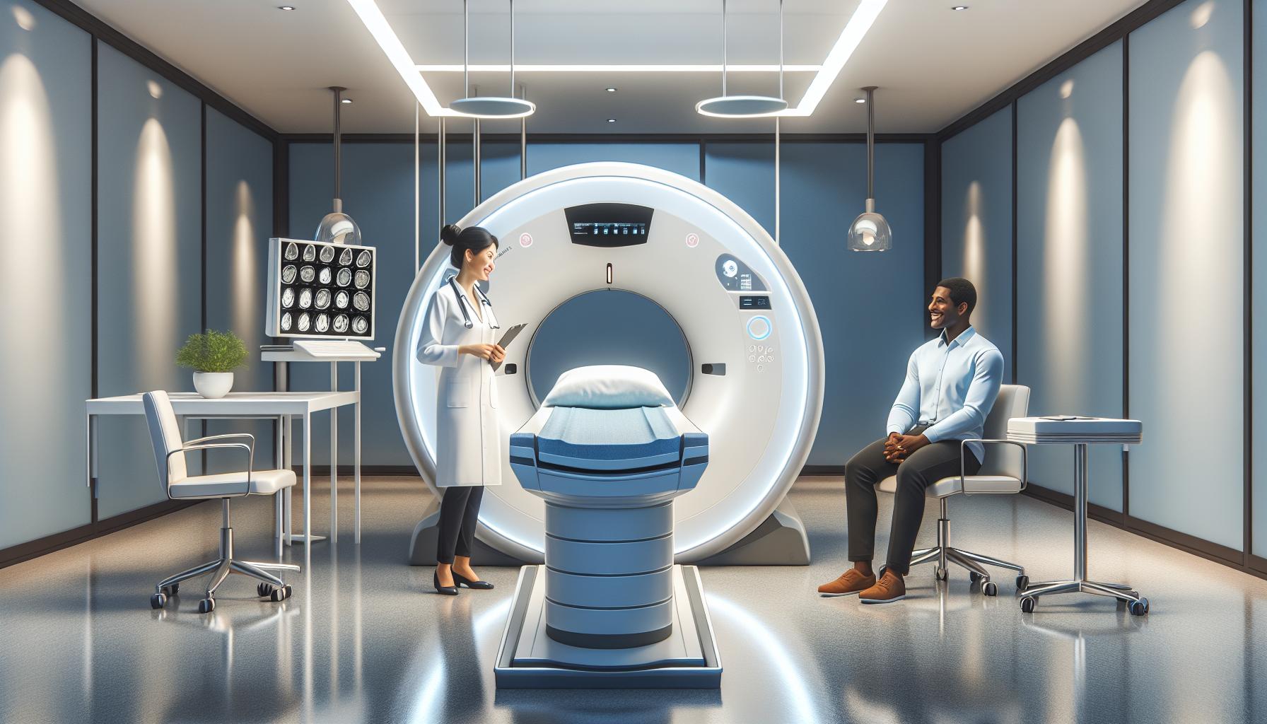
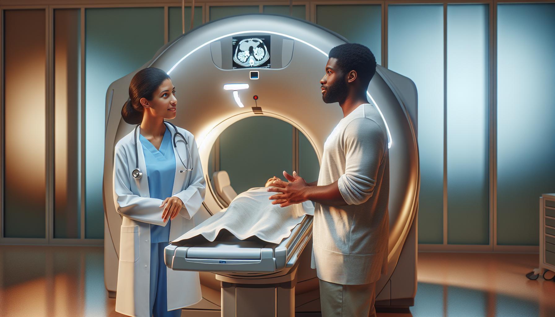
When it comes to medical imaging, confusion often arises between CT scans and MRIs. Both are crucial diagnostic tools used to visualize the body’s internal structures, yet they differ significantly in technology and application. Understanding these differences is vital for making informed healthcare decisions.
Did you know that while CT scans utilize X-rays to capture detailed images of organs and tissues, MRIs rely on magnetic fields and radio waves? This distinction can greatly affect the types of injuries or conditions diagnosed. As you navigate your healthcare options, it’s essential to grasp the unique benefits and limitations of each method.
Whether you’re preparing for a procedure or simply seeking knowledge for future reference, this article will clarify the key differences between CT scans and MRIs, empowering you with the information needed to discuss your imaging options confidently with your healthcare provider. Join us as we explore the essential insights that could impact your medical care.
Understanding the Basics: What Is a CT Scan?
A CT scan, or computed tomography scan, is a crucial imaging tool that uses X-ray technology to create detailed cross-sectional images of the body. This advanced technique allows healthcare providers to visualize internal organs, tissues, and structures in great detail, making it invaluable for diagnosing various medical conditions. In fact, CT scans can provide clearer images than standard X-ray examinations, particularly for complex areas such as the brain, chest, and abdomen.
During the CT scan procedure, patients lie on a table that slowly moves through a circular machine called a CT scanner. This machine takes a series of X-ray images from different angles, which a computer then processes to produce cross-sectional images-or “slices”-of the body. These images can reveal a wide range of issues, from tumors and internal bleeding to infections and bone fractures. Importantly, CT scans can often be completed quickly, making them vital in emergency situations where time is of the essence.
How It Works
The technology behind CT scans involves a series of steps that enhance patient safety and imaging accuracy:
- X-ray Generation: The CT machine uses X-ray beams to capture images from various angles.
- Image Processing: A computer processes the information to create detailed images of the body’s internal structures.
- Reconstruction: The images are reconstructed to create 2D or 3D views, giving doctors a comprehensive understanding of the areas being examined.
Patients may be required to stay still during the scan, and in some cases, a contrast material may be administered to enhance the visibility of certain tissues or blood vessels. While the procedure is generally safe, understanding the technology and its applications can help alleviate any anxiety about the experience.
CT scans play a vital role in modern medicine, providing essential information that can guide diagnosis and treatment. As with any medical imaging procedure, it’s important to discuss any concerns or questions with healthcare professionals, who can offer tailored advice based on individual health needs.
Unveiling MRI: How It Works and Its Uses
Magnetic Resonance Imaging (MRI) is a powerful non-invasive imaging technique that utilizes strong magnetic fields and radio waves to produce detailed images of organs and tissues within the body. Unlike CT scans, which harness X-ray technology, MRI provides exceptionally high-contrast images, particularly of soft tissues, making it an indispensable tool in medical diagnostics. With no known harmful effects from the magnetic fields used, MRI is especially favored for imaging the brain, spinal cord, muscles, and joints.
During an MRI scan, patients lie on a table that slides into a large, cylindrical magnet. It’s essential to remain as still as possible for the duration of the scan, which typically lasts between 20 to 60 minutes depending on the area being examined. In many cases, a contrast agent may be injected into the bloodstream to enhance the visibility of certain tissues. While some might feel apprehensive about the confined space of the MRI machine, it’s important to note that most machines are designed to be open or have a wider bore that can accommodate those who may experience claustrophobia.
One of the standout advantages of MRI is its ability to provide detailed images without the use of ionizing radiation, which is a concern with CT scans. Furthermore, MRI can capture multiple planes of the body in a single scan, giving medical professionals a comprehensive view of the area in question. This capability makes MRI an exceptional choice for diagnosing various conditions, such as tumors, brain disorders, and joint injuries.
As patients prepare for an MRI, they should wear loose, comfortable clothing without metal fasteners, and inform the medical staff about any implants, pacemakers, or other medical devices, as these can interact with the MRI’s magnetic field. Engaging in a conversation with healthcare providers can mitigate anxiety and clarify any questions about the procedure, enhancing the overall experience and ensuring a smooth imaging process.
Key Differences Between CT and MRI Scans
A critical aspect of medical imaging lies in understanding the distinct roles of CT scans and MRIs, as both techniques serve different purposes in diagnosing a range of conditions. CT, or Computed Tomography, utilizes X-ray technology to produce detailed cross-sectional images of the body. This method is particularly effective for quickly assessing injuries, detecting cancers, or visualizing complex fractures. The speed of a CT scan-often completed in minutes-makes it invaluable in emergency situations where immediate results are required.
In contrast, Magnetic Resonance Imaging (MRI) employs powerful magnets and radio waves to generate high-resolution images, especially of soft tissues. This makes MRIs exceptionally suited for evaluating neurological conditions, joint abnormalities, and other soft tissue diseases that may not show up as effectively on a CT scan. While MRI scans typically take longer and are more sensitive to patient movement, they provide clearer and more informative images without the risks associated with ionizing radiation.
When discussing safety, it’s important to note that CT scans expose patients to a small dose of ionizing radiation, which has been a point of concern, especially in repeated imaging studies. In contrast, MRI scans do not use radiation, making them a safer option for many patients. However, patients with certain metallic implants or pacemakers may not be eligible for an MRI due to the strong magnetic fields used in the scanning process.
Ultimately, the choice between a CT scan and an MRI often depends on the specific clinical question at hand, the area of the body being examined, and the patient’s medical history. Understanding these differences can help patients feel more informed and empowered when discussing imaging options with their healthcare providers. Always consult with a medical professional to determine the most appropriate imaging technique based on individual health needs.
Safety Considerations: Comparing Risks and Side Effects
The choice between a CT scan and an MRI often sparks questions regarding safety risks and side effects. Understanding how these imaging techniques function enables patients to make informed decisions about their health. CT scans, while crucial for swift assessments, use ionizing radiation to create detailed images. This exposure, although minimal in single instances, raises concerns about cumulative effects over time, particularly for individuals requiring frequent scans. It’s crucial for patients to discuss their imaging history with healthcare providers, ensuring a balanced approach considering the diagnostic benefits against potential risks.
On the other hand, MRIs employ radio waves and strong magnetic fields without any exposure to radiation, making them a compelling choice for many patients. However, certain safety precautions must be observed. Patients with metallic implants, like pacemakers or metal plates, might be at risk due to the powerful magnets used in the MRI process. In some cases, patients may experience claustrophobia during MRI scans, as the machine is typically enclosed. Providers often mitigate these feelings by offering open MRIs or using calming techniques to help patients relax.
It’s important to note that both imaging modalities have their unique risks but are generally safe when used appropriately. Potential side effects from CT scans may include allergic reactions to contrast dyes, whereas MRIs may cause discomfort from prolonged immobility and occasionally induce anxiety. Patients should prepare adequately, ensuring they communicate their medical history, allergies, and any current medications to their healthcare provider. This collaborative dialogue can help tailor the most appropriate imaging strategy while prioritizing patient safety and comfort.
In summary, patients should weigh the risks and advantages of both types of imaging techniques, maintaining open lines of communication with their healthcare team. Knowledge is empowering, and understanding these safety considerations can alleviate some anxiety associated with medical imaging. Always consult with a medical professional for personalized advice that reflects individual health needs.
Costs Involved: CT Scan vs. MRI Pricing Breakdown
The financial aspect of medical imaging can often be a source of concern for many patients facing the decision of whether to undergo a CT scan or MRI. Understanding the potential costs involved can alleviate some anxiety and aid in decision-making. Generally, CT scans are more affordable than MRIs, but the actual costs can vary significantly based on factors such as location, the facility’s pricing structure, and whether contrast material is needed.
Typically, the average cost of a CT scan can range from $300 to $1,500, depending on the complexity of the scan and the area of the body being examined. This cost may be influenced by additional factors, such as the need for contrast dye, which can increase expenses. MRIs, on the other hand, tend to be pricier, with a range from $400 to $3,500. The higher cost of MRI scans is often attributed to the more advanced technology required to produce detailed images without ionizing radiation.
Insurance and Payment Considerations
For many patients, insurance coverage plays a crucial role in determining out-of-pocket expenses. It’s vital to consult with your insurance provider to understand your specific coverage limits, deductibles, and how much they will reimburse for each type of imaging test. Some facilities also offer payment plans or financial assistance to help manage costs, which can be particularly beneficial for those facing multiple scans.
- Always check with your healthcare provider: Discuss the necessity of the imaging test and whether alternative options (that may be more cost-effective) are available.
- Ask about facility fees: Costs can vary between hospitals and outpatient imaging centers, so inquire whether there are differences in pricing.
- Consider the total cost: This includes not only the actual imaging but also potential follow-up appointments, additional imaging, or treatments based on the results.
With careful consideration of these factors, patients can better prepare financially for medical imaging procedures, reducing unexpected financial burdens. As always, prioritizing open discussions with healthcare professionals about medical needs and costs is essential in making informed and confident decisions.
Preparation Steps: Getting Ready for Your Scan
To ensure a smooth experience during your imaging procedure, preparing for a CT scan or MRI is essential. Understanding what each scan entails allows you to approach the process with confidence. Before your appointment, familiarize yourself with the specific requirements, as these can differ significantly between a CT scan and an MRI.
For a CT scan, it’s common to be asked to avoid eating or drinking for several hours prior to the exam, particularly if contrast material will be used. This contrast agent helps enhance the images and may be administered orally or through an injection. Be sure to discuss any allergies or health conditions with your healthcare provider, especially if you have a history of reactions to iodine or contrast materials. Additionally, wearing loose-fitting clothing is advisable; you may be given a gown to wear during the procedure to ensure there are no metal objects that could interfere with the imaging.
In contrast, MRI preparations may require you to remove all metal items, including jewelry and watches, as the strong magnetic field can attract these objects. Some facilities may also request that you wear a gown. Depending on the study, you may be required to remain still for an extended period; practicing relaxation techniques beforehand can be beneficial to reduce anxiety during the scan. Discuss any claustrophobia concerns with your provider, as some facilities offer open MRI machines that may be less confining.
Engaging with your healthcare team is crucial for a well-prepared journey. Don’t hesitate to reach out for answers to any lingering questions you may have about the exam or preparation steps. Your comfort and understanding of the process are paramount, as informed patients often experience less anxiety during their medical imaging procedures.
Interpreting Results: How CT and MRI Findings Differ
In the realm of medical imaging, understanding the nuances of how CT and MRI results are interpreted can significantly impact treatment decisions and patient outcomes. Both imaging modalities provide detailed views of the body’s internal structures, but they do so in fundamentally different ways, resulting in varying interpretations of findings.
CT scans utilize X-ray technology to produce cross-sectional images of the body. They are particularly effective in visualizing bone structures and detecting acute conditions such as fractures, internal bleeding, or tumors. The images generated by CT scans present a clear representation of the body’s anatomy, often allowing doctors to quickly identify abnormalities. For instance, a CT scan may reveal a fracture by highlighting discontinuities in the bone structure, while soft tissues can often be characterized using contrast agents that improve visibility. However, the interpretation of CT results must consider radiation exposure and potential false positives or negatives due to overlapping structures.
On the other hand, MRI scans employ strong magnetic fields and radio waves to generate detailed images, particularly of soft tissues, ligaments, and cartilage. MRI is unparalleled in assessing conditions like brain tumors, spinal cord injuries, or joint issues. The interpretation of MRI results can often involve looking for subtle changes in tissue characteristics, such as variations in water content, that indicate pathology. For example, a herniated disc may be identified through the displacement of spinal structures, which an MRI can visualize with remarkable clarity. Furthermore, because MRI does not use ionizing radiation, its findings may lead to a different risk-benefit analysis than CT results.
When it comes to clinical decision-making, the differences in interpretation bear significant weight. Medical professionals often rely on specific imaging techniques based on the suspected condition. A CT scan might be preferred for a rapid assessment of trauma, while an MRI would be more appropriate for chronic pain or soft tissue assessments. Ultimately, collaborating with a healthcare provider ensures that interpretations are not merely technical readings but are integrated into a patient’s broader health narrative.
In conclusion, while CT and MRI scans have distinct roles in medical imaging, understanding their interpretation is crucial for comprehensive patient care. Consulting with qualified healthcare professionals who can explain these differences in relation to individual health needs is key in navigating any medical imaging process. With knowledge comes empowerment, making it easier for patients to engage in informed discussions about their care path.
Common Conditions Diagnosed by Each Imaging Type
In today’s fast-paced medical environment, accurate diagnoses are crucial, and imaging technologies play a pivotal role in this process. Both CT and MRI scans are invaluable tools for healthcare providers, each uniquely suited to identify specific conditions based on their imaging strengths. Understanding the common conditions diagnosed by each modality can empower patients to engage with their healthcare provider effectively.
CT scans excel in assessing acute medical conditions, particularly those requiring rapid evaluation. They are often the first choice in emergency settings and can quickly identify:
- Fractures: CT scans provide detailed images of complex bone structures, making them ideal for identifying fractures that might not be visible in conventional X-rays.
- Internal Bleeding: The high-speed imaging of CT scans allows for quick detection of internal hemorrhages, particularly in trauma patients.
- Pulmonary Emboli: CT pulmonary angiography is a critical tool in diagnosing blood clots in the lungs.
- Tumors: CT scans effectively visualize the size and location of tumors in organs, aiding in cancer diagnosis and treatment planning.
Conversely, MRI scans shine when it comes to evaluating soft tissues and structures where subtle differences are paramount. MRI is often more suitable for conditions such as:
- Brain Tumors: MRI provides unparalleled detail of brain anatomy and is essential in diagnosing and staging brain tumors.
- Spinal Cord Injuries: MRI can assess soft tissue and the spinal cord, enabling clinicians to differentiate between various types of spinal injuries.
- Joint Disorders: Conditions such as torn ligaments or cartilage issues are better visualized with MRI, making it a preferred choice for orthopedic evaluations.
- Soft Tissue Masses: MRI is exceptionally good at distinguishing between normal and pathological tissues, assisting in diagnosing various types of soft tissue tumors.
In both cases, the choice between a CT scan and an MRI will depend on the patient’s specific symptoms, medical history, and the urgency of the situation. Consulting a healthcare provider can help clarify which imaging modality is appropriate based on individual health concerns and the suspected condition, ensuring the most effective diagnosis and subsequent treatment. Understanding these differences not only alleviates anxiety surrounding medical imaging but also fosters a proactive approach to one’s health.
When to Choose a CT Scan or MRI: A Guide
Choosing the right imaging technique can be pivotal in ensuring an accurate diagnosis. In many scenarios, the fast-paced nature of healthcare demands expedient decision-making, particularly in emergency situations. CT (computed tomography) scans are frequently employed in trauma cases where immediate analysis is essential. For instance, if there is a suspicion of internal bleeding from a car accident, a CT scan can quickly reveal the issue, allowing for prompt intervention.
On the other hand, MRI (magnetic resonance imaging) offers comprehensive details necessary for more nuanced assessments. Suppose a patient presents with unexplained headaches; an MRI can provide critical insights into potential brain tumors or soft tissue abnormalities, delivering a clearer picture of the brain’s structure compared to a CT scan.
Here are key considerations when deciding between CT and MRI:
- Nature of the Condition: If a condition is suspected to involve bones or is acute-such as fractures or bleeding-a CT scan is often preferred due to its speed and detail.
- Soft Tissue Evaluation: For conditions requiring examination of soft tissues, such as muscles, ligaments, and organs, an MRI is the superior choice due to its high contrast resolution.
- Patient Factors: Considerations such as a patient’s medical history, allergies (particularly to contrast agents used in CT), and claustrophobia can influence the decision.
- Urgency: In scenarios requiring rapid diagnosis and intervention, such as stroke or trauma cases, CT scans are typically employed first.
Ultimately, it is crucial to consult with a healthcare provider who can assess individual health concerns and tailor the imaging approach to best meet diagnostic needs. This collaborative decision-making process not only enhances the accuracy of diagnoses but also alleviates patient anxiety, ensuring a clear understanding of what to expect from each imaging modality.
Technological Advances: Innovations in Imaging
The landscape of medical imaging is rapidly evolving, with innovations enhancing both the quality and efficiency of CT and MRI scans. One significant advancement is the development of ultra-fast CT scanners, which can complete scans in a fraction of the time compared to traditional models. This not only reduces the duration of the procedure for patients but also minimizes motion artifacts, resulting in clearer images. These high-speed scans are particularly crucial in emergency situations where every second counts, allowing healthcare providers to make swift decisions in life-threatening conditions.
Another groundbreaking technology is the advent of dual-energy CT. This method utilizes two different energy levels of X-rays to improve tissue differentiation, providing enhanced contrast resolution. It allows radiologists to identify various tissue types more accurately and can even help in characterizing lesions or assessing vascular conditions effectively. This innovation exemplifies how CT technology is becoming capable of more sophisticated analyses that were previously only possible with MRI.
In the realm of MRI, one of the most notable advancements is the development of functional MRI (fMRI), which visualizes brain activity in real time by measuring changes in blood flow. This technology is invaluable for diagnosing neurological disorders, studying brain function, and planning surgeries with precision. Additionally, innovations like 7-Tesla MRI machines are pushing the boundaries of resolution, offering unprecedented clarity and detail, thereby allowing for earlier detection and treatment of conditions.
Patient comfort is also a growing focus in imaging technology. New MRI machines are designed with wider openings and quieter operation, addressing claustrophobia concerns that many patients experience. These enhancements not only improve the patient’s experience but also make them more likely to participate in necessary examinations, ultimately contributing to better health outcomes.
As these technologies continue to develop, they hold the promise of not just improving the quality of imaging but also fostering a stronger collaborative environment between patients and healthcare providers. Staying informed about these advancements empowers patients to engage in their healthcare decisions, ensuring they receive the most appropriate and effective imaging techniques tailored to their specific needs. Always consult your healthcare provider to understand how these innovations might apply to your situation, relieving any uncertainties surrounding the imaging process.
Patient Experiences: What to Expect During Your Scan
Undergoing a CT or MRI scan can feel daunting, but understanding what to expect can help alleviate any anxiety. Both procedures are vital diagnostic tools used by healthcare professionals to visualize the internal structures of the body. While CT scans utilize X-rays to create detailed cross-sectional images, MRI relies on magnetic fields and radio waves, producing remarkably clear images of soft tissues.
Preparation begins before you even arrive at the facility. Depending on the scan type, you might be advised to avoid eating or drinking for a few hours prior, especially for a CT scan where contrast materials might be used. Be sure to inform your healthcare team if you have any allergies, are pregnant, or are taking medications. It’s also beneficial to wear comfortable clothing without metal fasteners, as metal can interfere with both CT and MRI imaging.
When you arrive, you’ll check in at the reception and may fill out paperwork regarding your medical history. After that, a technician will explain the procedure, making sure you understand every step. For a CT scan, you’ll be positioned on a table that slides into the scanner. You might be asked to hold your breath for a moment while the images are captured-this is typically a quick process, lasting only a few minutes. In the case of an MRI, you lie on a table that moves into a large, tube-shaped magnet. It’s advised to remain still during the scan, and you may hear loud thumping noises from the machine, which is completely normal.
Throughout the process, it’s essential to communicate with your healthcare provider. If you feel discomfort or anxiety, let them know; they can provide guidance or support. Sometimes, headphones or relaxation techniques are offered to enhance comfort during the scan. In most cases, the procedure concludes within 30 minutes to an hour, after which you can resume normal activities.
The results will typically be reviewed by a radiologist, who will prepare a report for your doctor. Make sure to follow up with your healthcare provider to discuss the findings and any next steps based on the results. Being well-informed about the experience can significantly ease the process, turning a potentially stressful situation into a manageable and productive one.
Common Myths and Misconceptions About Imaging Techniques
Many people express concerns about imaging techniques like CT scans and MRIs due to various myths and misconceptions circulating in popular discourse. One prevalent belief is that these two diagnostic tools are interchangeable; while both are essential for imaging, they serve different purposes and use entirely different technologies. CT scans employ X-rays to create detailed anatomical images, particularly useful for visualizing bone structures and certain internal injuries, whereas MRIs utilize strong magnetic fields and radio waves to generate exquisite images of soft tissues, making them ideal for assessing brain, spinal cord, and joint conditions.
Another common myth is that CT scans are always dangerous due to radiation exposure. While it’s true that CT scans involve ionizing radiation, the amount is often comparable to the exposure from natural background radiation over a few years. Healthcare providers weigh the benefits against the risks, ensuring scans are performed only when clinically necessary. Furthermore, advancements in technology continually minimize radiation doses while maximizing image quality, so patients can feel reassured regarding the safety measures in place.
Many also believe that MRIs are not suitable for individuals with any metallic implants or devices. While it’s essential to inform your healthcare provider about such implants, many devices are now designed to be MRI-safe. This means that individuals with certain types of metal implants can still undergo MRI scans without significant risk. Nevertheless, some devices may still pose a concern, and it’s crucial to adhere to pre-scan protocols to ensure safety.
Lastly, there’s a misconception that both imaging techniques are painful or uncomfortable. In reality, while MRIs can involve some discomfort due to the enclosed space and noise of the machine, the scan itself is non-invasive. CT scans are often completed in just a few minutes with less discomfort involved. By understanding the truth behind these myths, patients can approach their imaging appointments with a clearer perspective, ultimately alleviating anxiety and improving their overall experience. Always consult with your healthcare professional about any concerns to receive personalized guidance tailored to your specific situation.
Q&A
Q: What are the advantages of using a CT scan over an MRI?
A: CT scans are faster, making them ideal for emergency situations, as they provide quick images of injuries or internal bleeding. They also excel in imaging bone structures and detecting certain cancers, providing detailed cross-sectional views. For more on the differences, see our section on Key Differences Between CT and MRI Scans.
Q: Are CT scans safer than MRIs?
A: Generally, MRIs do not use ionizing radiation, making them safer compared to CT scans that involve radiation exposure. However, CT scans can be necessary in certain situations where speed is essential. For safety considerations, refer to our Safety Considerations section for detailed insights.
Q: How does the cost of CT scans compare to MRIs?
A: CT scans are typically less expensive than MRIs. The cost can vary based on the facility and the complexity of the procedure. Detailed pricing breakdowns can be found in our Costs Involved section, helping you compare the two.
Q: How long does a CT scan take compared to an MRI?
A: A CT scan usually takes about 10 minutes or less, while an MRI can take anywhere from 30 minutes to over an hour, depending on the area being scanned. If time is a concern, refer to our preparation steps for insights on what to expect.
Q: What conditions are better diagnosed with a CT scan versus an MRI?
A: CT scans are better for detecting bone fractures, certain tumors, and internal bleeding, while MRIs are preferred for soft tissue evaluation, including brain, spine, and joint issues. Learn more about common conditions in our Common Conditions Diagnosed by Each Imaging Type section.
Q: Can MRI and CT scans be used together for diagnosis?
A: Yes, both imaging modalities can complement each other. A physician may use a CT scan to identify initial issues and an MRI for detailed soft tissue assessments. This approach maximizes diagnostic accuracy; see Interpreting Results for more detailed information.
Q: What should I wear during a CT scan or MRI?
A: For both scans, patients should wear comfortable, loose-fitting clothing without metal accessories. Most facilities provide gowns when necessary. Check our Patient Experiences section for more tips on what to expect during your scan.
Q: Can patients with metal implants have an MRI?
A: It depends on the type of implant. Most modern implants are MRI-safe, but certain implants could pose risks. Always consult with your physician and technician prior to the procedure. For additional guidance, refer to our preparation steps on getting ready for your scan.
In Summary
Understanding the differences between CT scans and MRIs is crucial for making informed decisions about your health. While both imaging techniques serve vital roles in diagnosing conditions, they operate differently and are used for varying purposes. If you still have questions or concerns about the right imaging option for your needs, don’t hesitate to explore our comprehensive guides on CT scan costs and preparation or MRI safety and procedures.
Ready to take the next step? Consider scheduling a consultation with a healthcare professional to discuss your specific circumstances. Sign up for our newsletter for the latest updates on medical imaging advancements and health tips. Your health journey is important, and knowledge empowers you to make the best choices. Share your thoughts or experiences in the comments below; we love hearing from you!


