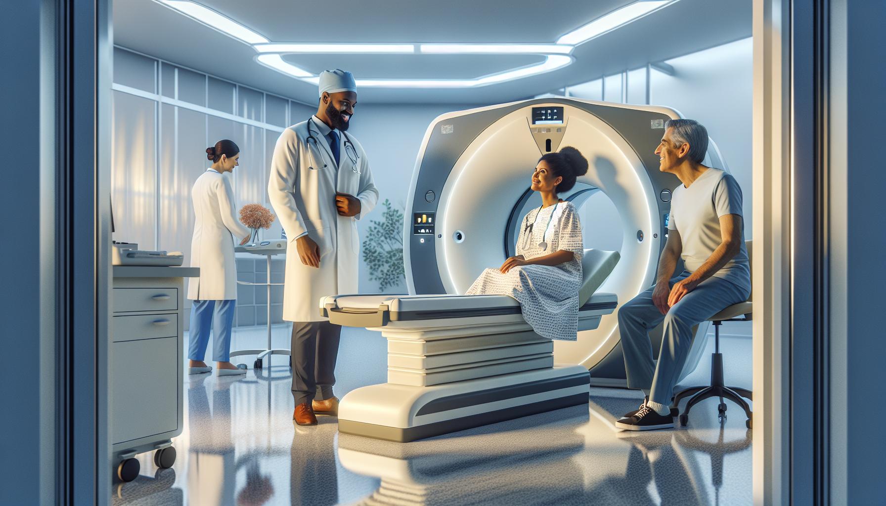
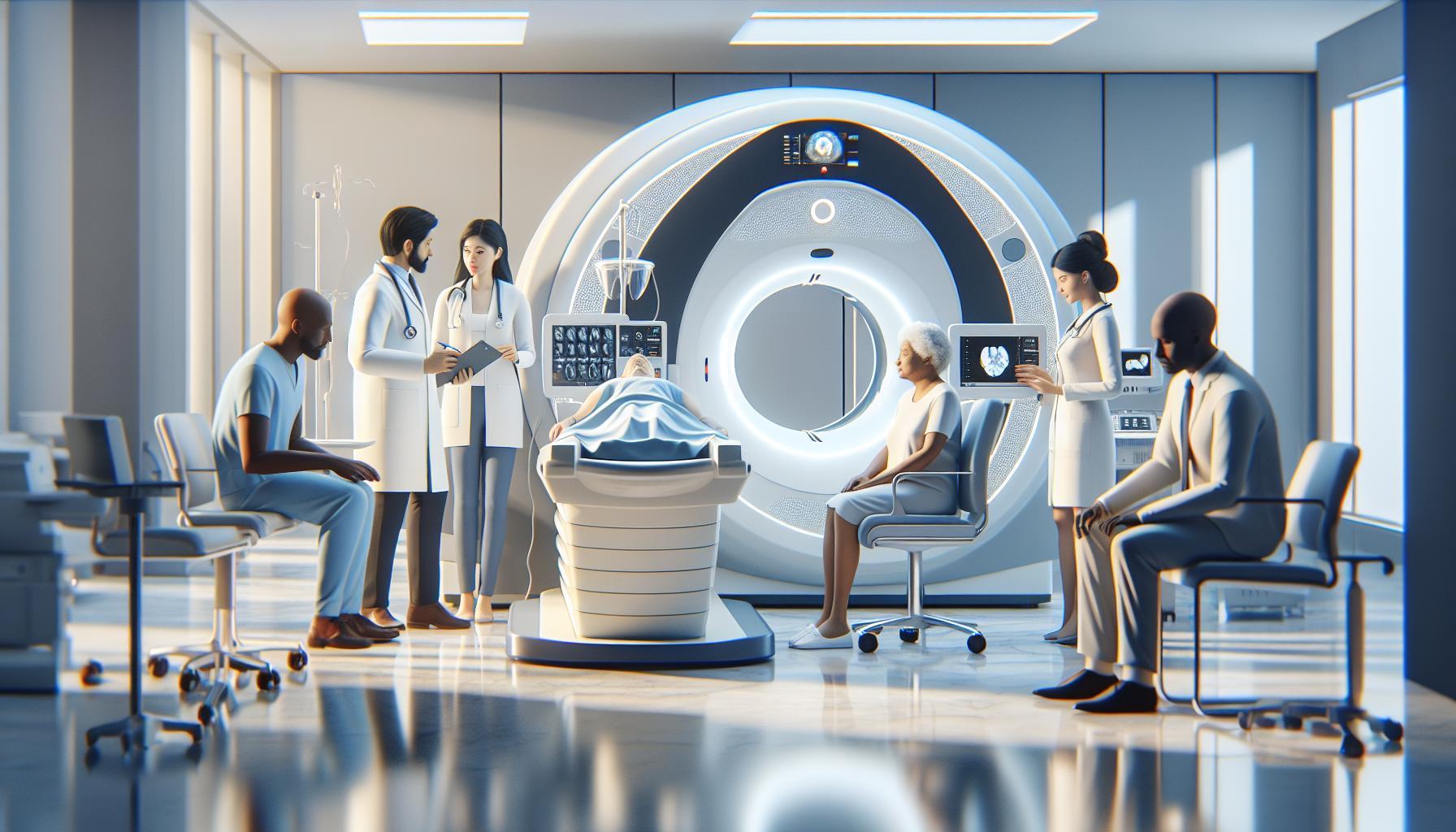
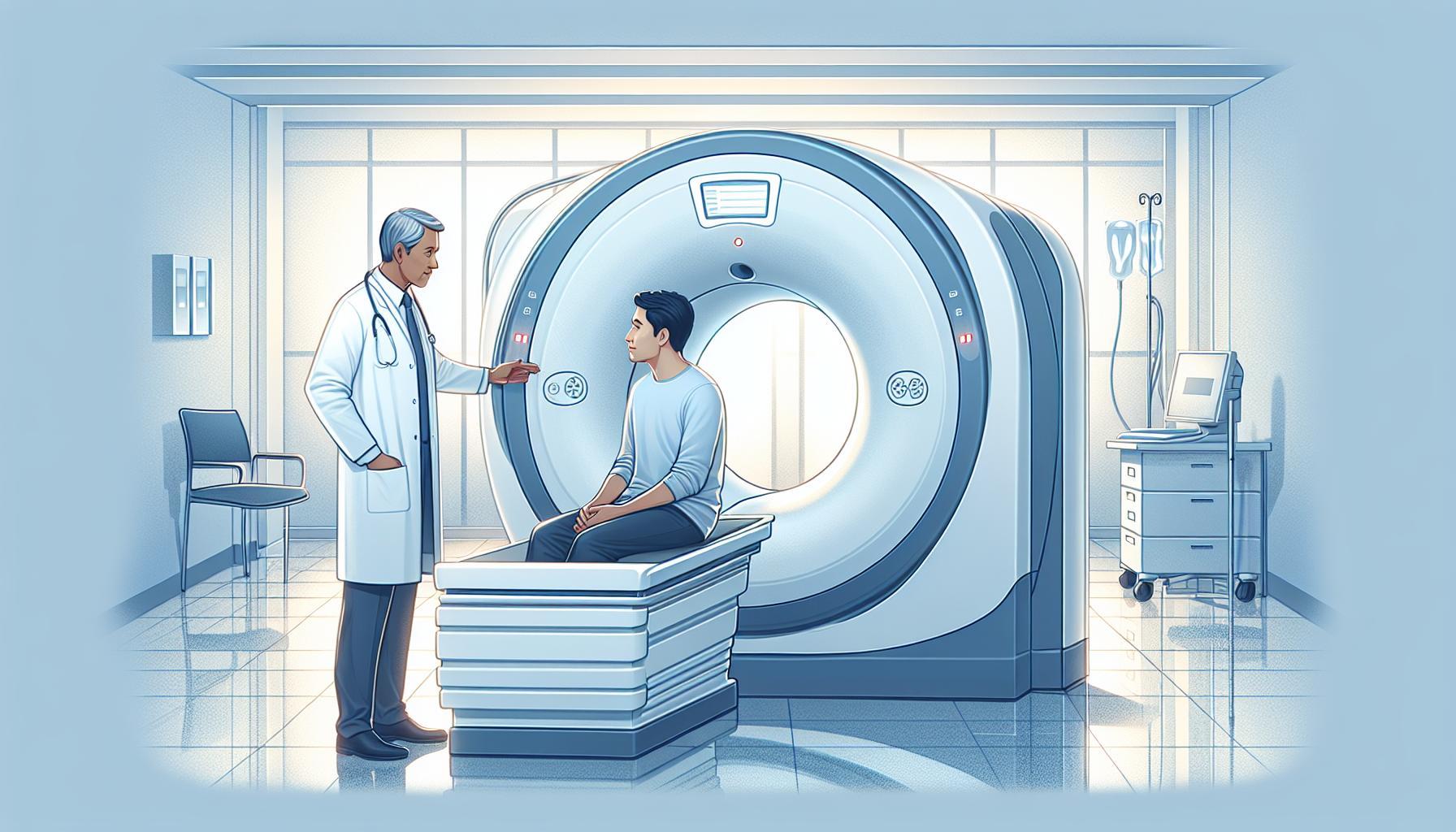
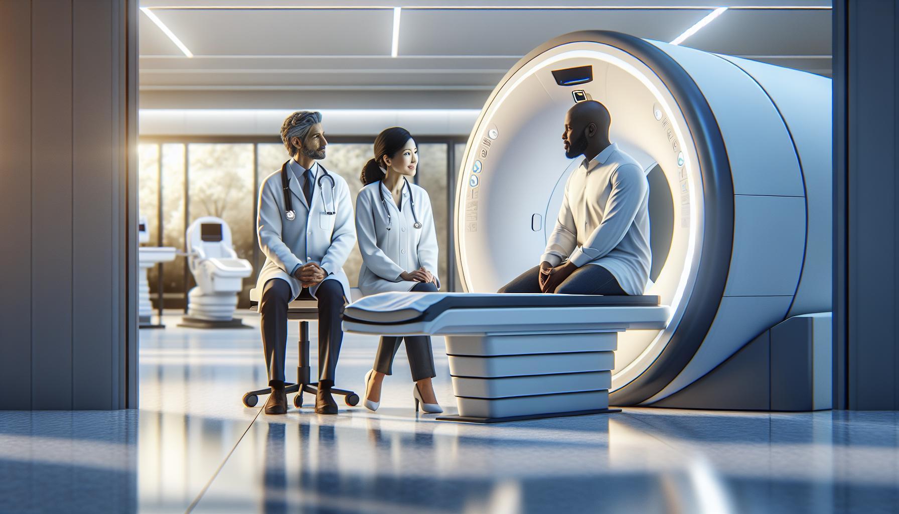
When faced with medical imaging options, understanding the differences between CT scans and PET scans can significantly impact patient care and outcomes. Both tests utilize advanced technology to visualize the body, yet they serve distinct purposes. A CT scan provides detailed cross-sectional images of the body’s internal structures using X-rays, making it invaluable for diagnosing injuries and diseases. In contrast, a PET scan reveals metabolic activity, offering insights into how tissues function, which is particularly useful in cancer detection.
As you navigate your healthcare journey, knowing the appropriate imaging tool for your specific needs can alleviate anxiety and empower informed decisions. Each method has its unique benefits and indications, making it essential to grasp their differences. Continue reading to explore a deeper comparison of CT and PET scans, helping you understand which may be best suited for your diagnosis or health concerns.
Understanding CT Scans: What to Expect
Undergoing a CT scan can be a vital step in diagnosing a range of medical conditions, and understanding what to expect can help ease any anxieties you may have. A CT scan, or computed tomography scan, uses advanced x-ray technology to create detailed images-or slices-of the inside of your body, including bones, organs, and soft tissues. This non-invasive imaging technique helps doctors gain critical insights into your health, from identifying fractures to locating tumors.
When you arrive for your CT scan, you will typically be asked to change into a hospital gown and may need to remove any metal objects such as jewelry that can interfere with image quality. Depending on the area being scanned, you may also be asked to drink a contrast dye or have it injected; this dye enhances the visibility of your internal structures. The actual scan is quick and painless, often lasting only a few minutes. During the procedure, it’s important to lie still while the scanner rotates around you; you may hear buzzing sounds as it works, but there is no discomfort involved.
After the scan, you can generally resume your normal activities immediately, although if you received contrast dye, you might be advised to drink plenty of fluids to help flush it from your system. The images will be reviewed by a radiologist, who will provide a report to your healthcare provider. They will discuss the findings with you and explain any necessary next steps, making it easy to understand your results. Emphasizing the significance of continual communication with your medical team can empower you to take charge of your health and address any concerns that may arise during the imaging process.
In summary, knowing what to expect during your CT scan can demystify the process and help you feel more prepared and at ease. If you have any questions or concerns, don’t hesitate to reach out to your healthcare provider for personalized guidance and support.
Exploring PET Scans: A Comprehensive Guide
Exploring the intricate world of imaging technology opens up significant insights into medical diagnostics, particularly with the advent of Positron Emission Tomography (PET) scans. These scans use small amounts of radioactive material to illustrate the function of organs and tissues within the body. Unlike CT scans, which primarily focus on the structure and anatomy by taking cross-sectional images, PET scans reveal how those structures are functioning. This vital distinction plays a crucial role in diagnosing and monitoring various conditions, particularly cancer, heart disease, and brain disorders.
When preparing for a PET scan, understanding the steps and implications can greatly alleviate concerns. The process typically begins with a consultation with your healthcare provider, who may discuss your medical history and any medications you are taking. Prior to the scan, you may be advised to refrain from eating or drinking for several hours to ensure accurate results. On the day of the scan, you will receive an injection of a radioactive tracer, which travels through your bloodstream and highlights areas of interest in your body. Depending on the type of scan, you may need to wait around 30 to 60 minutes after the injection to allow the tracer to accumulate in the target tissues.
During the actual PET scan, you’ll lie still on a comfortable table while the scanner rotates around you, capturing images of your body. The procedure itself is painless, though you might experience some discomfort from remaining still for an extended period. As the scan progresses, it generates real-time images that will assist your physician in evaluating how your organs and tissues are performing.
Post-scan, side effects are generally minimal, although some patients might experience slight drowsiness or warmth at the injection site. Drinking plenty of fluids afterward can help flush the radioactive material from your system more quickly. It’s important to maintain open communication with your healthcare providers throughout this process. They can help clarify any uncertainties about what to expect or discuss the results once available. By being well-informed, you empower yourself during this diagnostic journey, ultimately fostering a sense of control over your health and well-being.
Key Differences Between CT and PET Scans
Both CT (Computed Tomography) and PET (Positron Emission Tomography) scans serve essential roles in medical imaging, yet they do so by employing different technologies and focusing on distinct aspects of health assessment. Understanding these differences can empower patients to engage more confidently with their healthcare journey.
CT scans utilize a series of X-ray images taken from various angles and processed to create cross-sectional views of the body. This method is particularly useful for providing detailed anatomical information about organs, bones, and soft tissues, making it an effective tool for diagnosing structural conditions such as fractures, tumors, and diseases impacting organs. For instance, a CT scan can quickly reveal internal bleeding or damage from trauma, which is crucial in emergency situations.
In contrast, PET scans primarily highlight how organs and tissues are functioning rather than just their structure. By using a small amount of radioactive material as a tracer, PET scans enable the visualization of metabolic processes in the body. This technology shines in oncology, where it helps identify areas of high metabolic activity, often indicative of cancerous tumors. For example, whereas a CT scan may locate a tumor, a PET scan can provide insight into whether that tumor is active or responding to treatment.
Moreover, the preparation and execution of these scans differ markedly. Patients undergoing a CT scan generally do not require much preparation beyond removing metal objects and informing the technician about any allergies, particularly to iodine if contrast materials are used. Conversely, preparation for a PET scan may involve dietary restrictions to ensure that the uptake of the radioactive tracer is accurate, allowing for optimal imaging results.
While both imaging techniques are invaluable, the choice between them depends on the clinical situation. Combined, CT and PET scans can provide comprehensive views of both anatomical structures and physiological processes, allowing healthcare professionals to make well-informed decisions regarding diagnosis and treatment. Understanding these key differences empowers patients to participate actively in discussions with their healthcare teams, ensuring they receive the most appropriate imaging based on their specific health concerns.
How CT Scans Work: Step-by-Step Process
Undergoing a CT scan can be a straightforward process, designed to provide detailed images that aid in diagnosing various medical conditions. Understanding how this imaging technique works can alleviate concerns and empower patients to navigate their healthcare journey with confidence.
During a CT scan, the patient typically lies on a comfortable table that slides into a large, doughnut-shaped machine known as a CT scanner. The process begins with the operator positioning the patient correctly, ensuring the area of interest aligns with the scanner’s beam. For optimal imaging, the patient might be asked to hold their breath at certain moments while the scan is in progress. This helps to reduce movement and improve the clarity of the images.
Once properly positioned, a narrow beam of X-rays is emitted from the scanner and rotates around the body. Detectors capture the X-ray images from multiple angles, which are then processed by a computer to create cross-sectional slices of the body. Each slice provides a detailed view of specific organs, bones, and soft tissues, allowing healthcare providers to assess the area in question comprehensively. These cross-sectional images can be combined to generate a 3D representation of the internal structures, enhancing diagnostic accuracy.
Patients can generally expect the CT scan to take only a few minutes, though the preparation phase may take longer. Depending on the medical need, the use of a contrast material may be required, which involves either an injection or oral intake. This contrast enhances the visibility of specific areas, granting much clearer images. Importantly, the entire procedure is painless, and healthcare professionals are present to address any questions or concerns, ensuring a supportive environment throughout the process.
Understanding PET Scan Technology and Uses
The power of PET scans lies in their ability to visualize metabolic processes, providing unique insights into how organs and tissues function. Unlike CT scans, which primarily offer structural images through a series of X-rays, PET scans use a small amount of radioactive material to highlight areas of increased or abnormal activity within the body. This technique is invaluable for diagnosing conditions such as cancer, examining brain function, and assessing heart health.
When undergoing a PET scan, patients can expect to receive a radiotracer, typically injected into a vein, though it can also be ingested or inhaled. This tracer emits positrons, which collide with electrons in the body, releasing gamma rays that the PET scanner detects. The resulting images show metabolic changes, allowing healthcare providers to pinpoint areas of concern that may not be visible through conventional imaging. This is particularly effective in oncology, as cancerous cells often exhibit increased glucose metabolism.
In terms of preparation for a PET scan, patients are usually advised to refrain from eating for several hours beforehand to enhance the visibility of the radiotracer. It is also essential to share any medications or health conditions with your healthcare provider, as these factors can influence the scan’s results. For example, some diabetes medications may alter glucose metabolism, potentially skewing the findings.
Understanding how PET scan technology integrates with existing treatments is also crucial. In many cases, physicians will use PET scans in conjunction with CT scans to provide a comprehensive view of both the structure and function of tissues. This dual approach enhances diagnostic accuracy, aiding in everything from initial diagnosis to treatment planning and monitoring recovery. As always, if you have any concerns or questions regarding the procedure, discussing them with your healthcare team can provide reassurance and clarity.
Benefits of CT Scans in Medical Imaging
The advantages of computed tomography (CT) scans in medical imaging are substantial and can greatly enhance patient care, providing critical information that shapes diagnosis and treatment options. CT scans are renowned for their speed and precision, with the ability to produce cross-sectional images of the body’s internal structures in just a few minutes. This rapid imaging is essential in emergency settings, where quick assessment of injuries or infections can be life-saving.
One of the standout benefits of CT scans is their capability to create detailed images of various tissues, including bones, organs, and blood vessels, allowing healthcare providers to detect a multitude of conditions. Here are some critical uses of CT scans in medical imaging:
- Early Detection: CT scans can identify diseases at an early stage, particularly in cases like cancer where early intervention can significantly affect treatment outcomes.
- Comprehensive Evaluation: They provide a thorough view, making it easier to distinguish between healthy and diseased tissue, which is crucial for accurate diagnoses.
- Guidance for Procedures: CT scans are frequently used to help guide interventions, such as biopsies or the placement of catheters, ensuring precision in accessing the areas of interest.
- Monitoring Progress: For patients undergoing treatment for various conditions, CT scans can be instrumental in monitoring the effectiveness of therapies and detecting any changes over time.
The vast array of applications supported by CT technology provides clinicians with crucial insights, facilitating informed decision-making during patient care. Patients often express concern about the safety and radiation exposure associated with CT scans. However, advancements in CT technology have led to dose reduction strategies that minimize exposure without compromising image quality. Continuous dialogue with healthcare professionals can help address any worries patients may have and clarify the necessity of the procedure.
In conclusion, CT scans play a pivotal role in contemporary medical imaging, merging efficiency with diagnostic precision. Their relevance extends far beyond simplicity, influencing treatment pathways and patient outcomes substantively. Engaging patients in discussions about their imaging needs and procedures invites empowerment and assurance, allowing them to take an active role in their health journey.
Advantages of PET Scans for Diagnoses
The capability of positron emission tomography (PET) scans to provide insight into the metabolic processes of the body sets them apart as an invaluable tool in diagnostic imaging. Unlike CT scans, which focus on structural images, PET scans reveal how tissues and organs function, helping to identify issues such as cancer, heart disease, and neurological disorders at an earlier stage. This functional imaging plays a crucial role in informing treatment decisions and enhancing patient outcomes.
One of the primary advantages of PET scans is their ability to detect cancer at its earliest stages. By utilizing radioactive tracers that emit positrons, PET scans can identify abnormal metabolic activity often associated with tumors, even when structural changes may not yet be evident. This early detection can be vital in planning effective interventions and tailoring therapy to individual patient needs. The images produced can inform oncologists not only about the presence of cancer but also reveal how aggressively it is growing, which can significantly impact treatment choices.
Additionally, PET scans are essential in monitoring treatment effectiveness. Patients undergoing cancer treatment can receive PET scans to measure changes in tumor activity or monitor any signs of recurrence. This real-time evaluation allows healthcare providers to adjust treatment plans based on the patient’s response, ensuring that the most effective therapies are utilized. Furthermore, PET imaging has a role in assessing other conditions, such as Alzheimer’s disease, by allowing physicians to evaluate brain metabolism and identify changes that may indicate disease progression.
Patient understanding and comfort play a significant role in the success of these procedures. Before undergoing a PET scan, patients are often advised to avoid strenuous activities and limit certain foods, as these can affect the results. Knowing that these scans are non-invasive and provide essential information for their health journey can ease anxieties. Ultimately, open communication with healthcare providers about what to expect before, during, and after the procedure empowers patients, ensuring that they feel informed and supported through their diagnostic experience.
Patient Preparation for CT Scans Made Easy
Preparing for a CT scan doesn’t have to be a daunting experience. Understanding what to expect can significantly ease any anxiety you might have and help ensure the procedure goes smoothly. Unlike some imaging tests, such as a PET scan, CT scans primarily focus on capturing detailed images of your body’s internal structures using X-rays. This foundational knowledge can help frame your preparation process, making it more straightforward and less stressful.
Before your appointment, you may be required to avoid eating or drinking for a few hours, particularly if a contrast dye is to be used. This dye enhances the images produced during the scan, allowing for clearer visualization of particular organs or abnormalities. Ensure you follow any specific instructions provided by your healthcare provider regarding dietary restrictions or medications. It’s important to communicate openly about your medical history, including any allergies, especially to contrast materials, as these can influence both preparation and procedure.
Make sure to wear comfortable clothing, and ideally, avoid any garments with metal components like zippers, buttons, or belts. You may be asked to change into a medical gown to prevent any interference with the imaging. During the scan, you will lie down on a moving table that slides into the CT scanner, which looks like a large donut. You’ll need to stay still for a minute or two while the machine captures the images, which is crucial for obtaining the best results.
After the scan, you may resume your regular activities, but if you received a contrast dye, your healthcare provider may give specific aftercare instructions, such as drinking extra fluids to help flush the dye out of your system. Remember, the purpose of this imaging test is to provide important information about your health, so don’t hesitate to ask your healthcare provider any questions you might have about the process or the results. This proactive approach will empower you and help ensure your comfort throughout the entire procedure.
Preparing for Your PET Scan: A Complete Guide
Before undergoing a PET scan, understanding the preparation process can significantly alleviate any concerns you may have. Unlike other imaging tests, a PET scan not only pictures your internal structures but also provides functional information about your organs and tissues by using a small amount of radioactive material. This unique aspect of PET scans means that proper preparation is essential to ensure accurate results.
To begin the preparation, you will likely receive specific instructions from your healthcare provider about dietary restrictions. Generally, you may be asked to fast for several hours before the scan. This is crucial because eating can interfere with the distribution of the radioactive substance introduced into your body, affecting the quality of the images produced. It’s important to follow these guidelines closely, as they ensure that the scan results are as clear and informative as possible.
On the day of your PET scan, comfortable clothing is recommended; avoid wearing clothes with metallic accessories. You might be asked to remove jewelry and certain clothing items to eliminate any interference with imaging. Arriving a bit earlier than your appointment time can also help you complete any necessary paperwork and give you a moment to relax before the procedure.
During the scan itself, you will lie down on a special table that slides into the PET scanner. The process generally lasts about 30 to 60 minutes. While inside the machine, it’s important to remain still to ensure high-quality images are taken. After the scan, there may be no restrictions on your activities; however, it’s wise to hydrate afterward, helping your body process the radioactive material efficiently. Always feel free to ask your healthcare team any questions throughout this process-they’re there to support you and clarify any uncertainties you may have regarding your upcoming procedure.
Common Misconceptions About CT and PET Scans
Misunderstandings about imaging technologies like CT and PET scans are common and can lead to unnecessary anxiety for patients. One prevalent myth is that all imaging tests are the same; however, the differences lie in what they measure and how they provide insights into your health. CT scans utilize X-rays to generate detailed cross-sectional images of the body, focusing primarily on the anatomy. In contrast, PET scans help evaluate metabolic activity within cells, allowing for a more functional view, which can be particularly vital for detecting certain cancers and neurological conditions.
Another misconception is regarding the safety of these procedures. Many patients worry about radiation exposure. While both CT and PET scans involve radiation, the doses are carefully regulated and designed to be as low as reasonably achievable. It’s important to recognize that the benefits of accurately diagnosing conditions-and the subsequent medical interventions-often outweigh the risks associated with radiation exposure. It’s always advisable to discuss your concerns with your healthcare provider, who can provide specific information based on your medical history and the necessity of the scan.
Additionally, there is often confusion around preparation requirements. Some patients believe that they can eat normally before both types of scans, but this is not the case. For PET scans, fasting is usually required to ensure the best possible imaging results, while CT scans may have different preparation guidelines, particularly if contrast material is used. Understanding the specific requirements for each type of imaging can help alleviate frustration and ensure a smoother experience.
Lastly, the interpretation of the results can also be a source of anxiety. Many assume that if a scan shows ‘no abnormal results,’ then there is no issue. However, it’s crucial to acknowledge that the absence of findings does not always equate to the absence of disease. Each scan has its limitations, and sometimes further follow-ups or alternative imaging might be necessary for a comprehensive assessment. Establishing clear communication with your healthcare team is essential, as they can guide you through the results and what they mean for your overall health.
Costs and Insurance Coverage for Imaging Tests
Understanding the costs associated with CT and PET scans can ease some of the anxiety surrounding these important imaging tests. Both types of scans serve critical roles in diagnosing and managing medical conditions, but their costs can vary significantly based on multiple factors, including location, facility type, and whether the procedure is performed on an inpatient or outpatient basis. For instance, a CT scan may range from $300 to $3,000, whereas a PET scan may start around $1,000 and can go up to $6,000, depending on the complexity and the necessity of additional procedures.
When it comes to insurance coverage, it’s essential to check with your provider beforehand to understand what is included under your plan. Many insurance providers cover these imaging tests, especially if they are deemed medically necessary. This could include the diagnosis of conditions like cancer or serious diseases. However, some plans may require pre-authorization or have specific criteria that need to be met before coverage is granted. For those without insurance, there are often payment options available directly through medical facilities, such as financing plans or cash discounts, which can help manage out-of-pocket expenses.
Important Considerations
To help navigate the costs effectively, consider these practical steps:
- Verify Insurance Coverage: Before scheduling a scan, contact your insurance company to confirm what is covered and whether any pre-authorization is required.
- Ask About Payment Plans: Inquire with the imaging facility regarding financing options or plans that break down payments over time.
- Shop Around: Prices can vary widely between facilities. If time permits, comparing prices for similar services in your area can lead to significant savings.
- Discuss Necessity with Your Doctor: Talk to your healthcare provider about the necessity of the scan and potential alternatives, if applicable. This dialogue can clarify why a specific imaging test is required and whether it is the most cost-effective choice.
Being proactive about understanding the costs and insurance coverage can alleviate some of the stress surrounding CT and PET scans. Always feel empowered to ask questions and voice concerns with your healthcare provider to ensure you’re well-informed and comfortable with the decisions being made about your health.
Safety Considerations: Are CT and PET Scans Safe?
Both CT and PET scans are invaluable tools that help in diagnosing and managing various medical conditions, yet many patients feel anxiety regarding their safety. It’s essential to understand the underlying considerations that can reassure individuals about these imaging procedures.
CT scans utilize X-rays to create detailed cross-sectional images of the body, allowing physicians to detect abnormalities or diseases efficiently. While CT scans subject patients to ionizing radiation, advances in technology have significantly reduced the dose over the years, making modern CT scans safer than those performed in previous decades. Typical exposure from a singular CT scan is comparable to the radiation received from natural sources over several years. Medical professionals weigh the benefits of obtaining critical diagnostic information against the risks, ensuring that such scans are performed only when necessary.
Conversely, PET scans employ a small amount of radioactive material to highlight areas of metabolic activity in the body. This methodology is particularly useful in identifying cancerous tissues, monitoring heart conditions, and evaluating brain disorders. The amount of radiation involved is low and short-lived, posing minimal risks to most individuals. However, those who are pregnant or lactating should inform their healthcare provider about their condition prior to undergoing a PET scan to discuss alternative imaging strategies if needed.
Both imaging techniques have well-established safety protocols in place, ensuring patient comfort and health. Patients can take proactive steps to alleviate concerns. Before the procedure, it is advisable to discuss any questions about risks and benefits with the healthcare provider, who can provide guidance tailored to individual health needs. Moreover, remaining calm and following preparatory instructions can greatly enhance the experience and yield accurate results, ultimately aiding in effective treatment plans. Remember, prioritizing communication with health professionals empowers patients in their medical journey, ensuring informed decisions are made about their health.
Faq
Q: What are the main differences between CT and PET scans?
A: The main differences lie in their technology and purpose. CT scans use X-rays to create detailed images of the body’s structure, including bones and tissues, while PET scans use radioactive tracers to show metabolic activity and abnormalities in body functions, often used in cancer diagnosis.
Q: How does a CT scan work compared to a PET scan?
A: A CT scan works by taking multiple X-ray images from different angles and combining them to create cross-sectional views of the body. In contrast, a PET scan involves injecting a small amount of radioactive material that emits positrons, which are detected to visualize metabolic processes.
Q: When should a CT scan be used instead of a PET scan?
A: A CT scan is typically used for detailed imaging of injured bones or internal bleeding, while a PET scan is preferred for detecting cancer, monitoring treatment effectiveness, and assessing brain disorders. The choice depends on the medical question being addressed.
Q: Are there any risks associated with CT or PET scans?
A: Both CT and PET scans involve exposure to radiation. However, CT scans generally deliver higher radiation doses. Patients should discuss concerns with their healthcare provider to understand the necessity and risks associated with their specific situation.
Q: Can a CT scan and a PET scan be done together?
A: Yes, a PET-CT scan combines both imaging techniques to provide comprehensive information about the body. This combined approach allows for detailed anatomical and functional imaging, improving diagnosis and treatment planning, especially in oncology.
Q: What preparations are needed for a CT scan compared to a PET scan?
A: For a CT scan, patients may need to avoid food or drink for a few hours prior, especially if contrast material is used. For a PET scan, fasting for 6-8 hours before the scan is usually necessary to ensure accurate metabolic assessment.
Q: How long does each scan typically take?
A: A CT scan usually takes about 15 to 30 minutes, while a PET scan may take longer, ranging from 30 to 90 minutes depending on the protocol and the patient’s condition. Time can vary based on specific clinical indications.
Q: What are the costs typically associated with CT and PET scans?
A: Costs can vary widely based on location and insurance coverage, but typically, CT scans are less expensive than PET scans. Patients should contact their insurance provider for specific coverage details and out-of-pocket costs.
Wrapping Up
Understanding the differences between CT and PET scans is crucial for making informed decisions about your health and imaging needs. CT scans provide detailed anatomical images, while PET scans reveal functional processes, giving a comprehensive view of your condition. If you still have questions or are considering a scan, don’t hesitate! Explore our resources on how to prepare for a CT scan or learn more about the benefits and advancements in PET imaging to empower your healthcare journey.
For more insights, visit our articles on the benefits of medical imaging and patient preparation tips. Don’t forget to sign up for our newsletter to receive the latest updates on health technologies and healthcare practices directly in your inbox! Your health is paramount, and staying informed is the first step. Share your thoughts with us in the comments, and let’s continue the conversation!


