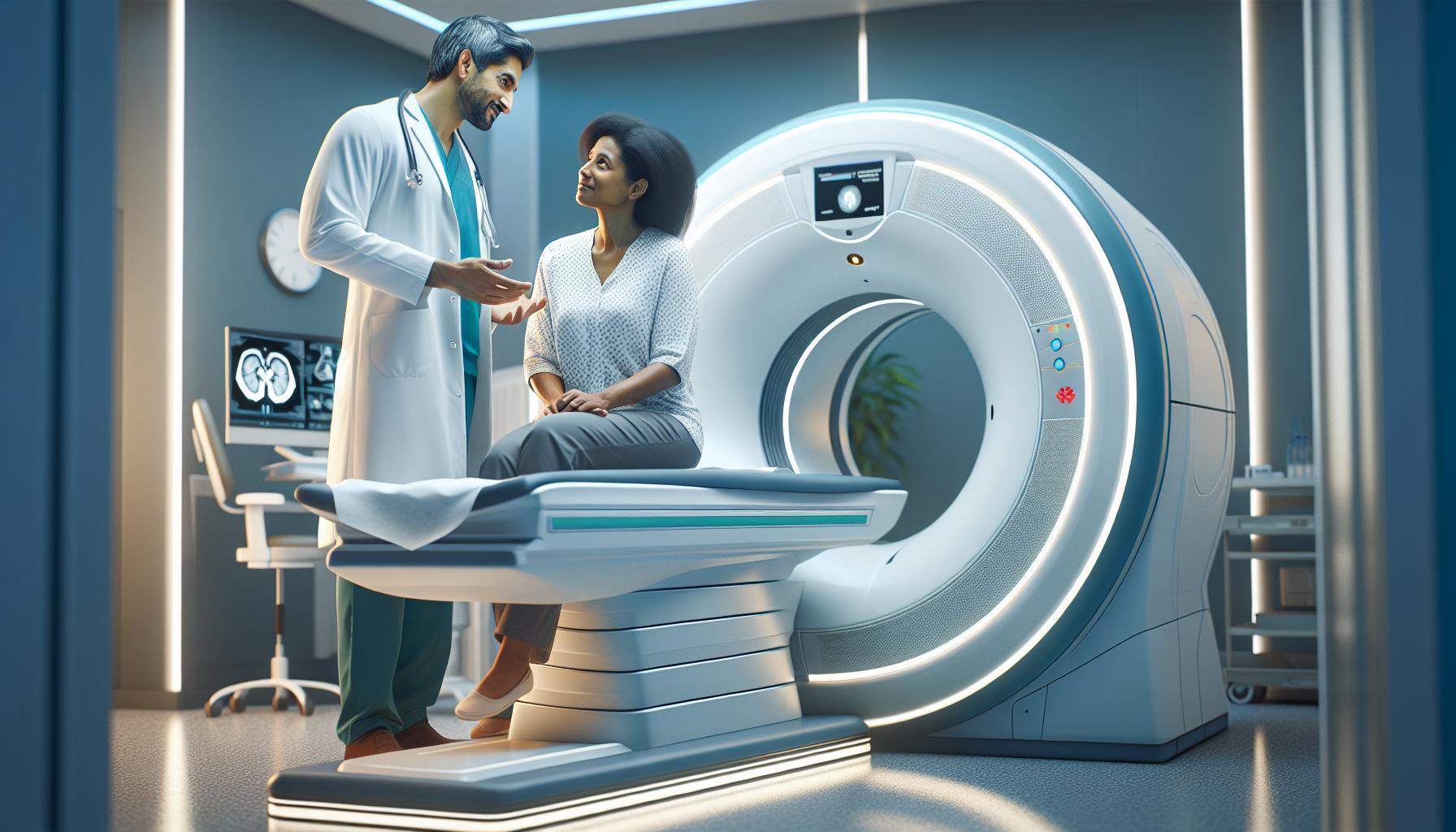
When it comes to strokes, timely detection is crucial for minimizing damage and potentially saving lives. A CT scan, or computed tomography scan, is a powerful imaging tool that can often reveal the critical signs of a stroke, particularly in its early stages. In fact, it’s one of the most common methods used by healthcare professionals to quickly assess a patient displaying stroke symptoms.
Understanding how a CT scan works and what it can reveal may ease your concerns if you or a loved one are facing this procedure. These scans can efficiently identify whether a stroke is caused by a blood clot or bleeding in the brain, providing vital information that guides urgent treatment decisions. As you explore this topic further, consider how knowing the role of a CT scan in stroke detection not only empowers you with knowledge but also emphasizes the importance of acting swiftly in medical emergencies. Stay with us to discover more about this life-saving diagnostic tool and what you can expect from the procedure.
Understanding CT Scans and Stroke Detection
A CT scan can be a critical tool in diagnosing strokes, often providing immediate and detailed images of the brain that help medical professionals determine the nature and extent of a stroke. Utilizing advanced imaging technology, a CT scan can quickly identify areas of the brain that may be underdeveloped or damaged due to a lack of blood flow, which is essential for effective and timely treatment. Within minutes, scans can reveal whether a stroke is ischemic, caused by a blockage of blood vessels, or hemorrhagic, due to bleeding in the brain.
Understanding how CT scans work is vital for patients. During the procedure, a series of X-ray images are taken from various angles and then processed by a computer, creating cross-sectional images of the brain. As you lie still on the table, the machine will rotate around you while you may hear whirring sounds. Although the thought of being scanned can be intimidating, it typically lasts only a few minutes. Patients are often urged to remain calm and still, as movement can affect the clarity of the images.
After the scan, healthcare professionals will analyze the results to assist in diagnosing conditions promptly. A key factor here is timing, as the sooner a stroke is diagnosed, the better the chances of minimizing damage and maximizing recovery. In many cases, CT scans can be completed in an emergency setting, leading to rapid intervention such as medication or surgical procedures that can save brain tissue and provide better outcomes for the patient.
It’s also essential to note that while CT scans are highly effective for stroke detection, they are just one part of a comprehensive stroke evaluation. Results can often be supplemented with other imaging techniques, like MRI scans or angiograms, depending on the specific circumstances of the case. If you or a loved one is experiencing stroke symptoms, seek medical help immediately; every second counts in stroke management.
How CT Scans Diagnose Different Stroke Types
A CT scan serves as a crucial diagnostic tool in the early detection of strokes, distinguishing between the two primary types: ischemic and hemorrhagic strokes. In cases of ischemic stroke, which accounts for about 87% of all strokes, blood flow to a portion of the brain is obstructed due to a blood clot or narrowing of arteries. The imaging produced by a CT scan can reveal areas of the brain that are experiencing reduced blood flow, allowing physicians to recognize not only the affected regions but also to gauge the overall extent of the injury as it develops and evolves.
Conversely, when it comes to hemorrhagic strokes, characterized by bleeding within the brain, a CT scan can quickly identify areas filled with blood, signaling the need for immediate medical intervention. The rapidity of this imaging process is critical, as timely diagnosis can significantly influence treatment decisions and patient outcomes. Given that the brain is highly sensitive to changes in blood supply, detecting the onset of a stroke during its early stages can empower healthcare providers to act swiftly, maximizing the potential for recovery and minimizing potential long-term damage.
The power of CT technology lies in its speed and efficiency. In emergency situations, where time is of the essence, imaging can often be completed within a matter of minutes. The results drawn from this diagnostic process not only assist in confirming a stroke diagnosis but also play a pivotal role in determining the subsequent steps in patient care. Healthcare professionals may use these images to initiate necessary treatments, ranging from medication to more invasive procedures aimed at restoring blood flow or addressing the source of bleeding.
Overall, CT scans stand as an indispensable ally in the toolkit of stroke management. Their ability to differentiate between stroke types allows for tailored and effective interventions that can save lives. However, while the capability of CT imaging is remarkable, patients are advised to remain cautious and consult their healthcare providers for a comprehensive evaluation and personalized medical guidance, as each case can present unique challenges and considerations.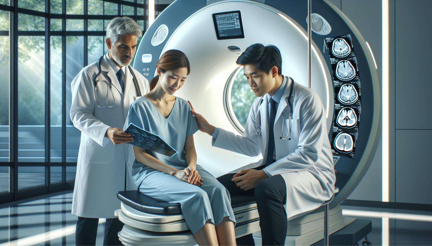
The Importance of Timely Stroke Diagnosis
The urgency surrounding stroke diagnosis cannot be overstated; every minute counts. In fact, research indicates that for every minute a stroke goes untreated, nearly two million brain cells die. This shocking statistic demonstrates the critical importance of rapid diagnosis and intervention, reinforcing the role of CT scans in emergency medicine. When a stroke is suspected, a timely CT scan can be the key to identifying the type of stroke-ischemic or hemorrhagic-and initiating appropriate life-saving treatment without delay.
The immediacy of CT imaging means that medical teams can quickly distinguish between different stroke types. For instance, in the case of ischemic strokes, where blood flow is restricted, timely identification can lead to the administration of clot-busting medications such as tPA (tissue Plasminogen Activator), which are most effective when administered shortly after symptom onset. Conversely, for hemorrhagic strokes caused by bleeding in the brain, swift CT scan results can facilitate rapid surgical intervention to control the bleed and relieve pressure on the brain. Every moment wasted could worsen brain damage and heighten the risk of long-term disability or even death.
Patients often feel anxious when facing diagnostic procedures, but understanding the process can alleviate some of that fear. During a CT scan, you will lie on a table that moves through the scanner, which will take images of your brain within minutes. No special preparation is required, and the procedure is typically painless. Knowing that this scan plays a vital role in determining your treatment plan can lend reassurance and a sense of control during a frightening time.
In summary, the timely diagnosis of a stroke is paramount to improving outcomes and reducing the risk of severe brain damage. Knowing the importance of CT imaging, both patients and their families can advocate for quick action in emergency settings. Healthcare professionals will use these critical images to guide decisions that will have a profound effect on recovery, reinforcing the idea that early intervention is often the best medicine.
Preparing for Your CT Scan: What to Expect
When facing a CT scan, it’s entirely normal to feel a mix of anxiety and anticipation. Understanding what to expect can ease these feelings and empower you to approach the procedure with confidence. CT scans are crucial in swiftly identifying possible strokes, enabling healthcare providers to administer timely and life-saving treatments. Knowing how to prepare for your scan can enhance your experience and ensure the process goes smoothly.
Before your CT scan, you may be advised to wear comfortable, loose-fitting clothing without any metal fasteners, as metal can interfere with imaging. It’s also essential to inform your healthcare provider of any medications you’re taking or if you have any implants, allergies, especially to contrast materials, or a history of kidney issues. Depending on the specific type of CT scan being performed, you may be required to refrain from eating or drinking for a few hours beforehand. Always follow your doctor’s instructions regarding this, as fasting protocols can vary.
During the scan, you’ll lie on a movable table that slides into the CT machine, which resembles a large donut. The scanner will rotate around you, taking images at various angles. Although you might need to hold your breath for a few seconds during the procedure, it generally lasts only about 10 to 30 minutes. The process is painless, and the only noticeable sensation may be the coolness of the table and the sounds of the machine. For some scans, particularly those involving contrast dye to enhance imaging, an intravenous line may be placed in your arm. You might feel a warm sensation or a metallic taste upon the dye injection, which is a normal reaction and typically subsides shortly after.
Post-scan, most people can resume their normal activities immediately unless advised otherwise. The results will be evaluated by a radiologist, who will send a report to your doctor to discuss the findings. Understanding these steps helps demystify the process and provides reassurance that you are taking a vital step in assessing your health, particularly in the context of stroke detection. Remember to communicate openly with your healthcare team about any questions or concerns you may have; their support is an integral part of your experience.
CT Scan Procedure: A Step-by-Step Guide
Undergoing a CT scan can be a pivotal moment in diagnosing a stroke, allowing healthcare professionals to visualize the brain and determine the presence of any abnormalities. Understanding how the procedure unfolds can alleviate anxiety and help you feel more prepared for what lies ahead.
Getting Ready for the Scan
Before your visit, it’s essential to prepare appropriately to ensure clear images. Here’s a simple checklist to guide you:
- Clothing: Wear comfortable, loose-fitting clothes. It’s best to avoid clothes with metal fasteners which may interfere with the scan.
- Medical History: Inform your technician about any medications you’re taking, allergies, and if you have any implants or a history of kidney problems.
- Fasting: Depending on the type of CT scan, you may need to refrain from eating or drinking for a few hours prior. Always follow your doctor’s specific instructions regarding fasting.
During the CT Scan
When you arrive, the technician will explain the procedure in detail. You’ll be asked to lie down on a movable table that gradually slides into the CT scanner-a large, donut-shaped machine. The process typically lasts between 10 to 30 minutes. As the scan begins, the machine will rotate around you, capturing images from various angles. You may hear some buzzing or clicking sounds, but the procedure is painless. Occasionally, you may be asked to hold your breath for a few seconds to ensure clearer images.
If your scan involves contrast material to enhance the images, an intravenous line will be placed in your arm. You might feel a warm sensation or an unusual metallic taste as the dye enters your bloodstream, which is completely normal. These feelings usually subside quickly.
Post-Scan Experience
Once the scan is completed, you can generally resume your normal activities immediately unless your doctor advises otherwise. The images will be analyzed by a radiologist, who will prepare a report for your physician. Understanding the results will be vital for determining the next steps in your care, particularly in the context of stroke management.
This straightforward approach to what happens during a CT scan can help transform apprehension into empowerment. Knowing what to expect allows you to advocate for your health confidently, ensuring that you’re well-informed and ready to discuss your results with your healthcare team.
Safety Considerations: Is a CT Scan Safe?
Undergoing a CT scan might seem daunting, and concerns about safety are entirely normal. It’s essential to understand that when medical professionals recommend a CT scan, they are doing so with a clear purpose: to obtain critical imaging that can significantly impact diagnosis and treatment, especially in urgent situations like strokes. The technology behind CT scans is designed to minimize risk while providing detailed images of internal structures.
Most importantly, CT scans involve a small amount of radiation to produce images. Although high doses of radiation can increase cancer risk over time, the exposure from a single CT scan is relatively low, generally considered safe for the vast majority of patients. The diagnostic benefits of a CT scan-such as quickly identifying a stroke or other serious conditions-often far outweigh the potential risks associated with radiation exposure. Always communicate openly with your healthcare provider; they can help you weigh the benefits against the risks based on your individual health history and circumstances.
Patients who are pregnant or suspect they might be pregnant should inform their healthcare team, as special considerations may be necessary to protect the developing fetus from radiation exposure. Additionally, if you have any allergies, particularly to contrast materials, or pre-existing conditions such as kidney problems, it’s crucial to discuss these before the procedure. Your medical team is there to ensure not only that the scan is effective but also that your safety is prioritized throughout the process.
After your CT scan, most individuals return to their normal activities immediately. If contrast dye was used, drinking plenty of fluids can help expedite its elimination from your body. Remember, while the prospect of a CT scan can cause anxiety, understanding the safety measures in place and the purpose of the scan can help ease your concerns. Always consult with your healthcare provider for tailored advice and information specific to your situation, allowing you to navigate this experience with confidence.
Interpreting CT Scan Results for Strokes
Recognizing the signs of a stroke can be a matter of life and death, and a CT scan plays a crucial role in the rapid diagnosis necessary for effective intervention. When interpreting CT scan results, healthcare professionals look for specific indicators that can confirm or rule out a stroke. These scans provide highly detailed images of the brain, revealing the presence of blood clots or bleeding, both of which can significantly affect treatment decisions.
In cases of an ischemic stroke, which occurs when a blood vessel is blocked, the CT scan may show a dark area where brain tissue has been deprived of blood. This loss of blood flow leads to cell death, and identifying this area promptly can help medical teams initiate treatment that restores circulation quickly. Conversely, a hemorrhagic stroke, caused by bleeding in the brain, is often indicated by the presence of bright areas on the scan, pointing to blood pooling in certain regions.
Understanding the timing of the CT scan is also pivotal. It should ideally be performed as soon as symptoms of a stroke are recognized. Generally, the sooner a stroke is diagnosed, the more effective the treatment options will be, ranging from clot-busting medications to surgical interventions like angioplasty. This urgency is one reason why emergency departments emphasize immediate imaging once a stroke is suspected.
Patient involvement in this process is essential; being aware of the signs of a stroke (such as sudden numbness, confusion, difficulty speaking, or visual disturbances) can lead to quicker action. For instance, recommending that someone memorize the FAST acronym-Facial drooping, Arm weakness, Speech difficulties, and Time to call emergency services-can empower individuals and their families to act swiftly in such critical moments. Ultimately, while CT scans are invaluable diagnostic tools, effective communication between patients and healthcare providers helps ensure that potential strokes are addressed effectively and promptly.
Comparing CT Scans to Other Imaging Techniques
CT scans are widely regarded for their speed and effectiveness in diagnosing strokes, but they are not the only imaging modality available. When emergency departments are faced with a suspected stroke, they often weigh the benefits and limitations of CT scans against other imaging techniques, such as MRI and ultrasound, to optimize patient outcomes.
One significant advantage of CT scans is their ability to quickly provide clear images of the brain, allowing for immediate assessment of both ischemic and hemorrhagic strokes. In contrast, while MRIs (Magnetic Resonance Imaging) provide more detailed images of brain tissue, they typically take longer to perform, which can delay crucial treatment. For instance, in acute stroke scenarios where every minute counts, a CT scan can identify a blockage or bleeding rapidly, setting the stage for timely interventions. MRIs, however, can be particularly useful in detecting subtle changes in brain tissue that may not be visible on a CT scan, especially in the context of early ischemic changes or prior strokes.
Another technique that complements CT is the use of ultrasound, specifically in the evaluation of blood flow in carotid arteries. There, ultrasound can help identify potential sources of emboli that may have led to a stroke. While it doesn’t visualize the brain tissue itself, it serves as a valuable tool in understanding risk factors and planning further management.
When considering the differing costs, availability, and patient factors, CT scans often emerge as the first-line imaging technique for acute strokes in emergency settings. The choice of imaging should be made collaboratively between the healthcare team and the patient, accounting for individual circumstances, medical history, and the urgency of the situation. Overall, understanding the nuances between these imaging modalities empowers patients and families to engage in informed discussions with their healthcare providers.
How Fast Can a CT Scan Reveal a Stroke?
In critical situations like a suspected stroke, time is of the essence. A CT scan can typically reveal a stroke within minutes, making it an invaluable tool in emergency medicine. Thanks to its rapid imaging capabilities, medical professionals can quickly assess the brain for signs of ischemic (caused by a blockage) or hemorrhagic (due to bleeding) strokes, allowing for expedited treatment decisions that can significantly impact patient outcomes.
When a patient arrives at the emergency department exhibiting stroke symptoms-such as sudden weakness, confusion, or trouble speaking-healthcare providers often prioritize a CT scan as part of their assessment protocol. The scan itself usually takes only about 5 to 10 minutes to perform, and images are available almost instantaneously. With the ability to view detailed slices of the brain, radiologists can identify areas of tissue damage or bleeding, often within as little as 10 to 20 minutes of the patient arriving for imaging. This speed is crucial; every minute lost can reduce the effectiveness of treatment options, such as clot-busting medications.
Another significant advantage of CT scans is their accessibility and the fact that they do not require the patient to remain still for an extended period, a key benefit for individuals who may be agitated or in distress. Moreover, in cases where the CT scan indicates a hemorrhagic stroke, immediate interventions-such as surgery-can be initiated without delay. These rapid diagnostic capabilities are essential in emergency settings, where quick decisions can lead to better recovery outcomes.
For patients and families who might feel anxious about the procedure, understanding that CT scans are designed to be efficient and relatively simple can be reassuring. During the scan, patients will lie on a table that slides into the CT machine, where they may hear a series of clicking sounds as images are captured. It’s important to remain as still as possible to ensure clear images, but healthcare providers are trained to guide patients through the process calmly.
In summary, the swift nature of CT scans allows healthcare professionals to determine the type of stroke quickly, which is critical for initiating the correct treatment plan. This rapid response not only enhances patient safety but also significantly improves the chances of a favorable recovery.
Real-Life Scenarios: CT Scans in Action
In the realm of emergency medicine, the ability to identify a stroke quickly can mean the difference between life and death. Imagine a middle-aged man experiencing sudden difficulty in speaking while watching television. His wife recognizes these alarming symptoms and calls 911 immediately. Upon arrival at the hospital, he is swiftly evaluated for a potential stroke. A CT scan is ordered; the speed of the process is remarkable, and within minutes, he’s moved into the imaging room. As the machine whirs to life, capturing cross-sectional images of his brain, the medical team prepares to make crucial decisions based on the results.
CT scans are invaluable tools in such critical situations. They can differentiate between ischemic strokes, which are caused by blockages in blood vessels, and hemorrhagic strokes, where bleeding occurs in or around the brain. For instance, in another scenario, a young woman arrives at the emergency room complaining of severe headaches and weakness on one side. The CT scan reveals a hemorrhagic stroke, allowing the medical team to initiate immediate intervention, potentially saving her life. These examples underscore how the rapid imaging capabilities of CT scans not only diagnose the type of stroke but also guide urgent treatment plans that can drastically improve outcomes.
For patients undergoing a CT scan, knowing what to expect can alleviate anxiety. As they lie still on the scan table, the clicking noises may seem unfamiliar but are simply part of the imaging process. Healthcare professionals are there to provide reassurance and explain each step. The scans generally take about 5 to 10 minutes, with results typically available soon after. This efficiency helps ensure that treatment can begin as quickly as possible, further emphasizing the critical role that CT scans play in emergency stroke care.
The advantages of CT scans extend beyond just speed; they are non-invasive and accessible, allowing healthcare providers to swiftly assess a patient’s condition without excessive delays. Recognizing the symptoms and acting promptly, such as calling emergency services or going to the hospital, can dramatically improve a patient’s chances of recovery. This collaborative effort between patients, their families, and healthcare providers highlights the life-saving potential of timely CT scan diagnostics in stroke management.
Understanding Costs and Insurance for CT Scans
When considering a CT scan, understanding the associated costs and how insurance works can significantly impact your experience and peace of mind. The average cost of a non-contrast CT scan typically ranges from $300 to $3,000, depending on factors such as the facility location, complexity of the scan, and whether contrast dye is used. It’s important to note that while some facilities may offer lower prices, the quality of care and the expertise of the radiologists should also be considered. Patients often benefit from asking their healthcare provider for recommendations on where to receive imaging services that balance both cost and quality.
Insurance coverage for CT scans varies widely among policies. Many health insurance plans cover CT scans when deemed medically necessary, particularly in emergency situations, such as stroke detection. To avoid unexpected expenses, it’s advisable to verify coverage details with your insurance provider prior to scheduling the scan. Specifically, inquire about your plan’s deductible, copay, and out-of-pocket maximums. In some cases, pre-authorization may be required, so being proactive can help streamline the process and minimize surprises.
Financial Assistance Options
For those without insurance or with insufficient coverage, there are options worth exploring. Many hospitals and imaging centers offer financial assistance programs or payment plans that allow patients to manage costs more effectively. Additionally, non-profit organizations may provide resources or funding for patients in need of medical imaging as part of their support for health-related issues.
In summary, navigating the costs and insurance details of CT scans can seem daunting, especially during a medical crisis. However, by understanding your financial options, communicating with healthcare providers, and utilizing available support resources, you can reduce the burden and focus on what truly matters-your health and recovery. It is always best to consult with your healthcare team for personalized advice and guidance tailored to your unique situation.
Next Steps After a Stroke Diagnosis
Following a stroke diagnosis, taking the right steps is crucial in the journey to recovery. Understanding what comes next can help alleviate fears and empower patients and their families to make informed choices about their care and rehabilitation journey. The first and foremost action is to discuss the results and their implications with your healthcare team. This conversation is essential, as it will provide clarity on the type of stroke, the severity, and potential treatment options. Engaging in these discussions can help you understand your condition better and what is needed moving forward.
Once a treatment plan is established, rehabilitation often begins as soon as possible. This may involve physical therapy, occupational therapy, and speech therapy aimed at regaining lost functions and adapting to any new challenges. Stay proactive by asking your healthcare provider about setting up an interdisciplinary care approach, which includes a range of specialists who can address various aspects of recovery. For instance, a nutritionist can help tailor a diet to support healing, while a psychologist or counselor can assist with the emotional toll of a stroke.
It’s also vital to monitor and manage any risk factors that could lead to a second stroke. This includes adherence to medications prescribed for blood pressure, cholesterol, or blood-thinning purposes. Working closely with your physician to regularly check in on these health conditions can make a significant difference. Moreover, adopting a heart-healthy lifestyle through regular exercise, a balanced diet, and avoiding tobacco can enhance recovery and reduce the risk of future strokes.
Lastly, building a support network is key. Engage family and friends who can provide emotional support, practical help, and accountability. Joining support groups, either in-person or online, can connect you with others going through similar experiences, offering a platform for sharing tips and encouragement. Remember, the recovery process can be gradual and varied for each person. Stay patient and maintain openness with your healthcare team to ensure your recovery pathway is as optimal as possible.
Faq
Q: Can a CT scan detect a stroke immediately?
A: Yes, a CT scan can detect a stroke quickly, often within minutes of the procedure. It is particularly effective in identifying hemorrhagic strokes, where bleeding occurs in the brain. Fast diagnosis is critical for initiating treatment and minimizing brain damage.
Q: What are the differences between CT and MRI for stroke diagnosis?
A: While both CT and MRI can detect strokes, CT scans are preferred for acute cases due to their speed and accessibility. MRIs offer more detailed images of brain tissue but take longer and are less suitable for immediate stroke situations.
Q: How long does it take to get CT scan results for a stroke?
A: Typically, CT scan results are available within 30 minutes to a few hours. This rapid turnaround is crucial in emergency settings, allowing for swift decision-making and treatment options to be implemented.
Q: What should I discuss with my doctor before a CT scan for stroke detection?
A: Before a CT scan, discuss your medical history, any allergies (especially to contrast dye), and medications you are taking. Understanding the procedure’s purpose and what to expect can help ease anxiety and ensure a smooth process.
Q: Is a CT scan safe for stroke detection?
A: Yes, CT scans are generally considered safe. They use X-rays to create images of the brain, but exposure to radiation is minimal. Your healthcare provider will weigh the benefits against any risks, especially in emergency settings.
Q: What happens during a CT scan for stroke diagnosis?
A: During a CT scan, you will lie on a table that moves through a large, doughnut-shaped machine. The procedure is painless and takes about 10-15 minutes. You may need to hold your breath for short periods while the X-ray images are taken.
Q: Can a CT scan show stroke signs even if symptoms are gone?
A: Yes, a CT scan can detect past strokes, including subtle changes in brain tissue that indicate previous ischemic or hemorrhagic strokes. However, it may not always show signs if the stroke occurred a long time ago.
Q: What are the next steps after a CT scan if a stroke is confirmed?
A: If a stroke is confirmed via CT scan, immediate treatment options will be discussed based on the type of stroke. Possible next steps include medication to dissolve clots, surgery to relieve pressure, or rehabilitation plans. Consult your healthcare team for personalized guidance.
To Wrap It Up
Understanding how a CT scan can detect a stroke is crucial for timely medical intervention and potentially saving lives. If you or a loved one is concerned about stroke symptoms, seeking medical advice promptly is essential. For further insights, check our articles on “Understanding Stroke Symptoms” and “CT Scans Explained: What to Expect” to empower yourself with knowledge regarding these critical medical procedures.
Don’t wait-if you suspect a stroke, contact a healthcare professional immediately. Remember, rapid response can make all the difference. Join our community by signing up for our newsletter for the latest updates on health awareness and medical imaging technologies. Your health is a priority; let’s navigate it together. Share this information with others to increase awareness, and leave a comment below with your thoughts or questions!


