Did you know that CT scans are a powerful tool in the early detection of various cancers? As anxiety around medical imaging grows, it’s vital to understand what these scans reveal about our bodies. Many patients wonder if a CT scan can genuinely identify cancer, which is a significant concern often linked to personal health and well-being.
In this article, we’ll explore the truth behind CT scans and their ability to detect cancerous growths. Understanding this topic is crucial, as it can empower you with knowledge about your health and the diagnostic processes your healthcare provider may recommend. By demystifying CT imaging, we aim to alleviate fears and provide clarity on how these scans work, what they can reveal, and what steps you should take if you’re considering one. Your health journey is important, and being informed can help you make confident decisions alongside your medical team.
Understanding CT Scans and Their Role in Cancer Detection
Advanced imaging techniques have revolutionized how we detect and manage cancer, with CT scans standing out as one of the most crucial tools. These scans provide detailed cross-sectional images of the body, allowing healthcare professionals to identify abnormalities that may indicate the presence of cancer. Particularly adept at visualizing soft tissues, CT scans play a vital role in detecting various types of tumors, often before symptoms arise, which can significantly enhance treatment options and outcomes.
When it comes to cancer detection, CT scans excel due to their high resolution and speed. Their ability to create comprehensive images of the body’s internal structures is fundamental for identifying not just cancerous tissues, but also for determining the extent of disease spread, known as staging. Typically, healthcare providers recommend CT scans when they suspect malignancies based on a patient’s symptoms or initial tests. In certain cases, CT scans are utilized for routine screening, particularly in high-risk populations, such as smokers or individuals with a family history of cancer.
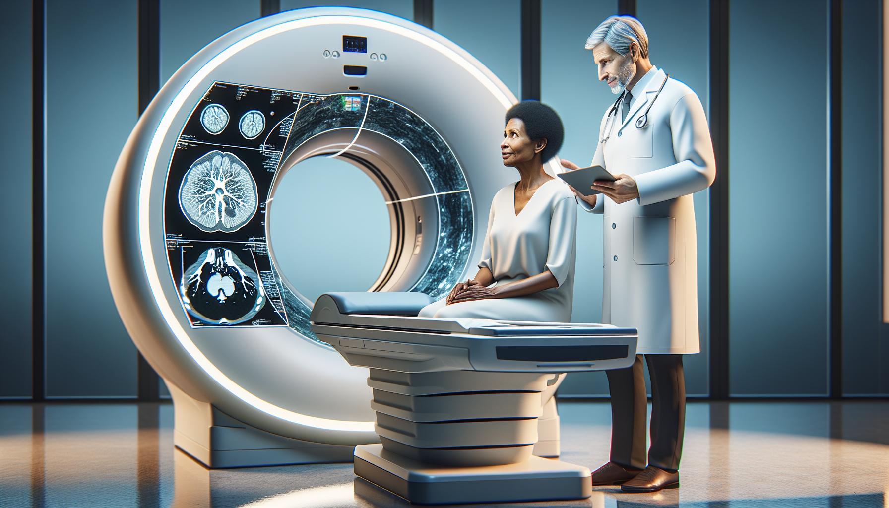
The interpretation of CT images involves looking for distinct features that may signal malignancy. For instance, irregularly shaped masses, enhanced density, or peculiar patterns of lymph node enlargement can all suggest cancer. However, it’s imperative to recognize that while CT scans are powerful diagnostic tools, they are most effective when combined with other diagnostic modalities and clinical evaluations. Thus, patients are always advised to discuss their results thoroughly with their healthcare providers to establish accurate diagnoses and actionable treatment plans.
In essence, understanding the role of CT scans in cancer detection not only empowers patients but also emphasizes the importance of early detection in improving survival rates. Engaging with healthcare professionals about the necessity and implications of CT imaging can help demystify the process and alleviate any concerns regarding its safety and purpose.
How CT Scans Work for Cancer Diagnosis
CT scans are a vital tool in the early detection and diagnosis of cancer, providing detailed images that allow healthcare professionals to delve deep into the body’s structure. By combining multiple X-ray images taken from various angles, CT scans generate cross-sectional views of internal organs, blood vessels, and tissues, which can reveal even the smallest abnormalities. This sophisticated imaging technique enables doctors to identify potential tumor lesions that may not be visible with traditional imaging methods such as standard X-rays.
In terms of functionality, a CT scan utilizes a rotating X-ray machine to capture images that are processed by a computer to create a 3D representation. This allows for an in-depth examination of areas such as the lungs, liver, kidneys, and lymph nodes. The clear resolution provided by CT imaging is particularly beneficial in differentiating between healthy and abnormal tissues, helping distinguish between benign and malignant growths. For instance, a tumor may appear as an irregularly shaped mass with distinct margins, which can signal a cancerous change.
Moreover, the speed and efficiency of CT scans are noteworthy, especially in emergency settings where rapid diagnosis is crucial. Their ability to quickly assess suspected cancers allows for timely interventions, improving the likelihood of successful treatment. While the scan captures images, a contrast material may be used to enhance visibility; this can further clarify the details of certain tissues or blood vessels, making it easier for radiologists to pinpoint areas of concern.
Patients are encouraged to discuss the results of their CT scans with their healthcare providers, as the interpretation of these complex images requires professional expertise. Radiologists look for specific patterns and details that indicate the presence of cancer, such as size, shape, and density of masses. Engaging in these conversations can empower patients, providing comfort and clarity about their health journey. Ultimately, CT scans represent a powerful ally in the fight against cancer, offering a comprehensive view that aids in diagnosis, staging, and treatment planning.
Key Factors Influencing CT Scan Accuracy
The accuracy of CT scans in detecting cancer is influenced by a variety of important factors, which can significantly affect the interpretation of imaging results. Understanding these factors can empower patients and help alleviate concerns as they prepare for this critical diagnostic tool.
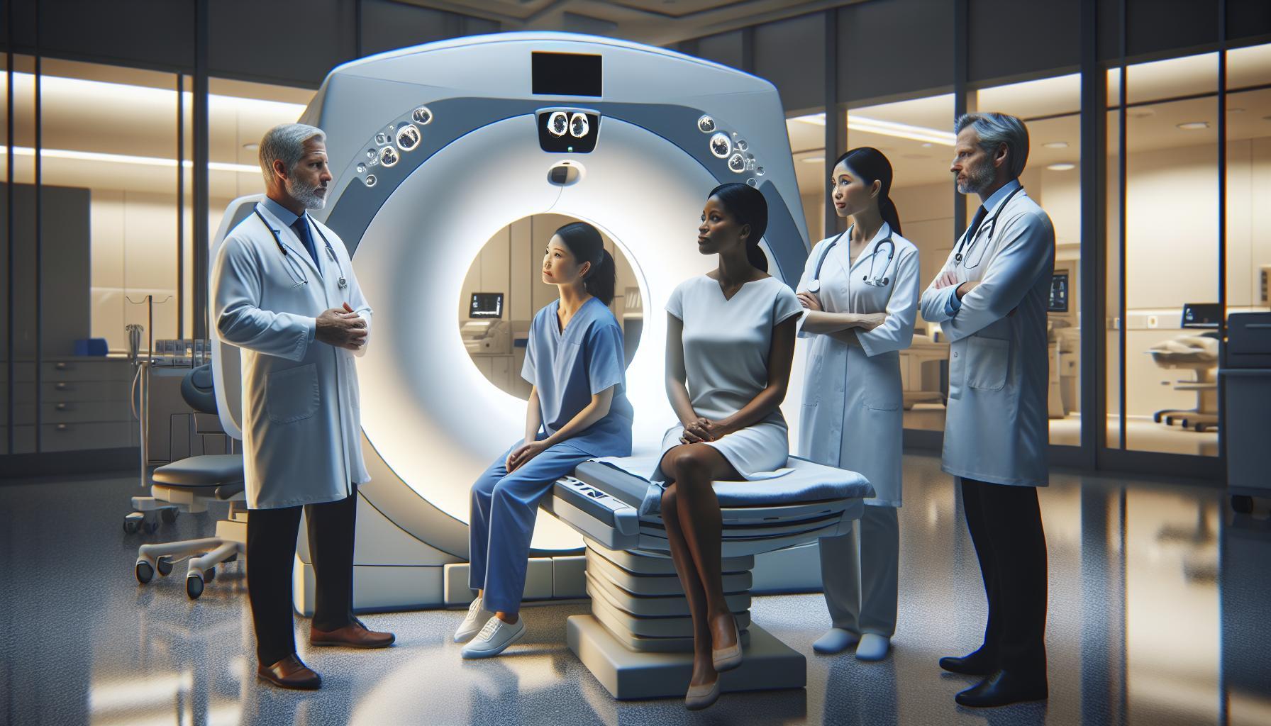
One primary influence is the quality of the CT equipment used. Advances in technology have led to high-resolution imaging, yet older machines may produce less detailed results. Moreover, the skill of the radiologic technologist performing the scan plays a crucial role. Proper positioning and technique ensure that images capture the necessary details for precise evaluation. Patients should not hesitate to ask about the equipment and the technologist’s experience, as these aspects can directly affect scan quality.
The timing of the scan is another critical factor. Concurrent medical conditions, such as infections or ongoing treatments, can alter the appearance of tissues, making it harder to differentiate between benign and malignant growths. Furthermore, a patient’s body composition can impact the clarity of images; for instance, excess adipose tissue may obscure the visualization of certain areas, potentially masking abnormalities. This is why prior medical history and discussions with healthcare providers are essential for tailored imaging strategies.
In addition, the use of contrast agents can enhance visibility, allowing for better delineation of tumors and different tissue types. It’s essential for patients to provide complete information about allergies and prior reactions to contrast materials, as this can influence the choice of agent and the scan’s success.
Finally, the interpretation of images by the radiologist is crucial. Variability in expertise and experience can lead to differences in diagnosis. Patients are encouraged to seek second opinions if there is uncertainty about scan interpretations. By understanding these factors, patients can approach their CT scans with greater confidence, knowing that they are equipped with knowledge that will aid in their discussions with healthcare professionals.
Common Cancer Types Detected by CT Scans
A CT scan is a powerful imaging tool that plays an essential role in the early detection and diagnosis of various cancers. By producing detailed cross-sectional images of the body, CT scans allow healthcare providers to visualize internal structures clearly, which can help in identifying tumors that might otherwise go unnoticed. Some of the most common types of cancer detectable by CT scans include lung cancer, colorectal cancer, pancreatic cancer, liver cancer, and kidney cancer.
Lung Cancer
CT scans are particularly effective in detecting lung cancer, one of the leading causes of cancer-related deaths. High-resolution images can reveal small nodules or masses that may indicate the presence of lung malignancies. Following established guidelines, such as those from the U.S. Preventive Services Task Force, individuals at high risk-such as heavy smokers-may be advised to undergo annual low-dose CT scans for early detection.
Colorectal Cancer
With a CT scan, physicians can efficiently assess the colon and identify polyps or tumors. While CT colonography is used as a screening tool, traditional colonoscopies remain the definitive method for diagnosis. However, if abnormalities are found during screening, CT scans can help determine the extent of disease spread and assist in surgical planning.
Pancreatic and Liver Cancer
Both pancreatic and liver cancers often remain asymptomatic until they are in advanced stages. CT imaging is vital in revealing tumors in these organs, which can be difficult to detect through other modalities. Features such as changes in the size or shape of the pancreas or liver and the presence of any metastatic lesions are crucial for accurate diagnosis and treatment planning.
Kidney Cancer
CT scans are also instrumental in the early detection of kidney cancer. They can help identify renal masses, assess their characteristics, and determine if cancer has spread to surrounding lymph nodes or other organs. The ability to visualize the kidneys in detail can lead to timely and effective intervention, significantly affecting patient outcomes.
The importance of early cancer detection through CT imaging cannot be overemphasized. Regular discussions with healthcare providers about screening options and personal risk factors can pave the way for appropriate imaging and timely diagnosis. Empowering oneself with knowledge can lead to proactive health management, underscoring the importance of staying informed and engaged in your own healthcare journey.
Distinct Features of Cancer on CT Images
Recognizing can significantly empower patients and inform their understanding of the diagnostic process. When healthcare providers analyze CT scans, they look for specific characteristics that may indicate the presence of malignancy. These features often reveal unique patterns that differ from normal tissue, offering insight into the type and stage of cancer.
One of the most critical indicators is the size and shape of lesions. Tumors typically present as masses that can be irregularly shaped or well-defined. Malignant tumors are often larger than benign ones and may demonstrate a growth pattern that looks different from surrounding tissues. In lung cancer, for example, analysis may show nodules with spiculated edges that suggest invasion into nearby structures, while benign nodules are usually round and smooth.
Another critical aspect is the density and enhancement of the lesions on the CT scan. Cancerous tissues often appear denser or can enhance differently after the administration of contrast material, depending on their vascularity and cellular composition. For instance, a hypervascular tumor may absorb more contrast, making it more visible against the background of surrounding normal tissue. Moreover, specific cancers like pancreatic cancer may demonstrate characteristic irregularities in outline and density, illustrating the malignancy’s aggressive nature.
The presence of metastatic disease is also an essential consideration. CT scans can reveal secondary tumors that have spread from their original site to nearby lymph nodes or distant organs. This spread is often visualized by examining the lymphatic pathway and surrounding structures for abnormalities, which can indicate the cancer’s progression. For example, liver metastases may appear as round lesions with varying degrees of enhancement compared to the liver parenchyma, providing essential information on the extent of disease spread.
Understanding these distinct features not only aids in early detection but also fosters a sense of active participation in one’s healthcare. Engaging in open discussions with healthcare providers about CT findings can elucidate concerns and clarify the next steps in diagnosing and managing cancer, ultimately reducing anxiety and empowering informed decision-making.
Preparation Steps Before a CT Scan
Before undergoing a CT scan, it’s essential to take several preparatory steps to ensure the procedure goes smoothly and the results are as accurate as possible. While the prospect of a CT scan can feel daunting, being well-prepared can help alleviate any anxiety and enhance the effectiveness of the imaging process.
First and foremost, your healthcare provider will guide you on whether you need to modify your diet prior to the scan. For some types of CT scans, particularly those involving the abdomen or pelvis, you may be instructed to avoid eating or drinking for several hours beforehand. This fasting helps to reduce any food or fluid interference that might obscure the images. It’s also important to inform your doctor about any medications you’re taking, as some may need to be paused or adjusted before the scan.
When you arrive at the imaging center, expect to fill out a form detailing your medical history, allergies, and any previous imaging studies. This information is crucial as it assists the radiology team in taking the best possible images tailored to your medical needs. If contrast dye will be used during your CT scan, often to enhance visualization of specific organs or tissues, be sure to mention any allergies to contrast materials, iodine, or shellfish, as this could pose a risk.
As you prepare for your CT scan, it may also be helpful to wear comfortable clothing without metal fasteners or zippers, as these can interfere with image quality. You might be provided with a gown to change into before the procedure. Lastly, bringing a family member or friend along can provide emotional support and help you recall any instructions given by medical personnel, ensuring you feel more at ease.
Overall, taking these preparation steps seriously is vital for maximizing the benefits of your CT scan and minimizing any potential risks. Always communicate openly with your healthcare team about any concerns you may have-they are there to support you through the entire process and ensure your comfort and safety.
Possible Risks and Safety Considerations
While CT scans are invaluable tools for diagnosing cancer, it’s essential to be aware of the potential risks and safety considerations associated with these imaging procedures. Understanding these factors can help alleviate concerns and empower you to make informed decisions about your healthcare.
One notable risk involves exposure to radiation. CT scans utilize X-rays, which involve ionizing radiation, unlike standard X-rays. While the amount of radiation in a single CT scan is generally considered safe and the benefits often outweigh the risks, cumulative exposure can raise concerns, especially for individuals who require multiple scans over time. The American College of Radiology recommends that CT scans be performed only when necessary and urges patients to discuss any previous imaging experiences with their healthcare provider to assess overall exposure.
Another safety consideration centers on the use of contrast materials during the procedure, which helps enhance the quality of the images. Some patients may experience allergic reactions to these substances, particularly iodine-based contrast dyes. Prior to undergoing a CT scan, it’s crucial to inform the medical team about any known allergies or previous adverse reactions to contrast agents. Additionally, patients with certain pre-existing conditions, such as kidney dysfunction, may be at an increased risk of complications from contrast use, necessitating further assessment or alternative imaging methods.
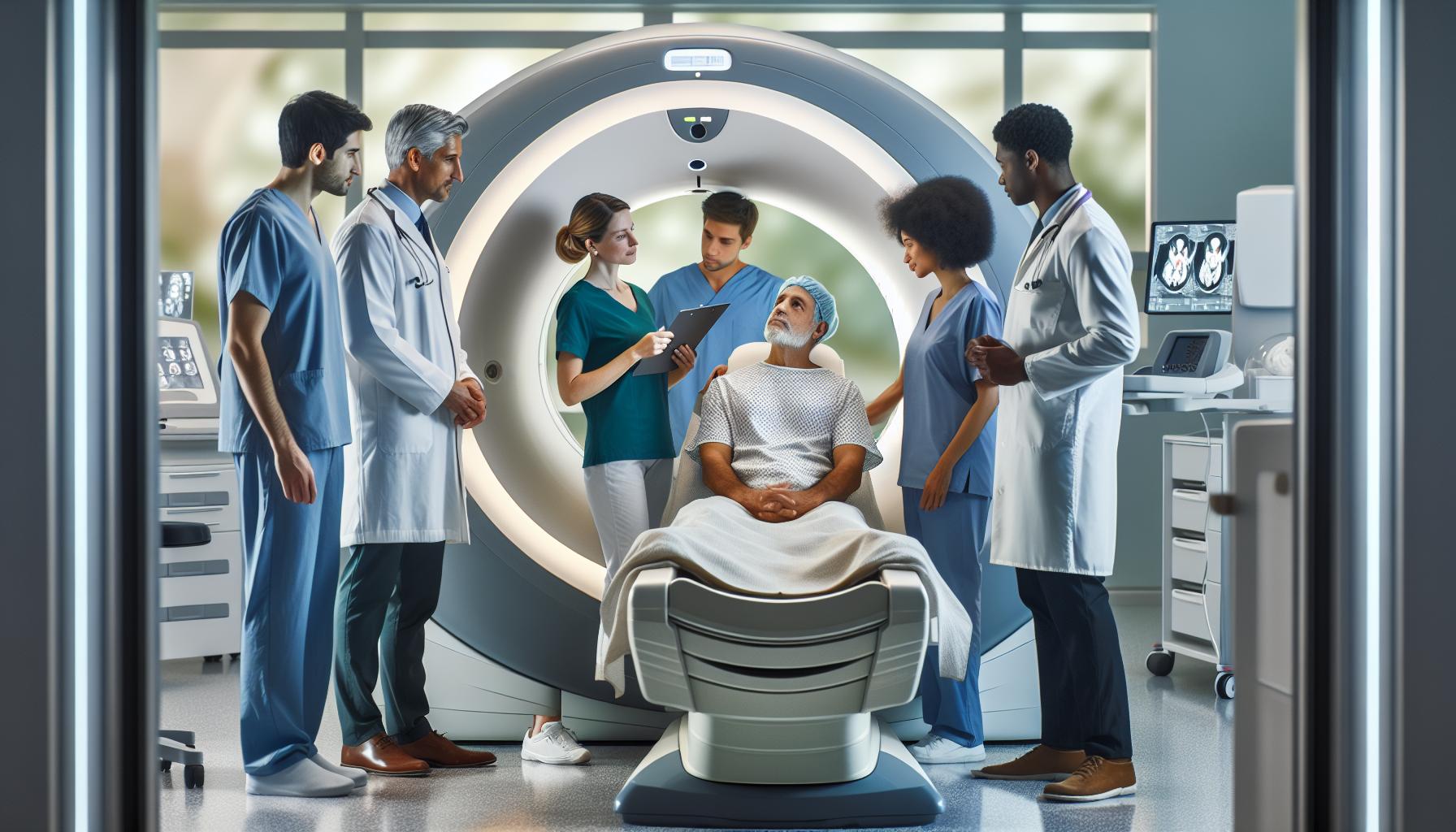
Finally, it’s important to recognize the psychological aspects of undergoing a CT scan. The anticipation of finding out whether cancer is present can create anxiety in many individuals. Open communication with your healthcare provider about these feelings can provide support and clarity. Your provider can walk you through the process, answer any questions, and provide reassurance about the steps taken to ensure your safety and well-being throughout the imaging procedure.
By remaining informed and communicating openly with your healthcare professionals, you can mitigate risks associated with CT scans and prioritize your health effectively.
What to Expect During a CT Scan
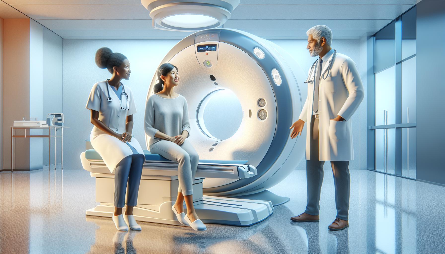
Having a CT scan can be a pivotal step in the journey of understanding your health, especially when it comes to diagnosing cancer. Preparing mentally and physically for this imaging procedure can make the experience smoother and more comfortable. As you enter the imaging room, it’s worth noting that the machine you will be positioned in is not as intimidating as it might seem. Often described as a large doughnut, the CT scanner uses a narrow beam of X-rays to capture detailed images of your body from various angles, allowing your doctor to see any abnormalities, including signs of cancer.
During the procedure, you’ll be asked to lie down on a motorized table, which will move you in and out of the circular opening of the CT scanner. It’s imperative to remain still while the images are being taken; even the slightest movement can blur results. The technician will communicate with you throughout the scan, reassuring you and directing you on breath control, if necessary-typically, patients are instructed to hold their breath for a few seconds. While the scan itself generally lasts about 10 to 30 minutes, the total time spent at the facility could be longer due to preparation and post-scan instructions.
After completing the scan, you may be advised to wait for the images to be reviewed for quality. This timeframe can vary, but don’t hesitate to express any concerns or ask questions at this point; your healthcare team is there to support you. Patients should also account for any specific instructions regarding hydration, especially if a contrast agent was used, as staying well-hydrated can help flush the contrast material from your system.
Ultimately, while the thought of a CT scan can be daunting, understanding what to expect during the process can significantly reduce anxiety. This imaging technique plays a crucial role in detecting cancer, and knowing the steps involved can help empower you on your health journey. Always consult with your healthcare provider if you have any lingering questions or concerns leading up to your scan.
Interpreting CT Scan Results: A Patient’s Guide
Understanding the results of a CT scan can be a critical component of your healthcare journey, particularly when screening for cancer. CT scans are powerful diagnostic tools that create detailed images of internal organs, allowing physicians to detect anomalies that might indicate the presence of cancer. However, interpreting these images is not straightforward and requires expertise. Knowing what to expect and how to approach the results can alleviate some of the stress and uncertainty.
Radiologists analyze the CT scan images, looking for subtle variations in tissue density and structure. Depending on the findings, they may note abnormalities such as masses, lesions, or changes in organ shape. It’s important to understand that not all abnormalities on a CT scan are cancerous; some may be benign or related to other conditions. Your healthcare provider will provide a comprehensive report detailing the findings and any recommendations for follow-up actions.
Key Aspects of CT Scan Results
When discussing your CT scan results with your doctor, consider focusing on the following aspects:
- Type of Findings: Be clear about what was noted on the scan-whether any masses were detected, their size, and location.
- Next Steps: Ask about recommended follow-up tests or procedures based on the findings.
- Context: Understanding the context is crucial. Discuss previous medical history, and correlate your symptoms with the CT scan results.
While waiting for the results can be stressful, it is essential to maintain open communication with your healthcare provider. They can help clarify the implications of the findings and the likelihood of various diagnoses, including whether further imaging or biopsies are necessary. Emphasizing the importance of follow-up care, having a supportive network, and engaging in healthy coping strategies can greatly improve your overall experience during this time.
Ultimately, being informed and prepared for discussions about your CT scan results can empower you to take an active role in your healthcare decisions. Remember, your healthcare provider is there to guide you through this process and provide the clarity you need.
Next Steps After CT Scan Findings
After receiving the findings from your CT scan, it’s essential to navigate this next phase with clarity and confidence. Your healthcare provider will typically discuss the results in detail, highlighting any abnormalities or areas of concern. This conversation can set the tone for your next steps, so it’s crucial to be prepared and proactive. Take this opportunity to ask questions about the findings, such as the nature of any detected lesions, their implications, and what they could mean for your health.
Most importantly, determine the recommended follow-up actions. This could involve additional imaging studies, such as MRI or PET scans, biopsies to assess the nature of a suspicious mass, or monitoring certain areas over time to track any changes. Documenting your questions and concerns in advance can help you feel more empowered during this discussion. Consider asking about the rationale behind each recommended step, as understanding the reasoning can provide reassurance and clarity amidst uncertainty.
In some cases, the results might indicate the presence of cancer, which can be a daunting realization. If this occurs, ensure that you have a robust support system in place. Emotional and psychological support, whether from friends, family, or mental health professionals, can be invaluable. Additionally, collaborating with your oncology team is essential for discussing treatment options and considering further tests required for a comprehensive evaluation of your condition.
Finally, remember that staying informed is your best ally. Educate yourself about potential next steps and treatment paths, whether surgical, chemotherapy, radiation, or a combination of approaches. Engaging in discussions with your healthcare team will allow for personalized care that aligns with your goals and preferences. Emphasizing open communication and informed decision-making will enable you to actively participate in your health journey and navigate the challenges ahead with confidence and understanding.
Advancements in CT Technology for Cancer Detection
The evolution of CT scan technology has significantly enhanced its effectiveness in cancer detection, offering patients and healthcare providers powerful tools for early diagnosis and treatment planning. Recent advancements have focused on improving image quality, reducing radiation dose, and integrating innovative techniques that bolster diagnostic accuracy.
Enhanced Image Quality
Modern CT scanners utilize advanced imaging algorithms that allow for higher resolution images with greater detail. These enhancements enable radiologists to observe subtle differences in tissue density that may indicate the presence of tumors or other abnormalities that could be overlooked by older machines. For instance, technologies such as iterative reconstruction techniques minimize noise and improve contrast resolution, facilitating better visualization of small lesions and providing a clearer picture of a patient’s anatomy.
Reduced Radiation Exposure
Patient safety remains a top priority, and innovations in CT technology have led to a considerable reduction in radiation exposure. Newer machines are designed to deliver the lowest possible dose while still producing high-quality images. Low-dose protocols and automatic exposure control systems adjust the radiation based on the patient’s size and the specific imaging requirements, ensuring that patients receive minimal radiation without compromising diagnostic quality.
Integration with Other Modalities
The integration of CT scans with other imaging modalities, such as PET (Positron Emission Tomography), offers a more comprehensive view for cancer detection. For example, PET/CT scans combine metabolic information from a PET scan with anatomical details from a CT scan, allowing for more precise localization of cancerous lesions. This fusion of data not only aids in identification but also provides valuable insights into the tumor’s activity, helping in the assessment of treatment responses over time.
As these technologies continue to advance, patients can expect even more accurate and safer imaging options for cancer detection. If you are considering a CT scan, discussing the latest technologies with your healthcare provider can ensure that you receive the best possible care tailored to your health needs.
When to Consider a CT Scan for Cancer Screening
A CT scan can be a crucial tool in the early detection of cancer, allowing for the identification of abnormalities that may not be visible through other imaging techniques. When considering whether to undergo a CT scan for cancer screening, various factors should be taken into account, including personal health history, risk factors, and specific symptoms. A discussion with a healthcare provider can help clarify these considerations.
Individuals at higher risk for certain types of cancer, such as long-term smokers or those with a family history of specific cancers, may benefit from regular screening with CT scans. For instance, the U.S. Preventive Services Task Force recommends annual low-dose CT scans for current and former smokers aged 50 to 80 who have a significant smoking history. This proactive approach aims to catch lung cancer at its most treatable stage.
It’s also important to consider symptoms you may be experiencing, such as unexplained weight loss, persistent cough, or chest pain, which could indicate underlying issues that might require further investigation. In such cases, a CT scan can provide detailed images that reveal the presence of tumors or other anomalies.
Keep in mind that while CT scans provide invaluable diagnostic information, they do involve exposure to radiation. Therefore, they should be appropriately timed, balancing the necessity of the scan against the potential risks. Ultimately, consulting with healthcare professionals, who can tailor recommendations based on individual health needs and circumstances, is key to determining when a CT scan is appropriate for cancer screening.
Frequently Asked Questions
Q: How effective are CT scans at detecting cancer?
A: CT scans are highly effective in detecting many types of cancerous lesions. They provide detailed images of the body’s internal structures, allowing for the identification of abnormal growths. However, their accuracy can vary based on factors like tumor size and location.
Q: Can CT scans detect early-stage cancer?
A: Yes, CT scans can detect early-stage cancer, particularly in areas like the lungs and abdomen. Early detection increases treatment effectiveness, making timely imaging essential for high-risk individuals.
Q: Are there any types of cancer that CT scans cannot detect?
A: CT scans may struggle with certain types of cancer, especially those located in areas like the pancreas or small intestines. In these cases, alternative imaging methods or procedures might be necessary for accurate diagnosis.
Q: How do doctors interpret CT scan results for cancer?
A: Doctors interpret CT scan results by examining the images for abnormalities, assessing their characteristics, and comparing them with previous imaging studies. They consider factors like size, shape, and location of any tumors to determine further action.
Q: What should patients do if their CT scan shows suspicious findings?
A: If a CT scan shows suspicious findings, patients should consult their healthcare provider for further evaluation. This may include additional imaging, biopsies, or consultations with specialists for comprehensive assessment and treatment options.
Q: What is the role of contrast agents in CT scans for cancer detection?
A: Contrast agents enhance the visibility of certain tissues and blood vessels during a CT scan. They can help differentiate between normal and abnormal structures, improving the accuracy of cancer detection.
Q: How often should someone at high risk for cancer get a CT scan?
A: Individuals at high risk for cancer should discuss a suitable scanning schedule with their doctor. Recommendations vary but may suggest annual or biannual scans depending on personal risk factors and medical history.
Q: Can lifestyle factors affect the accuracy of CT scans in detecting cancer?
A: Yes, lifestyle factors such as smoking and obesity can influence CT scan accuracy. These factors may create conditions that alter the appearance of tissues, potentially complicating the interpretation of scan results.
To Conclude
Understanding how CT scans play a role in detecting cancer can empower you on your health journey. While CT scans are a vital tool in identifying abnormalities, they are part of a broader diagnostic approach that includes consultations with healthcare professionals and potentially further testing. If you still have questions about the CT scan process or need to explore your options, consider visiting our detailed guide on preparing for a CT scan and understanding cancer screenings.
We encourage you to share your thoughts or experiences in the comments below-your insights may help others in similar situations. Don’t forget to check out our newsletter for ongoing updates on health topics like medical imaging and cancer detection, ensuring you remain informed and proactive about your health. Remember, while knowledge is power, consulting with your healthcare provider will give you personalized guidance suited to your situation.


