Understanding how to read a head CT scan can empower you to better interpret medical information, whether you’re a healthcare professional or simply curious about the process. CT scans are crucial tools in diagnosing conditions like strokes, tumors, and traumatic brain injuries. An estimated 1.7 million people suffer brain injuries each year in the U.S. alone, making the ability to understand these imaging studies all the more valuable.
In this guide, we’ll break down the fundamental steps of reading a head CT scan, highlighting key aspects to look for, including signs of bleeding or structural abnormalities. By equipping yourself with this knowledge, you can bridge the gap between raw data and clinical significance, enhancing your ability to engage in informed discussions with healthcare providers. Prepare to demystify the CT process and gain confidence in your understanding, ensuring you know what to look for and why it matters.
Understanding the Basics of Head CT Scans
Computed Tomography (CT) scans are a vital tool in modern medicine, particularly for evaluating the brain and head. Using a series of X-ray measurements, CT scans create detailed cross-sectional images of the head, helping to diagnose various conditions such as injuries, tumors, and stroke. This noninvasive imaging technique is instrumental in providing rapid and accurate assessments, particularly in emergency situations.
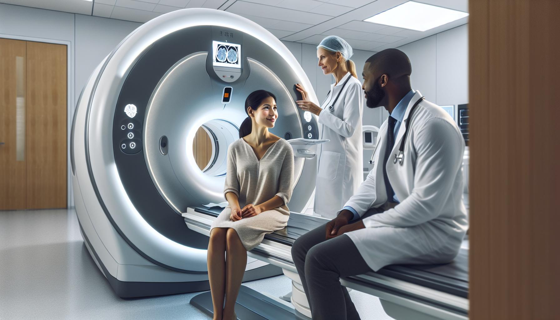
One of the critical benefits of a head CT scan is its speed. The process typically takes only a few minutes, making it an ideal choice for emergencies where time is crucial. Patients lie down on a sliding table that moves through a doughnut-shaped scanner. As the table shifts, the X-ray machine rotates around the patient, taking multiple images from different angles. These images are then processed by a computer to produce detailed 2D and 3D representations of the brain’s anatomy.
CT scans are particularly useful for evaluating symptoms like severe headaches, dizziness, and confusion, as well as for assessing the extent of head trauma. They can identify bleeding, fractures, and other critical factors that may require immediate medical attention. The clarity of the images produced helps healthcare professionals make informed decisions swiftly, often leading to better outcomes for patients.
While the prospect of undergoing a CT scan may induce anxiety, it’s essential to remember that this procedure is not only safe but also a critical step in understanding your health. Always consult with your healthcare provider for personalized information and guidance, ensuring you are well-informed and prepared for your upcoming imaging appointment.
Key Indicators for Head CT Imaging
When considering a head CT scan, several key indicators may prompt healthcare providers to recommend this imaging procedure. Understanding these indicators can help demystify the process and provide reassurance about the necessity and urgency of the scan.
First and foremost, head CT scans are often indicated for patients presenting with neurological symptoms such as severe headaches, confusion, dizziness, or loss of consciousness. These symptoms can point to potentially serious conditions, including bleeding in the brain or stroke. For instance, if a patient is involved in a fall or an accident and exhibits signs of head trauma, a CT scan can quickly assess for internal bleeding or fractures, allowing for immediate intervention if needed.
Additionally, specific medical conditions warrant the use of head CT imaging. Conditions like cerebrovascular accidents (strokes), tumors, and significant injuries can be effectively evaluated with a CT scan. The quick nature of the scan – typically completed within minutes – makes it extremely beneficial in emergency settings where rapid diagnosis and treatment are crucial.
Routine evaluations following known medical conditions, such as monitoring the progression of tumors or assessing the effectiveness of treatment, can also necessitate the use of a head CT. In these scenarios, both the patient and the healthcare provider need clear, detailed images to make informed clinical decisions regarding ongoing management.
To sum up, the key indicators that lead to a head CT scan often revolve around the need for fast and clear imaging to diagnose serious health concerns. If you ever find yourself questioned about whether a CT scan is right for your situation, don’t hesitate to communicate openly with your healthcare provider. They can assess your symptoms and medical history, ensuring you receive the appropriate care.
Preparing for Your Head CT Procedure
Preparing for a head CT scan can often feel daunting, but being well-informed can alleviate much of the anxiety surrounding the procedure. Understanding your preparation steps is crucial to ensuring that your scan is effective and that you have a smooth experience at the imaging facility.
To start, it is essential to follow your healthcare provider’s specific instructions regarding food and drink. For many head CT scans, particularly those performed without contrast, you may be advised that it is safe to eat a light meal before the scan. However, in cases where a contrast agent is used, it is typically recommended to avoid eating or drinking for at least two hours prior to your appointment. Always make sure to clarify with your medical team based on the specifics of your procedure.
Dressing appropriately will also contribute to your comfort during the scan. Opt for loose-fitting clothing that is free of metal objects, such as zippers or buttons. For women, wearing a top that buttons or zips in the front can be particularly helpful since you may need to remove your top for the scan. If you have any accessories that may contain metal, it is best to leave them at home to expedite the process.
Lastly, upon arriving at the facility, allow yourself plenty of time to complete any required paperwork or pre-procedure screenings. Communicate openly with your technician about any allergies, especially to contrast materials, and any prior reactions you may have had related to imaging procedures. For patients with concerns about claustrophobia or anxiety, discussing these feelings beforehand can lead to recommendations for comfort measures during the scan.
By preparing thoughtfully and embracing the opportunity to ask questions, you can approach your head CT scan with confidence and ease. Your healthcare team is there to support you and ensure you receive the highest level of care throughout the process.
What to Expect During a Head CT
Undergoing a head CT scan can feel intimidating, but understanding the process can alleviate much of that anxiety. Once you’re settled into the imaging facility, you’ll be greeted by a radiologic technologist who specializes in performing such scans. They will review your health history briefly to ensure that everything is set for a safe and effective imaging session. If you are receiving a contrast agent, they may ask you more questions about any potential allergies to contrast materials.
Once you’re ready, you’ll lie down on a motorized table that slides into the CT scanner-a large, donut-shaped machine. It’s important to remain as still as possible during the scan since movement can blur the images. The technologist may ask you to hold your breath for short periods as the scans are taken, typically lasting only a few minutes. The scanner will make a rhythmic whirring sound, which is completely normal and part of the imaging process.
While you’re in the scanner, it’s natural to feel a bit uneasy, especially if you have concerns about claustrophobia. Some imaging centers offer options to help with anxiety, such as playing calming music or using a wider scanner that feels less confined. Additionally, a family member or friend may be allowed to accompany you in the waiting area to provide support you may need before and after the scan.
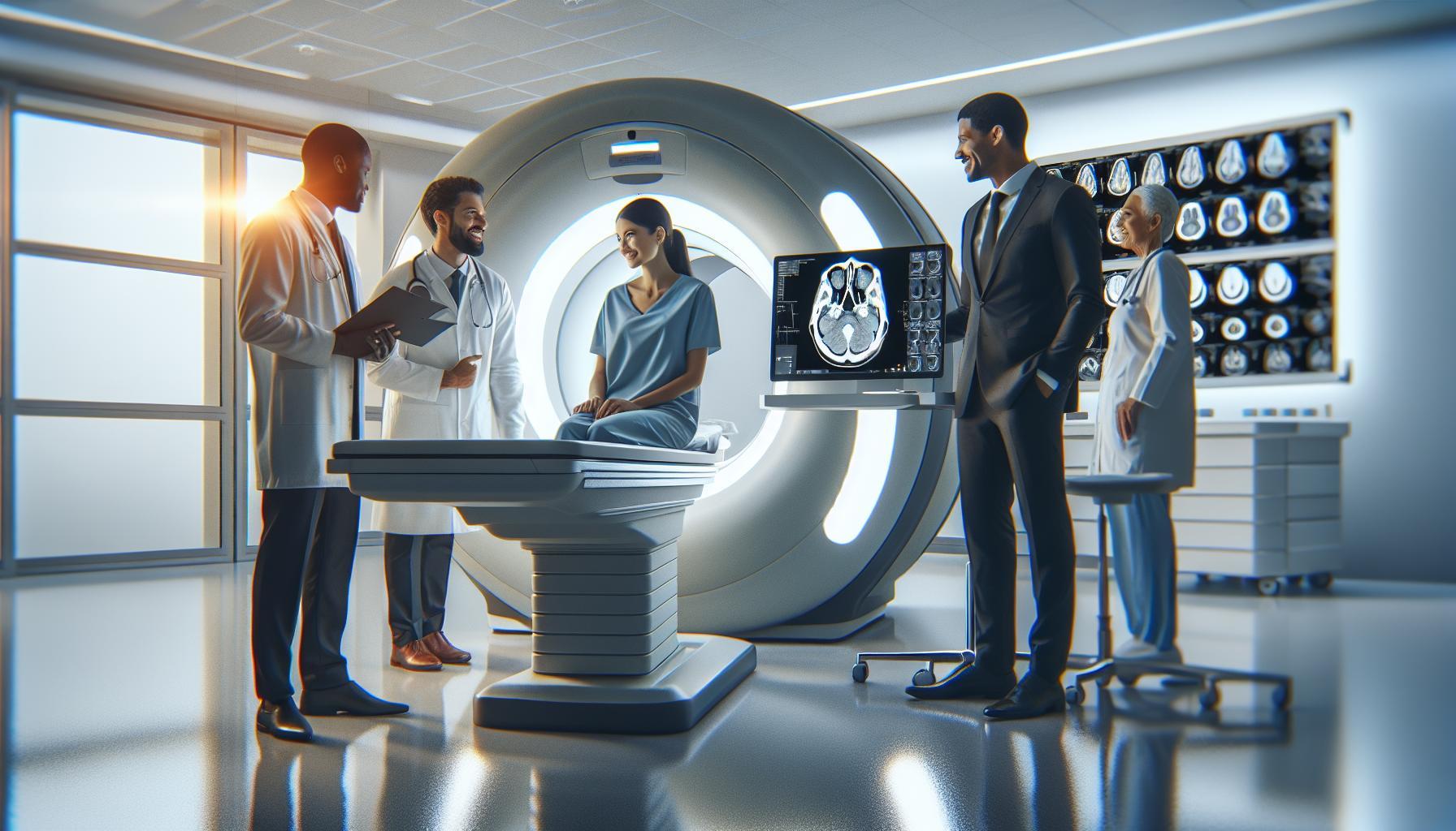
Finally, once the images are captured, you’ll be able to leave the imaging facility, often without any restrictions to your usual activities, especially if no contrast was used. The radiologist will analyze your scans and send the results to your referring doctor, who will interpret the findings and discuss them with you. This entire process usually takes less than an hour, making it a quick but essential step in diagnosing or monitoring your health condition. Understanding and preparing for scan can empower you to approach the process with confidence, paving the way for clearer communication with your healthcare providers about your results and next steps.
Interpreting CT Scan Results: A Step-by-Step Guide
Interpreting a CT scan can seem daunting, but breaking the process into manageable steps can make it more accessible. Understanding the key elements can greatly assist you in grasping what your scan results may indicate about your health. When reading a head CT scan, start by familiarizing yourself with the anatomy visible in the scan. This includes recognizing the brain’s various regions, cranial structures, and adjacent tissues, which is essential for identifying abnormalities effectively.
To begin the interpretation, follow this structured approach:
Step 1: Assess the Basics
Start with a general overview of the scan. Look for any obvious anomalies such as asymmetry, mass effects, or significant deviations from normal anatomical structures. Common points of focus include:
- Brain Size and Shape: Are there signs of swelling (edema) or atrophy?
- Cisterns and Sulci: Are these spaces normal or showing abnormal fluid levels?
- Ventricles: Check for enlargement, which may indicate conditions like hydrocephalus.
Step 2: Check for Blood
Utilize the mnemonic “Blood Can Be Very Bad,” which highlights that blood is generally a critical finding. Examine areas for:
- Intracerebral Hemorrhage: Look for areas of hyperdensity that indicate bleeding.
- Subarachnoid Hemorrhage: Assess for blood in the basal cisterns.
- Subdural and Epidural Hematomas: Identify crescent-shaped or lenticular hyperdense areas, respectively.
Step 3: Evaluate Bone
Examine the skull for fractures or degenerative changes. Look for linear or depressed fractures, and assess if there are any suspicious bone lesions.
For beginners, keep in mind that CT scans capture images with varying levels of density, so understanding how to differentiate normal variations from pathology is crucial. Textures and colors on the scan display can inform you about tissue types and conditions: for instance, fat appears dark, while bone appears bright due to its density.
Step 4: Consult with Professionals
While these steps can guide your interpretation, remember that a radiologist’s expertise is invaluable. They will provide a comprehensive analysis and context beyond what is visible in the images. Always discuss findings with your healthcare provider to get a tailored explanation of how these results relate to your health.
In conclusion, while interpreting CT scan results involves specific steps and knowledge, remaining calm and seeking expert advice will empower you in understanding what the images convey about your health.
Common Findings on Head CT Scans
When examining a head CT scan, you may encounter a variety of findings, some of which are quite common and typically warrant little concern, while others may indicate a need for further investigation. Understanding these common findings can help demystify the imaging process and alleviate some of the anxiety that often accompanies medical procedures.
Among the frequent discoveries on head CT scans, hemorrhages are particularly noteworthy. Intracerebral hemorrhage, for instance, manifests as areas of high density on the scan and may suggest trauma or vascular disorders. Subdural and epidural hematomas can appear as crescent-shaped or lenticular hyperdense areas, respectively, depending on their location and underlying cause. These findings often prompt immediate attention due to their potential implications on brain function and integrity.
Another aspect to consider includes brain morphology. Variations in brain size and shape, such as signs of swelling (edema) or atrophy, can provide clues to underlying conditions. Additionally, examining the ventricles is crucial; enlarged ventricles may suggest hydrocephalus, a condition characterized by an accumulation of cerebrospinal fluid. Changes in the cisterns and sulci, which are the spaces and grooves in the brain, may indicate abnormal fluid levels, requiring further evaluation.
In addition to these abnormalities, changes related to skull integrity may also be present. Fractures or degenerative changes in the skull can be evident in the CT images, indicating past trauma or age-related alterations. Recognizing these conditions fosters a better understanding of the imaging results and their clinical significance, reinforcing the importance of discussing all findings with your healthcare provider for tailored interpretation and guidance.
Ultimately, while common findings on a head CT scan can be reassuring, they are best understood within the context of comprehensive medical care. Always consult your healthcare professional to discuss your specific results, as they can provide the context needed to understand what these findings mean for your health and well-being.
Identifying Abnormalities in Head CT Images
is a crucial skill that can offer significant insights into a patient’s neurological health. The intricacies of these scans may seem daunting at first, but with an understanding of what to look for, it becomes easier to discern areas of concern. Many head CT images display variations that may not warrant alarm, yet recognizing specific abnormalities can guide healthcare professionals towards appropriate interventions when needed.
One common abnormality that arises is the presence of hemorrhages. These may appear as hyperdense areas on the scan, which signal potential issues such as trauma or chronic vascular conditions. For instance, an intracerebral hemorrhage typically manifests as a localized area of increased density, prompting immediate clinical evaluation. Similar attention is warranted for subdural and epidural hematomas, which appear in distinct shapes – crescent-shaped and lenticular, respectively. Each of these findings can suggest varying pathology and thus necessitates further assessment.
In addition to hemorrhages, changes in brain morphology can also indicate underlying issues. Signs of swelling, known as edema, or brain atrophy can be detected, shedding light on chronic conditions such as neurodegeneration. Important landmarks to monitor include the size of the ventricles, where enlargement may hint at conditions like hydrocephalus. It’s essential to assess the surrounding cisterns and sulci; abnormal changes here can indicate fluid imbalances that may require intervention.
Key Points for Identifying Abnormalities
- Examine for Hemorrhages: Look for hyperdense areas indicating potential bleeding.
- Monitor Brain Morphology: Observe signs of edema or atrophy that can signal chronic conditions.
- Check Ventricles: Enlarged ventricles may suggest cerebrospinal fluid issues.
- Review Surrounding Structures: Changes in cisterns and sulci can indicate fluid imbalance.
Recognizing these abnormalities does not replace the need for professional interpretation; rather, it empowers you to engage in informed discussions with healthcare providers. For optimal health outcomes, always consult with your physician regarding any findings. This approach not only enhances your understanding but allows for tailored medical guidance that considers your unique health profile.
The Role of Contrast in Head CT Imaging
The use of contrast material in head CT imaging significantly enhances the clarity and detail of the images, allowing healthcare providers to make better-informed decisions. Contrast agents, typically iodine-based, are injected into a vein before the scan to highlight blood vessels and various tissues, facilitating the identification of abnormalities that might otherwise go undetected. For instance, administering contrast can help differentiate between different types of tissue and highlight areas of concern, such as tumors or areas of bleeding.
When using contrast in head CT scans, it is important to understand that this additional step may be necessary for certain indications. For example, in cases where a clinician suspects a brain tumor or evaluates the vascularity of a lesion, contrast may be particularly useful. This enhanced imaging enables a more precise assessment of the location and effect of any abnormal growths, helping guide treatment options.
Before undergoing a procedure involving contrast, patients are usually advised to hydrate well, which helps flush the contrast material out of their system more efficiently post-scan. Though reactions to contrast materials are rare, it’s essential to discuss any allergies or previous experiences with contrast dyes with your healthcare provider, ensuring that appropriate precautions are taken.
The addition of contrast material not only contributes to more comprehensive imaging but also assures patients and doctors alike that they have the most accurate and complete view of the brain’s condition. This helps alleviate anxiety by fostering a collaborative and informed approach to diagnosis and treatment. As always, discussing any questions or concerns with your healthcare professional can help ensure a smooth and understanding experience throughout the imaging process.
Safety Considerations for Head CT Scans
Although head CT scans are invaluable diagnostic tools, understanding the safety considerations involved can greatly alleviate concerns and promote an informed approach to undergoing such procedures. It’s well-known that CT scans expose patients to radiation, and this is a common concern. However, the amount of radiation from a single CT scan is relatively low when compared with the cumulative risk of exposure from other sources. For most individuals, the theoretical risk of developing cancer from a single head CT scan is minimal, estimated to be less than 0.05%[[1]](https://www.mskcc.org/news/scan-safety-radiation-reality-check). Therefore, when considering a head CT scan, the benefits often outweigh the risks, especially if it is key to diagnosing a serious medical condition.
To further ensure safety during a head CT scan, it’s crucial to disclose your entire health history to your healthcare provider, particularly any previous allergic reactions to contrast materials or existing health issues. Patients should also inform their doctor if they are pregnant or may be pregnant, as this information is critical for assessing potential risks and determining the necessity of the scan. Hydration can be important if contrast material is used, as good hydration may help reduce the likelihood of potential side effects and assist in flushing the contrast material from your body post-examination.
During the procedure, patients are typically under minimal discomfort. However, maintaining open communication with the medical staff about any anxiety or discomfort can enhance the experience. They can provide reassurance and adjust the procedure as needed. By understanding safety protocols and consulting with healthcare professionals about any concerns, patients can feel empowered and at ease when preparing for their head CT scans. This proactive approach in discussing safety allows for a clearer understanding of the procedure and reinforces the collaborative relationship between patient and provider, ultimately fostering a more positive healthcare experience.
Frequently Asked Questions About Head CTs
Understanding how to read and interpret head CT scans can often raise many questions among patients and their families. It’s important to have a clear understanding of what to expect, not only from the scanning process but also in terms of results and implications. Here are some common questions that frequently arise regarding head CTs:
What is a head CT scan used for?
A head CT scan is primarily used to diagnose a variety of conditions affecting the brain. These conditions can include traumatic brain injuries, strokes, tumors, and other neurological disorders. The detailed images provided by a CT scan allow healthcare professionals to assess the brain’s structure and detect any abnormalities that may require intervention.
How should I prepare for a head CT scan?
Preparing for a head CT scan is generally straightforward. If your scan involves a contrast medium, you may be advised to refrain from eating or drinking for a few hours beforehand. Always communicate any allergies to contrast material or medications with your healthcare provider. Staying well-hydrated prior to the scan can also be beneficial.
Will I feel any discomfort during the scan?
Most patients find that a head CT scan is painless and quick, typically lasting only a few minutes. You may be asked to remain still and hold your breath for short periods, which can be uncomfortable for some. However, it’s vital to inform the technician if you experience any anxiety or discomfort, as they can provide solutions to improve your experience.
What happens after the scan?
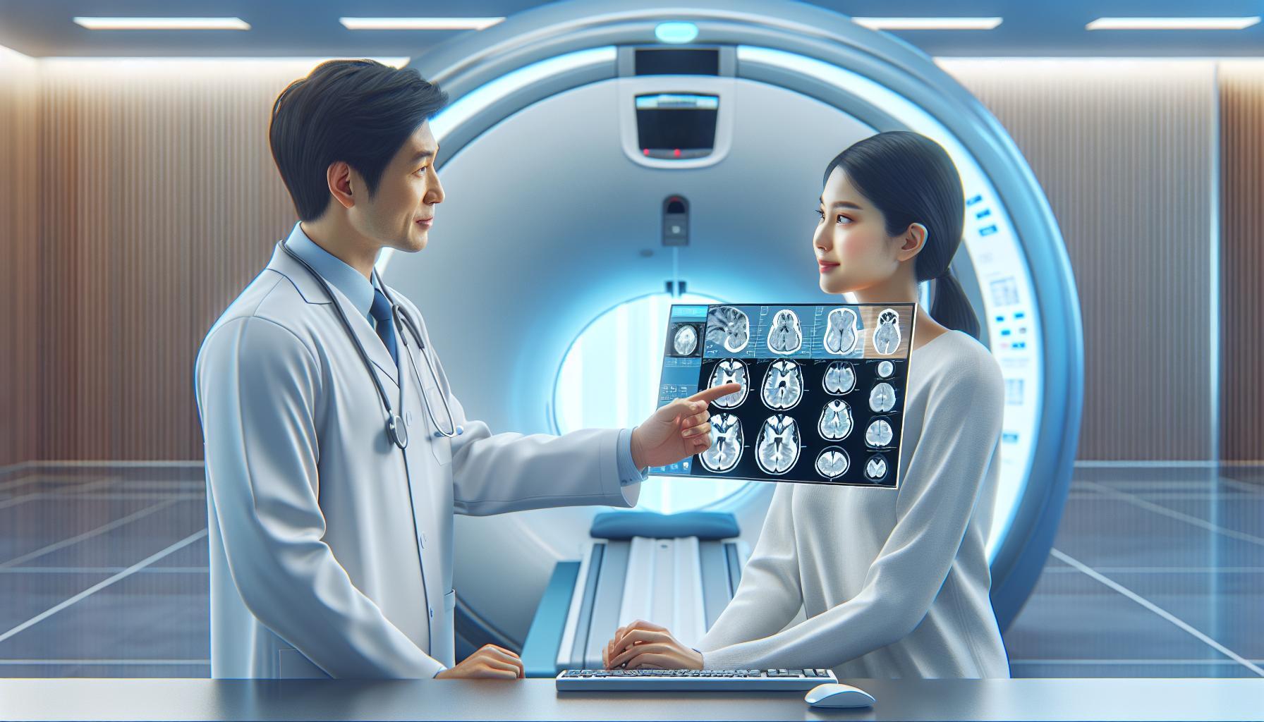
Once the CT scan is complete, the images will be reviewed by a radiologist who will compile a report for your physician. Your doctor will discuss the results with you and what they mean for your health. It’s a good opportunity to ask questions about any findings and the next steps in your care.
Is there any radiation risk associated with CT scans?
CT scans do involve exposure to radiation, but the levels are typically low and the risk of harm is minimal for most individuals. The benefits of accurately diagnosing and treating potential medical issues usually outweigh these risks. If you have concerns about radiation, discuss them with your healthcare provider, who can provide further information based on your specific situation.
Understanding these aspects of head CT scans can help alleviate anxiety and foster a supportive environment for making informed health decisions. Communication with healthcare professionals is key; always feel empowered to ask questions and express concerns about any part of the process.
Advanced Techniques in Head CT Interpretation
Interpreting head CT scans has evolved significantly with advanced imaging techniques that enhance clarity and diagnostic capabilities. For instance, utilizing multiplanar reconstruction allows radiologists to view the images in various planes beyond the standard axial orientation. This capability facilitates a comprehensive assessment of complex anatomical structures, ensuring that subtle abnormalities are not overlooked. By transforming traditional flat images into detailed three-dimensional representations, physicians can pinpoint lesions or other pathologies more accurately.
Another notable advancement is the application of automated image analysis tools, including artificial intelligence algorithms, to assist in identifying stroke, hemorrhage, and tumors. These technologies can expedite diagnosis and potentially reduce human error. For instance, machine learning models are training to recognize specific patterns indicative of certain neurological conditions, which helps radiologists prioritize cases based on urgency and complexity. Although these tools are incredibly beneficial, they serve as supplements to professional evaluation, reinforcing the crucial role of trained clinicians in confirming diagnoses.
Incorporating the use of contrast agents has also proven essential in enhancing CT scan interpretations. Contrast enhancement illuminates blood vessels, highlighting areas of abnormal perfusion that could indicate tumor presence or vascular issues. Understanding when to apply contrast is vital; for example, in suspected cases of cerebral ischemia or to delineate between tumor types.
With advancements in technology, radiologists are better equipped than ever to deliver accurate interpretations that can inform treatment decisions. While this progress is promising, it’s essential to maintain an open dialogue with healthcare providers regarding the implications of scan results, potential follow-up procedures, and overall health management. Ultimately, the synthesis of technology and human expertise ensures a meticulous approach to interpreting head CT scans, facilitating improved patient outcomes.
Tips for Discussing Results with Your Doctor
When facing the results of a head CT scan, it’s normal to feel a mix of anxiety and curiosity. Understanding how to navigate this conversation with your doctor can empower you and ease some of the uncertainty. To ensure a productive discussion, consider coming to your appointment prepared with specific questions. This approach not only clarifies your concerns but also demonstrates your engagement in your own healthcare journey.
Prepare Your Questions
Before your appointment, jot down any questions or thoughts you have about the scan results. Focus on areas such as:
- What do the results indicate about my condition?
- Are there any follow-up tests needed?
- What treatment options are available, and what do they entail?
- Are there lifestyle changes I should consider?
- What are the possible side effects of the suggested treatments?
Having these questions ready can help guide the conversation and ensure you cover all your concerns.
Clarify Medical Terminology
Medical jargon can sometimes feel overwhelming. If your doctor uses terms or phrases that are unclear, don’t hesitate to ask for clarification. Phrases like “benign,” “malignant,” or “hemorrhage” can evoke strong emotions, so it’s crucial to understand what they mean in your specific context. For instance, asking, “Can you explain what this finding means for my overall health?” can lead to a better understanding and more relevant information.
Involve a Support Person
Bringing a family member or friend to your appointment can be incredibly beneficial. They can provide emotional support and help you remember details discussed during the visit. Furthermore, they may think of questions you hadn’t considered. This shared experience can help alleviate some anxiety by allowing you to process the information together.
Effective communication with your healthcare provider about CT scan results not only enhances your understanding but also fosters a collaborative relationship that is critical for your health journey. Remember, your questions are valid, and it’s essential to advocate for clarity and comprehensive understanding in your healthcare discussions.
Frequently Asked Questions
Q: What is a head CT scan used for?
A: A head CT scan is primarily used to assess head injuries, detect strokes, identify brain tumors, and investigate other neurological conditions. It provides detailed images that help doctors diagnose bleeding, swelling, and other abnormalities effectively.
Q: How does a head CT scan differ from an MRI?
A: A head CT scan uses X-rays and computer algorithms to create images, making it faster and often preferred for emergency situations. In contrast, an MRI uses magnetic fields and is better for soft tissue details but takes longer and isn’t always suitable for acute issues.
Q: Can you read a head CT scan without medical training?
A: While basic knowledge might allow for some general understanding, interpreting a head CT scan accurately requires medical training. Healthcare professionals are trained to identify subtleties and abnormalities that are crucial for diagnosis and treatment.
Q: What common conditions can be identified on a head CT?
A: Common conditions visible on a head CT include traumatic brain injuries, hemorrhages, tumors, swelling, and signs of stroke. Detailed imaging helps in differentiating these conditions and guiding treatment.
Q: What steps should be taken after receiving head CT results?
A: After receiving head CT results, it’s essential to discuss them with your doctor to understand implications and next steps. They can explain findings and recommend additional tests or treatments as needed.
Q: Are there any risks associated with a head CT scan?
A: While head CT scans are generally safe, they involve exposure to radiation, which, although minimal, can be a concern if multiple scans are performed. Always discuss potential risks with your healthcare provider before the procedure.
Q: How can I prepare for a head CT scan?
A: Preparation for a head CT scan typically involves informing your doctor of any medications or allergies. You may also be asked to refrain from eating or drinking for a few hours if contrast material is used. Always follow specific instructions given by your healthcare provider.
Q: What does it mean if contrast is used during a head CT scan?
A: If contrast material is used during a head CT scan, it enhances the visibility of blood vessels and specific areas in the brain. This allows for a more detailed assessment of conditions such as tumors or vascular abnormalities, aiding in more accurate diagnosis and treatment planning.
To Conclude
By now, you have a clearer understanding of the essential steps to read a Head CT scan effectively. Remember, while this guide empowers you with knowledge, consulting a healthcare professional remains vital for accurate interpretations and personalized insights. Don’t hesitate to explore more on our site, such as “Understanding CT Scan Safety” and “Common Conditions Diagnosed with CT”, to further enhance your knowledge.
Take the next step-subscribe to our newsletter for the latest updates in medical imaging, and join our community of informed patients and caregivers. Your engagement not only helps you stay informed but also supports others seeking reliable information. If you have questions or want to share your experiences, please leave a comment below. Together, we can demystify the complexities of medical procedures and empower one another with knowledge.


