In today’s world, where health concerns can arise unexpectedly, understanding medical imaging is crucial for many. A CT scan, or computed tomography scan, utilizes advanced technology to create detailed images of the body’s internal structures, making it a vital tool for detecting tumors and other abnormalities. But how effective is a CT scan at spotting tumors? For those facing uncertainty about their health or the implications of a potential diagnosis, knowing what a CT scan can reveal can provide reassurance and clarity.
As you navigate the complexities of medical evaluations, the ability of CT scans to detect tumors may greatly influence your healthcare journey. Whether you’re preparing for a scan, supporting a loved one, or simply seeking knowledge to be proactive about health, understanding the detection capabilities of CT scans helps empower your decisions. Join us as we explore the nuances of CT imaging, its effectiveness in tumor detection, and the factors that can affect the results.
Understanding the Role of CT Scans in Tumor Detection
A CT scan, which stands for computed tomography, plays a crucial role in the detection of tumors across various parts of the body. By producing detailed cross-sectional images, CT scans enable healthcare professionals to visualize internal organs, tissues, and structures with remarkable clarity, leading to better diagnosis. Studies indicate that CT imaging can reveal tumors that may not be identified through traditional X-rays, making it a vital tool in oncology and other medical fields.
The capability of a CT scan to detect a tumor hinges on several factors, including the size and location of the tumor, as well as the specific imaging techniques employed. For instance, smaller tumors, particularly those less than one centimeter, may sometimes evade detection, especially in complex areas like the abdomen or pelvis. However, advancements in technology and the use of specialized algorithms have enhanced the sensitivity of CT scans, enabling the identification of even subtle abnormalities. Furthermore, healthcare professionals often use contrast agents to improve the visibility of certain tissues, ensuring that tumors appear clearly against surrounding structures.
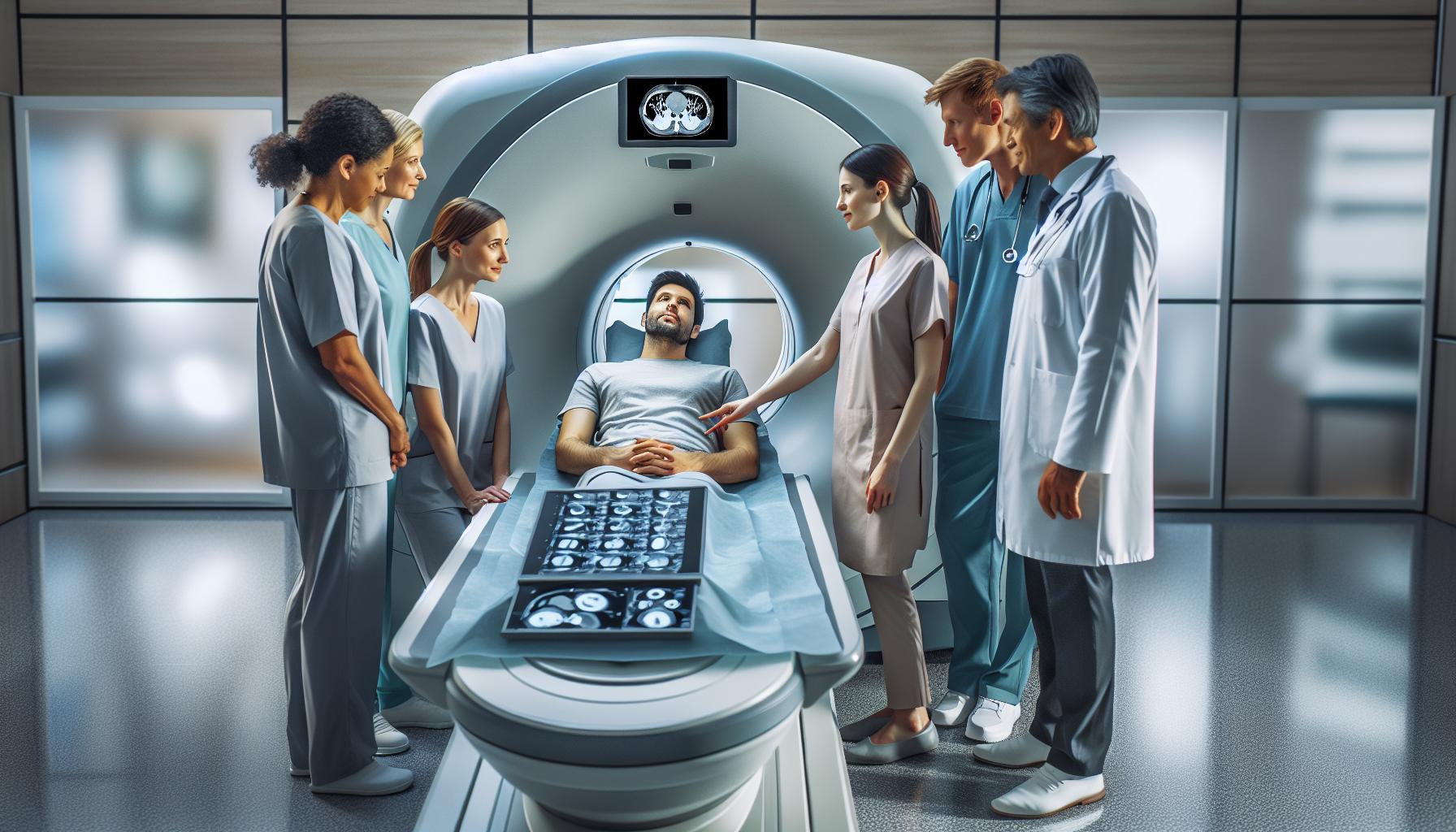
Being aware of how a CT scan functions can alleviate some anxiety surrounding the procedure. Most scans are quick and painless, typically lasting only a few minutes. During the process, the patient lies on a table that slides into a circular machine, where X-ray beams rotate around the body, capturing multiple images that a computer processes into cross-sectional views. These detailed images provide essential information that influences treatment plans and therapeutic decisions. In practice, a patient might undergo a CT scan to evaluate unexplained symptoms, monitor ongoing cancer treatment, or assess the effectiveness of previous interventions.
In summary, CT scans serve as an indispensable tool in the early detection and diagnosis of tumors, profoundly impacting patient outcomes and treatment pathways. The clarity and detail they provide allow for comprehensive assessments, paving the way for timely medical interventions. If you have concerns about a CT scan or its implications for your health, discussing them with your healthcare provider can provide personalized insights and reassurance.
How CT Scans Work: A Step-by-Step Guide
The technology behind computed tomography (CT) scans allows for remarkable insights into the human body, often revealing details that can be crucial for diagnosing conditions like tumors. Understanding how these scans work can help ease any anxiety patients may feel about the procedure and empower them with knowledge regarding their health.
In essence, a CT scan utilizes a series of X-ray images taken from different angles around the body. Here’s a closer look at the step-by-step process:
- Preparation: Before the scan, healthcare providers may give instructions regarding dietary restrictions or medication adjustments. If a contrast agent is to be used, patients might be advised to refrain from eating or drinking for a few hours.
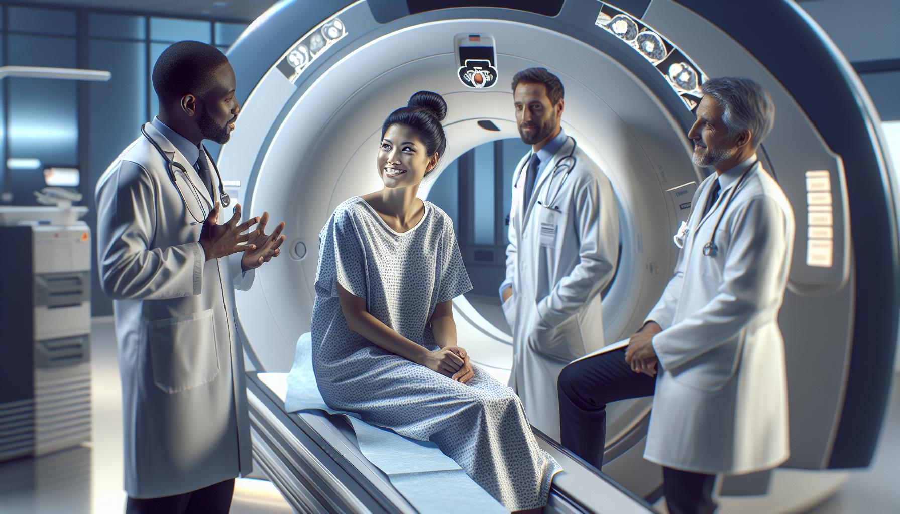
- Positioning: Once at the facility, the patient changes into a hospital gown and lies on a movable table. The technician ensures the patient is comfortable but in a position that allows for optimal imaging.
- Imaging Process: As the table slides into the CT machine, the patient will need to remain still to ensure clear images. X-ray beams rotate around the body, capturing multiple images which a computer compiles into detailed cross-sectional views. This process typically lasts only a few minutes.
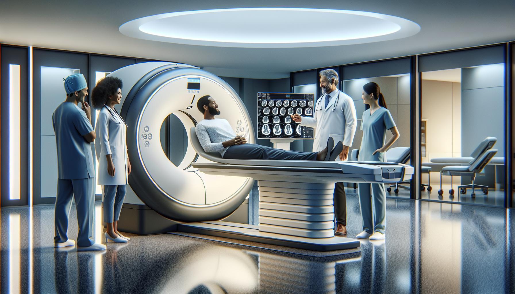
- Post-Scan Evaluation: After the scan, the images are reviewed by a radiologist, who looks for abnormalities such as tumors. The findings will then be discussed with the healthcare provider, who will explain the results and the next steps.
Benefits of CT Scans
During this imaging process, the use of advanced algorithms enhances the ability to detect even small tumors, thereby increasing the chances of early diagnosis. This can be particularly valuable in cases where symptoms are vague or not easily associated with a particular condition.
Ultimately, understanding each step of the CT scan can alleviate concerns about the procedure and highlight its importance in tumor detection. Consulting with healthcare providers throughout the process can provide additional reassurance, as they can tailor recommendations and prepare patients for what to expect.
Key Factors Influencing Tumor Visibility on CT Scans
Certain critical factors play a significant role in the visibility of tumors on CT scans, affecting the likelihood of achieving an accurate diagnosis. Understanding these factors can help alleviate concerns and clarify expectations for patients undergoing imaging procedures.
Firstly, the size and type of tumor are primary considerations. Larger tumors are generally easier to detect, while smaller tumors or those located in challenging anatomical areas can be missed. Tumors can vary widely in density, composition, and growth patterns-some may be solid, while others might be cystic or have fluid-filled components. These characteristics influence how they appear on the scan, affecting visibility. For instance, a solid tumor may present as a well-defined mass, while a cyst may require particular attention to detail in interpretation.
Another element influencing tumor detection is the patient’s body composition and anatomy. Factors such as obesity can impact the clarity of the images due to increased tissue density, potentially obscuring tumor visibility. Additionally, the presence of surrounding structures-like fat, organs, and tissues-can either enhance or diminish the clarity with which a tumor is seen. Variability in individual anatomy means that what can be clearly visualized in one patient might not be as apparent in another.
Moreover, the technique used during the scan is crucial. The radiologist’s experience and the specific settings chosen for imaging, such as slice thickness and contrast enhancement protocols, can make a difference in tumor detection rates. Using a contrast agent can help differentiate between tumors and nearby structures, thereby improving visibility. For instance, vascular tumors or those with significant blood supply often become more evident after contrast administration, providing clearer definitions against non-vascularized tissues.
Finally, the timing and frequency of scans should be considered. Regular imaging follow-ups can reveal changes in tumor size or characteristics that indicate growth, which may not always be apparent in initial scans. Patients are encouraged to discuss any concerns with their healthcare providers and consider tailored follow-up protocols based on individual risks and tumor characteristics.
By understanding these factors, patients can feel more empowered and informed as they navigate their diagnostic journeys. Maintaining open communication with healthcare professionals is essential, ensuring that any questions and concerns are addressed throughout the imaging process.
Common Types of Tumors Detected by CT Imaging
CT imaging plays a crucial role in detecting a wide variety of tumors throughout the body, offering a non-invasive method to provide detailed images that assist healthcare providers in diagnosis and treatment planning. There are several common types of tumors that are frequently detected via CT scans, including:
Solid Tumors
Solid tumors are among the most common types identified through CT imaging. These can occur in various organs, such as the lungs, liver, kidneys, and pancreas. For instance, lung cancer often appears as irregular masses or nodules, while liver tumors might present as distinct lesions that differ in density from surrounding tissue. The well-defined edges of solid tumors can often make them easier to detect than other types, especially in later stages.
Cysts
Cysts are fluid-filled sacs that can occur in many areas of the body, such as the kidneys and ovaries. While many cysts are benign and require no treatment, CT scans can determine their size, shape, and location, helping physicians establish a diagnosis. For instance, a renal cyst may be identified during a routine scan, prompting further observation to ensure it does not lead to complications.
Lymphoma
Lymphomas, which are cancers of the lymphatic system, can also be effectively visualized using CT scans. Enlarged lymph nodes and masses can be detected in areas such as the neck, abdomen, or chest. The ability to assess the size and distribution of these lymphoid tissues helps oncologists formulate treatment strategies and monitor disease progression.
Brain Tumors
CT scans are particularly valuable in assessing brain tumors, providing critical information about tumor size, location, and any associated complications such as swelling or bleeding. The imaging allows healthcare providers to differentiate between types of brain tumors and is often complemented by MRI scans for more detailed evaluation.
Metastatic Disease
One of the significant advantages of CT imaging is its ability to detect metastatic tumors-cancer that has spread from the primary site to other parts of the body. For example, breast cancer frequently metastasizes to the lungs or bones, and CT imaging can reveal the extent of this spread, which is essential for treatment planning.
By understanding these common types of tumors and how they appear on CT scans, patients can feel more informed and empowered as they navigate their healthcare journeys. Remember, any concerns regarding tumor detection should be discussed with a healthcare provider, who can provide personalized insights based on individual medical history and needs. Regular communication and follow-ups are vital for comprehensive cancer care.
Limitations of CT Scans in Tumor Detection
While CT scans are a powerful tool for detecting tumors, they are not without limitations. One of the primary concerns is the reliance on the tumor’s size and density. Smaller tumors, especially those less than a few millimeters, may not be easily detectable. The resolution of CT imaging varies, and tiny lesions can remain hidden, particularly in the early stages of cancer when tumors are often too small to show up distinctly. Additionally, some lesions may overlap with normal structures, making them difficult to differentiate on the scan.
Another important factor is the nature of the tumor itself. Certain types of tumors, such as those that are cystic or have fluid-filled spaces, may appear less conspicuous on CT scans. Furthermore, benign conditions, like inflammatory masses, can mimic malignant tumors, leading to possible misinterpretations. The characteristics of these growths may blend in with surrounding tissues, complicating diagnosis.
Using contrast agents can enhance visibility but introduces its own set of potential issues. Not all patients can tolerate these agents, and there’s a risk of allergic reactions. Additionally, some tumors may not uptake the contrast material as expected, resulting in unclear images. This might lead to the need for further imaging modalities, such as MRI or PET scans, for better visualization and confirmation.
It’s also crucial to consider how the patient’s condition affects scan results. Movement during imaging can create blurring, and variations in breathing or other physiological processes can obscure fine details. Moreover, the interpretation of CT scans is highly dependent on the radiologist’s expertise. An experienced radiologist may spot subtle changes that others might miss, highlighting the importance of a thorough review by skilled professionals.
In light of these challenges, it’s essential for patients to have open discussions with their healthcare providers. Understanding the limitations of CT scans strengthens awareness and aids in developing a comprehensive diagnostic approach that may include additional imaging techniques or biopsies for conclusive diagnosis. Your medical team is there to guide you through the process, ensuring that you receive the best possible care tailored to your individual needs.
The Importance of Contrast Agents in CT Scans
Using contrast agents in CT scans significantly enhances the visibility of potential tumors and other abnormalities. These agents work by altering the way X-rays interact with different tissues in the body, allowing radiologists to differentiate between normal and abnormal structures more effectively. For instance, when a contrast agent is introduced into the bloodstream, it preferentially accumulates in areas with increased blood flow-such as tumors-thereby making these areas appear brighter and more distinct on the images generated by the CT scan.
Understanding the role of contrast agents can alleviate concerns regarding their use. These substances, often iodine-based, are administered either through an injection into a vein or via oral intake. As a result, they help to delineate organs, blood vessels, and the boundaries of tumors, which can be particularly important when assessing complex anatomical regions such as the abdomen or pelvis. By enhancing the clarity of images, contrast agents can reveal the size, shape, and characteristics of tumors that may otherwise be obscured, assisting healthcare providers in making more accurate diagnoses and treatment plans.
Despite their benefits, it’s essential to approach the use of contrast agents with careful consideration. Some patients may experience allergic reactions, while others might have pre-existing conditions that affect how their bodies react to these substances. Communicating your medical history, including any previous adverse reactions to contrast materials, is crucial during your evaluation before the scan. If you have kidney issues or are pregnant, your healthcare provider will review the necessity and safety of using contrast agents in your particular case.
In addition to enhancing image quality, advanced formulations of contrast agents are continuously being developed to minimize potential side effects and improve outcomes. Engaging in a conversation with your healthcare team before the procedure can empower you with knowledge and ease anxiety. They can provide detailed insights into what to expect during the scan, including the process for administering the contrast agent and the reassurance needed to manage any fears you may have about the procedure. By understanding how contrast agents contribute to CT imaging, you can participate more actively in your healthcare journey.
Patient Preparation for Optimal CT Scan Results
Preparing for a CT scan is a crucial step in ensuring that the results are as clear and informative as possible, especially when looking for potential tumors. Understanding the logistics and requirements of your upcoming scan can alleviate anxiety and enhance the quality of the images produced, thereby assisting your healthcare team in making a more accurate diagnosis.
Before the procedure, your healthcare provider will give specific instructions tailored to your individual needs. Hydration is essential, so drink plenty of clear fluids in the days leading up to your scan unless advised otherwise. If your scan requires a contrast agent, your provider may ask you to fast for a few hours beforehand, typically around 4-6 hours, to optimize the imaging results. If you have diabetes or other health conditions, don’t hesitate to discuss your usual routine with your doctor, who may provide additional instructions.
Understanding Dietary Restrictions
It’s important to follow any dietary restrictions provided by your medical team. Here’s what you may typically expect:
- Solid Foods: Avoid eating solid foods for several hours before your scan.
- Liquids: Clear liquids are usually permitted. You can consume water, clear broths, or black coffee (without cream).
- Medications: Take prescribed medications as usual, unless directed otherwise. If you’re unsure, confirm with your healthcare provider.
In addition, inform your doctor about any allergies, especially to iodine or shellfish, as this can affect the use of contrast agents. Be sure to mention any pre-existing medical conditions, particularly related to kidney health, which could influence the decision to use certain substances during your scan.
Feeling nervous before a medical procedure is completely normal. To help ease your worries, consider bringing a comfort item, such as a family photo or a calming playlist on your phone. Many facilities also provide IV sedation for anxious patients; this is something you can discuss with your provider.
Ultimately, being well-prepared not only aids in obtaining clear images but also ensures that you feel supported and informed throughout the process. Always take the opportunity to address any questions you may have with your healthcare provider before your appointment, so you’re fully ready for your CT scan. The more comfortable you are, the better the quality and clarity of the results, helping to guide your next steps in healthcare effectively.
Understanding Scan Results: What to Expect
The anticipation of receiving results from a CT scan can be overwhelming, particularly when the reason for the scan is the search for tumors. Understanding what to expect from these results can help alleviate some of the anxiety that accompanies the waiting period. When the radiologist reviews the images, they look for any unusual masses or abnormalities that could indicate the presence of a tumor, taking into consideration the shape, size, density, and location of any detected anomalies. This process helps in determining whether further action, such as additional imaging or a biopsy, may be necessary.
Typically, your healthcare provider will communicate the findings of the scan to you. If a tumor is identified, the provider will explain its characteristics and the next steps in your care plan. This might include additional tests to assess the tumor’s type or stage, or discussing treatment options. If nothing abnormal is found, it can also provide a sense of relief, yet it’s essential to continue monitoring any ongoing symptoms and to maintain open communication with your healthcare team.
While the radiologist’s report is a crucial component in defining the next steps, it’s also important to understand that imaging findings must be interpreted in the context of your overall health, medical history, and symptoms. Your healthcare provider may need to coordinate with specialists if further analysis is required. Therefore, discussing the results as a collaborative process can offer a more comprehensive understanding of your health status and ensure that appropriate steps are taken moving forward.
Patience is key during this time. Results may take time due to the meticulous process of interpretation and reporting. If there are any concerns or questions about the findings, don’t hesitate to reach out to your healthcare provider. They are there to support you, clarify uncertainties, and guide you through what these results mean for your health journey.
When to Consider Follow-Up Imaging or Biopsy
Receiving the results of a CT scan can provoke a whirlwind of emotions, especially when the purpose is to check for tumors. If a suspicious area is identified, your healthcare provider may recommend follow-up imaging or a biopsy. These steps are crucial in providing clarity about the findings and helping determine the best course of action. Understanding when and why these additional procedures are necessary can empower you in navigating your health journey.
Follow-up imaging might be suggested when initial CT scans reveal an abnormality that needs further examination to clarify its nature. For instance, if a lesion is detected but its characteristics are inconclusive, a repeat scan after a certain period could indicate whether it is growing or changing. This can help differentiate between benign and malignant conditions. In some cases, advanced imaging techniques such as MRI or PET scans may be employed for deeper insights. On the other hand, a biopsy may be warranted when there’s a pressing need to obtain a tissue sample for definitive diagnosis. This is often the case if imaging shows a mass that appears suspicious for cancer. Your doctor will explain the type of biopsy needed-whether it be needle biopsy, surgical biopsy, or endoscopic biopsy-offering guidance on what to expect and how to prepare.
In processing these recommendations, it’s essential to consider various factors, such as your symptoms, overall health status, and the location and size of the tumor. Asking your healthcare provider questions about the rationale for choosing follow-up imaging or biopsy can enhance your understanding and ease concerns. Remember, the goal of these additional procedures is to obtain a clear picture of your health situation, allowing for informed decisions regarding treatment options.
Ultimately, remaining engaged in your healthcare process makes an impactful difference. Each step taken-whether through follow-up imaging or a biopsy-brings you closer to understanding your condition. This collaborative approach with your healthcare team ensures that you receive the personalized care necessary for your unique situation, transforming anxiety into actionable insight.
Advancements in CT Technology for Improved Detection
Recent advancements in CT technology have significantly enhanced tumor detection capabilities, making this imaging technique a vital tool in modern medicine. Innovations such as improved detector technology and advanced reconstruction algorithms have resulted in clearer, more detailed images, enabling radiologists to identify tumors at earlier stages and distinguish them from normal tissue with greater accuracy. These enhancements not only contribute to more precise diagnoses but also facilitate better treatment planning.
Key Innovations in CT Technology
- High-Resolution Detectors: New generation CT scanners utilize advanced detector materials that capture x-rays more efficiently, yielding sharper images. This increased resolution is crucial for detecting small tumors that may have gone unnoticed in previous generations of equipment.
- Spectral and Dual-Energy CT: These innovative CT methods provide additional information by analyzing different energy levels of x-rays. This technology allows for the differentiation of tissue types and enhances the contrast between tumors and surrounding healthy tissues, leading to improved diagnostic accuracy.
- Fast Scanning Techniques: The ability to perform scans more quickly reduces patient exposure to radiation and improves comfort, particularly for those who may have difficulty remaining still during procedures. Rapid scanning is especially beneficial for patients with larger body sizes or those who require multiple imaging sequences.
- AI Integration: Artificial intelligence is increasingly being integrated into CT imaging workflows to assist radiologists in detecting tumors. AI algorithms can automatically highlight areas of concern, streamline the analysis process, and even suggest possible diagnoses based on imaging patterns, helping clinicians make more informed decisions.
Patient-Centric Considerations
It’s important for patients to understand that while these technological advancements enhance detection, they do not eliminate the need for careful evaluation and follow-up consultations with healthcare providers. Always engage in discussions about scan results and any additional diagnostic steps that may be necessary. This collaborative approach ensures that both patients and providers are aligned in their understanding of health status and next steps.
As technology continues to evolve, patients can feel reassured that their healthcare teams are equipped with the best tools available for tumor detection. This partnership not only empowers patients with knowledge but also emphasizes the importance of regular check-ups and monitoring, ensuring that health concerns are addressed promptly.
Real-Life Scenarios: Interpreting CT Scans with Tumors
When interpreting CT scans, it’s essential to understand that not all tumors present identically and various factors can influence their visibility. One compelling case is that of a 55-year-old patient who presented with unexplained abdominal pain. A CT scan revealed a small renal cell carcinoma that was just a few centimeters in size, nestled near the kidney’s outer cortex. Despite its size, the tumor was detected due to the advanced resolution of the CT technology utilized, showcasing how modern imaging can uncover abnormalities that might be missed by less precise methods.
Similarly, consider a scenario involving a patient undergoing routine screening for lung cancer. The CT scan displayed a 3 mm nodule in the lung, prompting a follow-up protocol due to its potential significance. In this case, the quick detection was crucial, as smaller nodules have a higher likelihood of being malignant. The radiologist’s expertise played a key role in recognizing the suspicious nature of the nodule, leading to further investigation and timely intervention.
Different types of tumors can exhibit various characteristics on CT scans. For instance, a benign mass, such as a lipoma (a fatty tumor), may appear well-defined with a consistent density, contrasting sharply with malignant tumors that often present with irregular borders and variable densities. Understanding these distinctions allows healthcare providers to make informed decisions towards diagnosis and treatment. However, it’s crucial for patients to remain mindful that while technological advancements greatly aid in tumor detection, the interpretation of images can still be complex and should always involve a thorough conversation with their healthcare provider to accurately assess findings and consider next steps.
In all cases, the potential for additional imaging or biopsies may arise based on the nature of the findings. This underscores the important role of ongoing communication between patients and their medical teams, ensuring that the journey toward diagnosis and treatment is as clear and supportive as possible. Recognizing what’s at stake can help to alleviate anxiety and promote a proactive approach to health care.
Consulting Your Healthcare Provider: Next Steps After a CT Scan
After undergoing a CT scan, the journey towards understanding your health status continues with an essential conversation with your healthcare provider. Discussing the results is not just a formality; it’s an opportunity to clarify any uncertainties and outline potential next steps tailored specifically to your situation. It’s important to approach this meeting armed with a few key questions that can help guide the discussion, ensuring you feel informed and empowered about your health decisions.
First, consider asking about the specific findings of the scan. What were the notable observations, and do they indicate the presence of a tumor? Understanding the radiologist’s interpretations and the conclusions drawn from the images is crucial. It’s also important to inquire about the implications of these findings: Do they necessitate further tests, routine monitoring, or immediate intervention? In many cases, a follow-up imaging study may be needed to track changes over time, especially if a suspicious area was identified. If a biopsy or additional diagnostic procedures are advised, asking about the reasons for these decisions can help you appreciate their urgency and importance.
A compassionate provider will take the time to explain not just what the results mean, but also discuss the emotional impact such news can have. It’s perfectly normal to feel anxious or overwhelmed, and expressing these feelings can facilitate a more supportive dialogue. Many healthcare teams understand the significance of mental wellness in the context of serious health discussions, and they can provide resources or referrals for counseling services if needed.
In terms of practical considerations, it’s useful to ask about what to expect moving forward, including any preparations for follow-up appointments or tests. Knowing the timeline for results, how to manage physical symptoms, and what lifestyle adjustments might be beneficial can foster a sense of control in a potentially uncertain period. Remember, this is a collaborative process; your insights and concerns are just as significant as the medical findings, and maintaining open communication is key to navigating the path ahead effectively.
FAQ
Q: Can a CT scan detect all types of tumors?
A: No, a CT scan may not detect all types of tumors. While it is effective for many cancers, some tumors, particularly small or early-stage ones, may not be visible. Always consult your healthcare provider to discuss imaging options for specific concerns.
Q: How accurate are CT scans in identifying tumors?
A: CT scans have high accuracy in detecting tumors, especially larger ones. However, the accuracy can vary based on tumor type, location, and size. It’s essential to combine CT results with other diagnostic methods for a complete evaluation.
Q: What should I do if a CT scan suggests a tumor?
A: If a CT scan indicates a tumor, consult your healthcare provider promptly. They may recommend further tests, such as MRI or biopsy, to determine the nature of the tumor and establish a treatment plan.
Q: How are tumors classified based on CT scan results?
A: Tumors on CT scans are often classified by size, shape, and density. Radiologists evaluate these characteristics to identify whether a mass is benign or malignant. Further imaging or pathology tests may be needed for definitive classification.
Q: Do certain contrast agents improve tumor detection on CT scans?
A: Yes, using contrast agents can enhance tumor visibility on CT scans. These agents help highlight differences between normal and abnormal tissues, making tumors more distinguishable. Discuss with your healthcare provider whether a contrast agent is necessary for your scan.
Q: Are there any risks associated with CT scans for tumor detection?
A: While CT scans are generally safe, they involve exposure to radiation, which carries a small risk of cancer over time. The benefits of accurate tumor detection often outweigh these risks. Always talk to your doctor about concerns related to radiation exposure.
Q: How long does it take to get CT scan results for tumors?
A: CT scan results are typically available within a few hours to a few days, depending on the facility. The radiologist will analyze the images and send a report to your healthcare provider, who will then discuss the findings with you.
Q: What follow-up is needed after a CT scan shows a tumor?
A: Follow-up may involve additional imaging like MRI or PET scans, biopsies, or consultations with specialists. Your healthcare provider will outline the necessary steps based on the CT scan findings to ensure appropriate diagnosis and treatment.
In Retrospect
Understanding the capabilities of CT scans in detecting tumors can empower you in your healthcare journey. Remember, while CT scans are valuable tools, discussing your results and next steps with your healthcare provider is crucial for personalized care. If you’re feeling uncertain or have more questions, don’t hesitate to explore our detailed guides on “Preparing for Your CT Scan” and “Understanding Your Imaging Results” to enhance your knowledge and readiness.
We encourage you to subscribe to our newsletter for the latest in medical imaging news, ensuring you stay informed and empowered. If you found this information helpful, share it with others who might benefit as well! Continue your journey by exploring more on our site, where your health and understanding come first. Your health matters-take the next step today!


