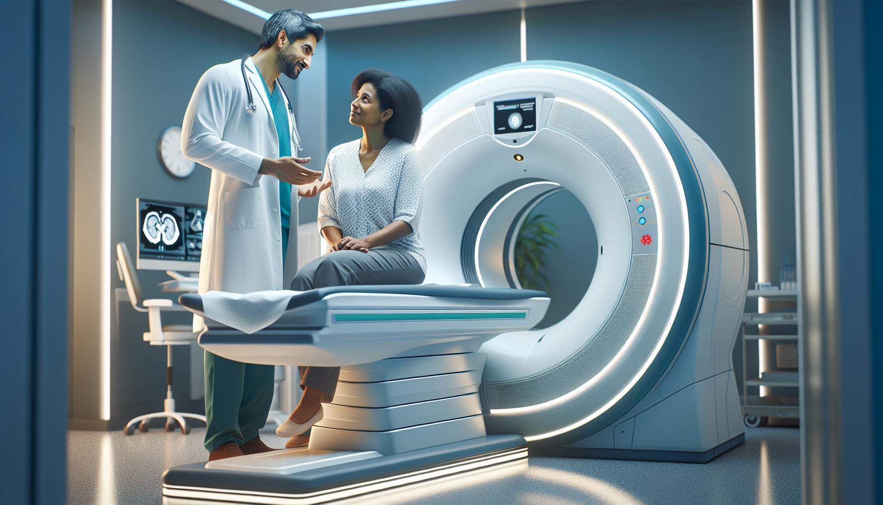
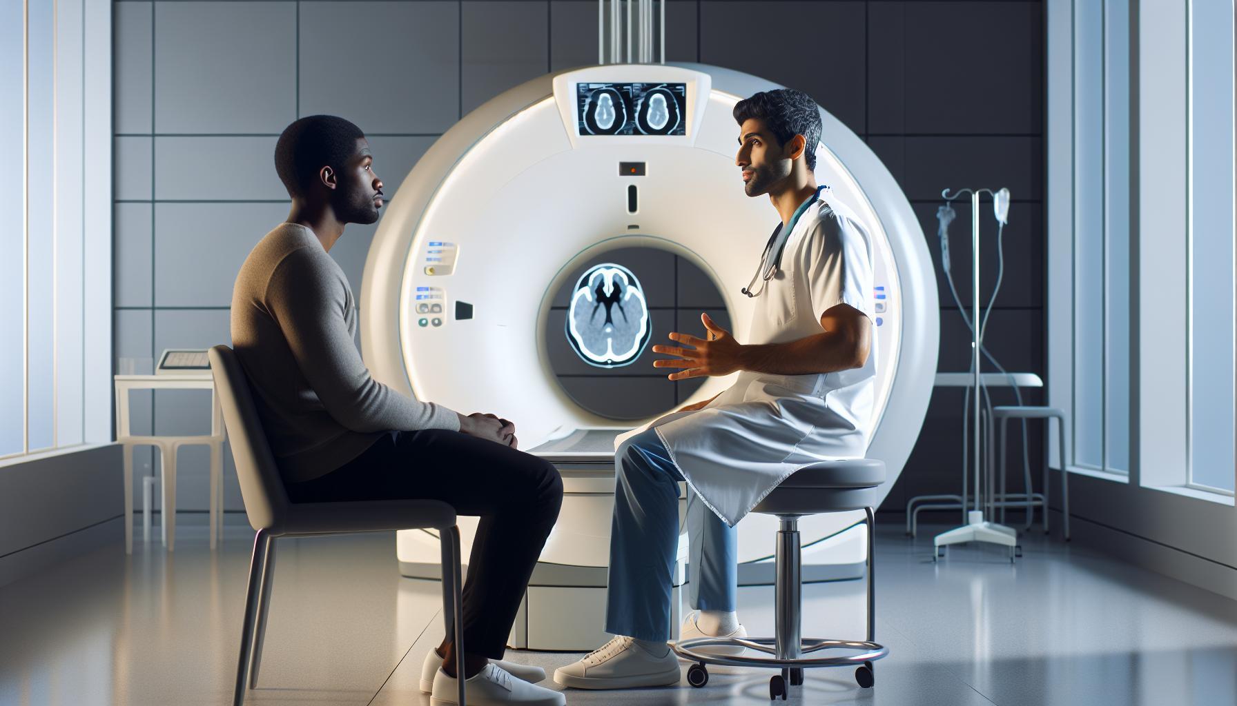
Did you know that over 30 million adults in the U.S. suffer from digestive tract disorders at some point in their lives? A CT enterography is a crucial imaging technique that allows healthcare professionals to examine the small intestine in detail. By utilizing advanced computed tomography technology along with specialized contrast material, it provides high-resolution images that can reveal underlying conditions, guiding effective treatment plans.
Understanding what a CT enterography entails can alleviate some of the concerns patients might have about this procedure. It not only offers a non-invasive way to visualize bowel health but also helps in diagnosing various gastrointestinal disorders, providing a clearer picture of one’s overall health. Whether you’re dealing with abdominal pain or simply seeking clarity on a gastrointestinal issue, this imaging technique is an important tool in modern medicine. Keep reading to explore how CT enterography works, preparation tips, and what to expect during the process.
What Is CT Enterography and How Does It Work?
CT enterography is an advanced imaging technique specifically tailored to visualize the small intestine with remarkable clarity. During this procedure, a computed tomography (CT) scanner captures multiple cross-sectional images by utilizing sophisticated X-ray technology. These images are then assembled by a computer to produce highly detailed pictures of the bowel and surrounding structures, allowing healthcare providers to assess gastrointestinal conditions effectively. One of the standout features of CT enterography is its ability to provide comprehensive evaluations of inflammation, strictures, and other abnormalities in the small intestine, making it invaluable in clinical settings.
To prepare for a CT enterography, patients typically consume a special oral contrast agent that enhances the visibility of the small bowel during imaging. This contrast may require fasting for a few hours prior to the test to ensure that the stomach is clear, which helps in obtaining the best quality images. During the scan, patients lie on a motorized table that slides into the CT machine. While the procedure is generally quick and painless, the experience allows healthcare professionals to capture dynamic images of the digestive tract, providing critical insights into various conditions such as Crohn’s disease or small bowel obstructions.
In terms of function, CT enterography surpasses traditional imaging modalities, especially for the small bowel, due to its non-invasive nature and superior visualization capabilities. This technique not only complements other investigative methods, such as capsule endoscopy and MRI enterography, but it also yields rapid results, thus allowing for timely decision-making in urgent clinical scenarios. While it is a robust diagnostic tool, understanding the specifics of how it works and what to expect can ease patient concerns regarding the procedure and enhance their overall experience.
Benefits of CT Enterography for Bowel Imaging
CT enterography offers significant advantages when it comes to imaging the small intestine, a region often challenging to visualize effectively with other diagnostic methods. For individuals experiencing gastrointestinal issues, the ability to obtain clear, detailed images of the small bowel can lead to timely diagnoses and better treatment options. Unlike traditional imaging techniques, CT enterography provides comprehensive assessments of inflamed tissues, strictures, and other abnormalities that may not be apparent in regular X-rays or ultrasounds.
One of the standout benefits of CT enterography is its non-invasive nature. Patients can generally expect a quick procedure that doesn’t require incisions or anesthesia, which significantly reduces recovery time and associated risks. The use of a specialized oral contrast agent not only enhances the visibility of the small intestine but also helps to delineate any problematic areas, allowing for precise evaluation of conditions such as Crohn’s disease, small bowel obstructions, or tumors. This diagnostic tool empowers healthcare providers to make informed decisions regarding further tests or treatment plans based on clear assessments from the imaging results.
Moreover, the speed at which CT enterography produces results is crucial for effective patient care. While some imaging modalities may take days for results to be reported, CT enterography can often deliver findings shortly after the scan, facilitating quicker treatment initiation. This rapid turnaround time is especially beneficial in urgent cases where timely interventions can vastly improve patient outcomes.
Incorporating CT enterography into the diagnostic process offers patients peace of mind, knowing that they are receiving one of the most advanced imaging techniques tailored specifically for small bowel evaluation. By understanding the benefits of this procedure, patients can engage in meaningful discussions with their healthcare providers about their symptoms and the role that CT enterography may play in their overall treatment.
Preparing for Your CT Enterography Procedure
To ensure a successful CT enterography procedure, adequate preparation is essential for optimal imaging results. Unlike other imaging modalities, CT enterography requires specific steps to be taken beforehand, primarily involving the use of a contrast material that helps visualize the small intestine. Understanding the preparation process can alleviate concerns and enhance the effectiveness of this advanced diagnostic tool.
Start by discussing any medications you are currently taking with your healthcare provider. Some medications, particularly those that affect intestinal motility, may require adjustments before the procedure. Typically, patients are advised to refrain from eating for a few hours prior to the scan. Your doctor will provide specific fasting guidelines based on the timing of your appointment. This ensures that the intestines are clear and allows for better visualization during the imaging process.
In preparation for the contrast agent, you will need to drink a specialized solution, usually about an hour before the scan. This oral contrast material is crucial as it enhances the visibility of the small intestine on the CT images. It’s essential to follow instructions on how to consume this solution, and some patients might find it helpful to chill it to improve taste. Staying well-hydrated leading up to the procedure will also aid in the effectiveness of the contrast.
Before you arrive for the scan, be sure to wear comfortable, loose-fitting clothing, as you may be asked to change into a hospital gown. Remove any metal accessories, such as jewelry or belts, as these can interfere with the imaging process. Arriving a bit early allows for any necessary paperwork and gives you a moment to relax before the procedure. At the facility, healthcare professionals will guide you through each step, answer any questions you may have, and provide reassurance, ensuring you feel comfortable throughout the process.
By being well-prepared, you can approach your CT enterography with confidence, knowing that you are taking active steps towards understanding and managing your gastrointestinal health. Always remember that your healthcare provider is your best resource for personalized instructions and support tailored to your unique situation.
What to Expect During a CT Enterography Scan
Undergoing a CT enterography scan can be a significant step towards understanding and improving your gastrointestinal health. It’s normal to feel a bit anxious before the procedure, but knowing what to expect can alleviate these concerns and help you approach the experience with confidence and calm.
Once you arrive at the imaging facility, you’ll likely be greeted by friendly medical staff who will guide you through the process. After checking in, you may need to change into a hospital gown. It’s advisable to wear comfortable clothing for your visit, but typically removal of any metal items such as jewelry or belts is required, as these can interfere with the imaging quality. At this stage, don’t hesitate to ask any questions you might have; the staff is there to support you.
Before the scan begins, you will drink a specialized oral contrast solution, which helps highlight the small intestine during imaging. This solution is crucial for ensuring that your area of interest is clearly visible on the CT images. The procedure itself is quick and relatively simple. You’ll lie down on a movable table, which will gently slide into the CT scanner. You’ll be instructed to hold your breath briefly while the machine takes the images-this typically lasts only seconds. It’s important to stay as still as possible during this phase, but the process will not take long.
During the scan, you might hear whirring sounds and the machine may emit some noise, but this is completely normal. Most patients find the scan to be painless, and afterwards, you can return to your regular activities without any restrictions. Bringing along a support person can also help ease your apprehension and provide comfort throughout the experience. Knowing that healthcare professionals are watching and guiding you every step of the way should provide further reassurance that you are in good hands. After the scan, the results will be interpreted by a radiologist, and your healthcare provider will discuss them with you in due course, ensuring you understand the findings and the next steps for your health.
Interpreting Your CT Enterography Results
Interpreting the results of your CT enterography can be an enlightening experience, revealing crucial insights into your digestive health. Once the scan is completed, a specialist radiologist will review the images generated during the procedure. This process involves analyzing detailed visual data, which can illuminate various aspects of your small bowel, such as abnormalities, inflammation, or blockages. Radiologists are trained to identify even the most subtle changes in the tissue, allowing them to provide a comprehensive report based on their observations.
Typically, the findings from your CT enterography will be categorized into distinct areas, such as the presence of inflammatory diseases (like Crohn’s disease), tumors, or structural abnormalities. As you wait for your results, it’s natural to feel a spectrum of emotions, ranging from anxiety to anticipation. To alleviate some of this uncertainty, consider having a list of questions ready for your healthcare provider when discussing your results. Important points to clarify may include:
- What specific findings were noted? Understanding the details will help you comprehend the implications for your health.
- Are additional tests required? Knowing if further investigation is needed can prepare you for next steps.
- What are the treatment options? If issues are detected, being informed about your choices empowers you to take an active role in your care.
Once your healthcare provider has discussed the findings with you, they will likely recommend a personalized care plan that aligns with your specific diagnosis and health needs. Remember, while the results of a CT enterography can provide crucial insights, they are just one piece of the puzzle in your overall health journey. Continuous dialogue with your medical team is paramount, as they can adjust your treatment plan based on your ongoing assessments and experiences.
Comparison with Other Imaging Techniques
When considering options for imaging the bowel, it’s essential to understand the various techniques available and how they compare. CT enterography stands out as a powerful tool, providing detailed images of the small intestine. However, other imaging modalities also have unique strengths. For instance, traditional x-rays and magnetic resonance imaging (MRI) are commonly used, each with its own advantages and limitations.
CT enterography offers higher resolution images of soft tissues compared to standard x-rays, which are primarily useful for detecting obstructions or foreign bodies in the abdomen. It also excels in visualizing inflammatory bowel diseases like Crohn’s disease or ulcerative colitis, capturing the intricate details that might be missed by simpler imaging methods. On the other hand, MRI is particularly advantageous for patients allergic to iodinated contrast material used in CT scans, as it employs a different contrast agent that typically poses fewer risks. Additionally, MRI does not involve ionizing radiation, making it a preferred option for pediatric patients or those requiring repeated imaging.
Another option is ultrasound, which is non-invasive and does not use radiation. While effective for evaluating certain abdominal conditions, ultrasounds may not provide the same level of detail as CT enterography, especially for evaluating the small intestine. Ultrasound can, however, be a useful first step for assessing children or in emergency situations due to its rapid execution and lack of radiation.
In summary, while CT enterography remains a cornerstone for in-depth analysis of bowel disorders, weighing the pros and cons of each imaging technique is crucial. Collaborating closely with healthcare providers allows for a tailored approach to diagnosis, ensuring the selected method aligns with individual health needs and circumstances. This way, patients can feel more secure in their diagnostic journey, knowing each imaging option serves a specific purpose in their overall care strategy.
Safety Considerations and Risks of CT Enterography
CT enterography is an advanced imaging technique that provides critical insights into the small intestine, but like all medical procedures, it comes with certain safety considerations and risks. One common concern is exposure to ionizing radiation. While CT enterography generally delivers a higher radiation dose compared to traditional x-rays, the risk is typically outweighed by its diagnostic benefits, especially when investigating conditions such as Crohn’s disease or tumors that might not be well visualized with other imaging modalities. Healthcare providers take precautions to minimize exposure, applying the principle of “As Low As Reasonably Achievable” (ALARA) to ensure patient safety.
Another aspect to consider is the use of contrast material, often iodine-based, which enhances the visibility of the intestines. Some patients may experience allergic reactions to this contrast media, ranging from mild reactions such as hives or nausea to more severe anaphylactic responses. Prior to the scan, it’s essential to discuss any history of allergies or previous reactions with your healthcare provider, which allows for appropriate pre-medication or alternative imaging strategies if needed. For those with kidney issues, there’s also a rare but serious condition known as contrast-induced nephropathy, highlighting the importance of a comprehensive health review before undergoing the procedure.
Post-procedure, patients are typically monitored for any immediate reactions to the contrast medium and encouraged to hydrate adequately to help flush it from their system. It is advisable to reach out to your healthcare provider if you experience any unusual symptoms, such as persistent abdominal pain or significant changes in urination, following the scan.
Overall, while the benefits of CT enterography in diagnosing and managing bowel-related conditions are significant, awareness of the potential risks and active communication with healthcare professionals can empower patients to make informed decisions about their care. Understanding these safety considerations not only alleviates anxiety but fosters a more supportive relationship between patients and providers, ultimately contributing to better healthcare experiences.
Cost of CT Enterography: What to Expect
The financial aspect of a CT enterography can often be a source of apprehension for patients, given the intricacies involved in medical imaging costs. Understanding what to expect can not only alleviate anxiety but also facilitate better planning for the procedure. Typically, the cost of CT enterography varies widely based on several factors including geographic location, healthcare facility, the complexity of the imaging required, and whether the patient has insurance coverage. On average, patients might expect to pay between $1,000 to $3,000 for a CT enterography, which can fluctuate based on the inclusion of the contrast material and any additional services.
Patients with insurance coverage should check with their provider about specific benefits related to a CT enterography, including copays, deductibles, and any pre-authorization requirements. It is prudent to inquire whether the facility where the imaging will be performed is in-network to minimize out-of-pocket costs. For those without insurance, discussing potential financial assistance or payment plans with the medical facilities can be beneficial. Some centers may offer self-pay discounts or sliding scale fees based on income.
To help provide clarity and transparency, here’s a breakdown of potential costs associated with a CT enterography:
| Cost Component | Estimated Range |
|---|---|
| CT Enterography Scan | $1,000 – $2,000 |
| Contrast Material | $100 – $500 |
| Facility Fees | $200 – $800 |
| Radiologist Fees | $100 – $500 |
Ensuring that you have a comprehensive understanding of these costs empowers you in navigating the logistics of your healthcare. Moreover, it’s essential to discuss any financial concerns with your healthcare provider before the procedure. They can offer guidance on next steps and provide resources for managing costs effectively. By approaching the financial aspect with knowledge, you can focus more on your health and less on the stress of potential expenses.
When Should You Consider CT Enterography?
When you experience persistent abdominal pain, unexplained weight loss, or changes in bowel habits, discussions about diagnostic imaging may arise. CT enterography is often an excellent choice for evaluating suspected small bowel disorders due to its ability to visualize the entire bowel wall and detect extraenteric involvement effectively. This imaging modality is particularly useful in diagnosing conditions like Crohn’s disease, small bowel tumors, or complicated diverticulitis, where detailed information about the bowel’s structure and function is crucial for appropriate treatment.
Several factors can prompt your healthcare provider to recommend a CT enterography. If you have a history of inflammatory bowel disease, this technique can help assess disease activity or complications such as strictures or abscesses. Additionally, for patients who have undergone previous surgeries, CT enterography can evaluate any post-surgical changes and detect the formation of adhesions or obstructions that may be contributing to ongoing symptoms. It is also valuable in triaging patients with intestinal bleeding where traditional endoscopic techniques may not provide sufficient visualization.
Before proceeding, it’s essential to consult your healthcare provider to determine if CT enterography is the right diagnostic tool for your symptoms. They can conduct a thorough evaluation of your medical history, current health status, and specific concerns, ensuring that this imaging method aligns with your overall care plan. Empowering yourself with knowledge about your condition and asking questions will not only help alleviate anxiety but will also enable you to make informed decisions about your health.
Real-Life Applications and Success Stories
The effectiveness of CT enterography is beautifully illustrated through various real-life applications that resonate with patients experiencing gastrointestinal challenges. One particularly compelling story comes from a young woman diagnosed with Crohn’s disease, a condition that often leaves patients grappling with debilitating symptoms and uncertainty. After enduring months of unexplained abdominal pain and weight loss, her healthcare provider recommended CT enterography. The scan provided a comprehensive view of her small intestine, revealing areas of inflammation and strictures that were not visible through traditional imaging methods. This diagnosis empowered her medical team to tailor a specific treatment plan, ultimately leading her toward a more manageable and hopeful path in her journey with Crohn’s.
Moreover, CT enterography is invaluable for identifying complications following surgeries, as seen in the case of a middle-aged man who had undergone bowel resection. He experienced persistent discomfort post-surgery, leading his doctor to suspect adhesions. A CT enterography examination confirmed this suspicion, allowing the surgical team to address the adhesions directly through another minimally invasive procedure. Patients in such situations often express relief, knowing that advanced imaging has clarified the root cause of their symptoms and streamlined the path to recovery.
Broader Impact on Healthcare
The role of CT enterography extends far beyond individual cases; it actively shapes treatment protocols in clinics and hospitals. As healthcare providers gain more experience using CT enterography, its applications continue to grow. It has proven especially useful in managing intestinal bleeding, where traditional methods may fall short. For instance, a patient with obscure gastrointestinal bleeding underwent CT enterography, which successfully pinpointed the source of the issue, facilitating a swift treatment approach.
As we consider these stories, it becomes clear that CT enterography not only aids in diagnosis but also enhances patient care by guiding treatment decisions. For those facing similar fears or uncertainties about their gastrointestinal health, CT enterography represents a vital tool that can provide clarity, helping to bridge the gap between symptom presentation and effective management. Always consult with your healthcare professional to determine if this imaging technique is right for your specific situation; their expertise is crucial in navigating your health journey.
Understanding the Limitations of CT Enterography
While CT enterography is a powerful imaging tool for diagnosing gastrointestinal issues, it’s essential to understand its limitations to set realistic expectations. One significant drawback is that while the procedure offers detailed images of the small intestine, it may not effectively visualize certain areas of the gastrointestinal tract, such as the esophagus and colon. This limitation could lead to incomplete evaluations, especially if symptoms suggest involvement beyond the small bowel.
Furthermore, CT enterography relies on the use of contrast agents, which can pose challenges for some patients. Allergic reactions to the contrast material, though rare, can occur and could range from mild to severe. Additionally, individuals with kidney issues may have an increased risk of contrast-induced nephropathy. This emphasizes the importance of thorough pre-screening by healthcare professionals to determine suitability for the procedure.
In certain cases, CT enterography might not be the most definitive imaging modality for specific diagnoses. For example, while it is effective in identifying inflammatory bowel diseases, conditions like irritable bowel syndrome (IBS) may not be accurately assessed through this imaging type. Each patient’s unique clinical situation dictates the most appropriate imaging technique; sometimes alternative methods like MRI or endoscopy might be indicated for clearer, more comprehensive views of the intestinal lining and potential abnormalities.
Lastly, it’s important to recognize the exposure to radiation that comes with CT scans. While the benefits often outweigh the risks, especially in assessing significant conditions, medical professionals should always strive to minimize radiation exposure by adhering to the principle of ALARA (As Low As Reasonably Achievable). Engaging in thorough discussions with your healthcare provider can provide personalized insights on the risks and benefits associated with CT enterography, ultimately empowering patients to make informed decisions about their healthcare journey.
Frequently asked questions
Q: What conditions can CT enterography diagnose?
A: CT enterography can diagnose various gastrointestinal conditions, including Crohn’s disease, small bowel obstruction, tumors, and diverticulitis. It provides detailed images of the small intestine, enabling healthcare providers to assess the severity and extent of these diseases.
Q: How does CT enterography differ from a standard CT scan?
A: Unlike a standard CT scan, CT enterography specifically focuses on the small intestine using a contrast material to enhance visualization. This technique improves the clarity of the bowel wall and any associated abnormalities, providing more precise diagnostic information.
Q: Is CT enterography safe for everyone?
A: While generally safe, CT enterography may not be suitable for individuals with certain conditions, such as severe kidney disease or allergies to contrast materials. Always consult your healthcare provider to evaluate the benefits and risks based on your specific health situation.
Q: How should I prepare for a CT enterography?
A: Preparation typically includes fasting for several hours before the procedure and drinking a specific contrast solution to fill the intestine. Your healthcare provider may provide detailed instructions tailored to your needs for optimal results.
Q: Can I eat or drink before CT enterography?
A: Usually, you will be instructed to fast for several hours prior to the CT enterography. This may involve avoiding food and drinks, including water, to ensure clear images during the scan. Always follow the specific guidelines provided by your healthcare provider.
Q: How long does a CT enterography take?
A: A CT enterography procedure generally takes about 30 to 60 minutes. However, the overall visit could be longer, allowing for preparation, administration of contrast, and post-scan observation if necessary.
Q: What should I expect after a CT enterography?
A: After a CT enterography, you may need to drink plenty of fluids to help flush out the contrast material. Most people can resume normal activities immediately, but discuss any concerns with your healthcare provider, especially if you feel unwell.
Q: How is the accuracy of CT enterography compared to other imaging methods?
A: CT enterography is often more accurate than traditional X-ray imaging for evaluating small bowel disorders. It offers superior visualization of the bowel wall and surrounding tissues, making it a preferred method for diagnosing certain gastrointestinal conditions.
For more details, you can refer to specific sections in the article that cover preparation and interpretation of results.
Wrapping Up
As we’ve explored in this guide, CT enterography is a vital imaging technique for diagnosing bowel conditions, offering clarity and precision in assessing your gastrointestinal health. If you have more questions or worries about the procedure, remember that speaking with your healthcare provider can provide personalized insights tailored to your needs. Don’t hesitate-taking that next step can lead to better health outcomes.
For more in-depth information, check out our articles on preparing for a CT scan and understanding the costs associated with medical imaging. These resources can help ease your mind and provide additional guidance as you navigate your healthcare journey. To stay updated on crucial health topics and tips, consider signing up for our newsletter. Your health matters, and being informed is the best way to take proactive steps for your well-being. Keep exploring, and empower yourself with knowledge today!

