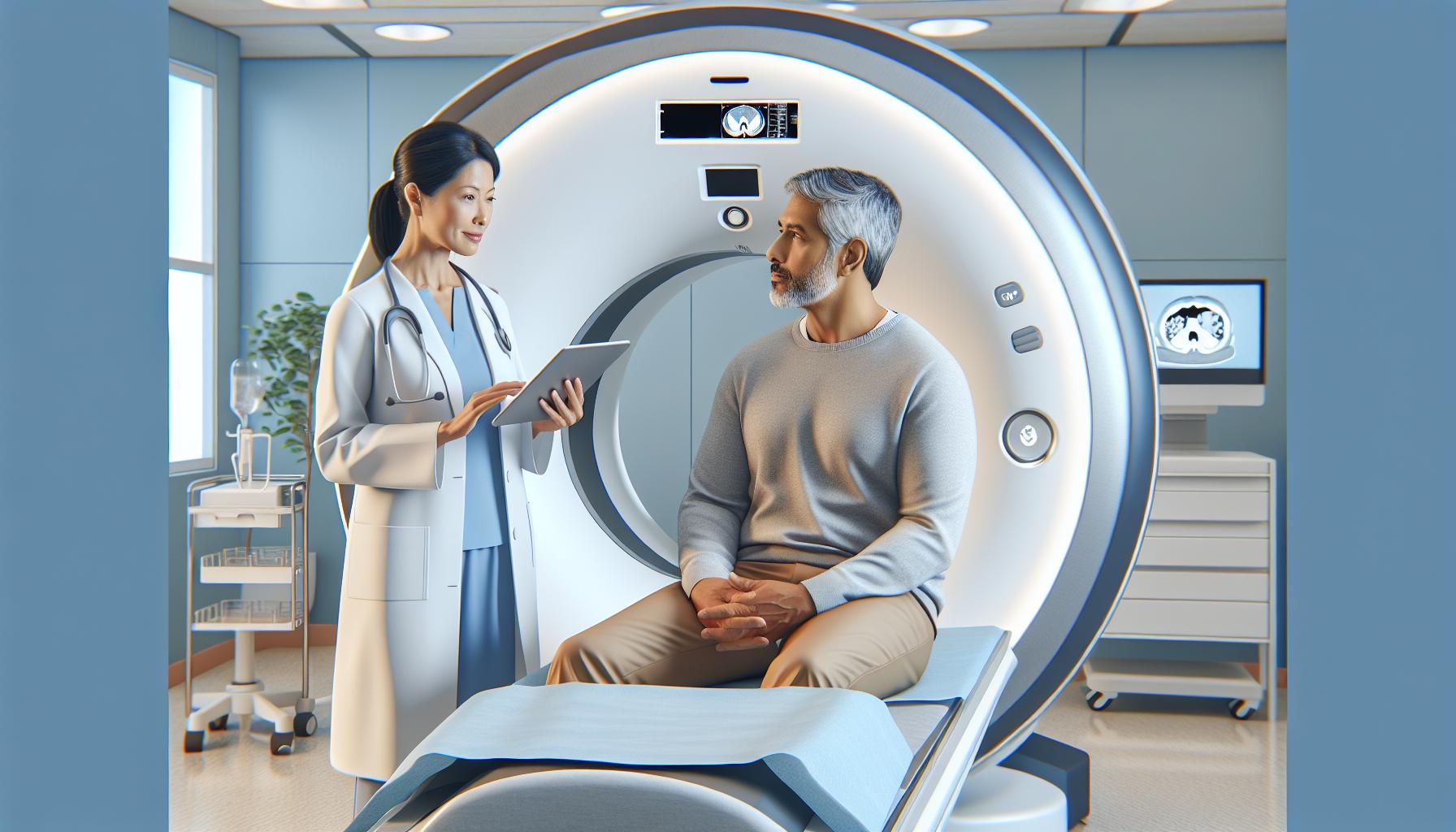
Understanding whether bladder cancer can be detected by a CT scan is crucial for those concerned about their health. CT scans are commonly used to visualize internal organs and can reveal suspicious growths that may indicate bladder cancer. With bladder issues affecting many individuals, the fear of cancer can understandably loom large.
By learning how CT scans work in detecting potential tumors and the importance of early detection, you empower yourself with knowledge that can lead to better outcomes. As you read on, we will delve into how this imaging technology functions, what to expect during the procedure, and why consulting with healthcare professionals is vital for addressing any concerns regarding bladder health and cancer detection. Your health matters, and understanding these detection facts could be the first step towards peace of mind.
Does Bladder Cancer Show on a CT Scan?
Bladder cancer can indeed be visible on a CT scan, offering a crucial tool in the diagnostic process. This imaging technique uses advanced x-ray technology to create detailed cross-sectional images of the body, which can help detect tumors within the bladder as well as any potential spread to surrounding tissues or lymph nodes. A CT scan is often employed when a healthcare provider suspects bladder cancer, as it provides clearer images than traditional x-rays, making it easier to identify abnormalities.
When evaluating bladder health, radiologists typically look for signs of tumors or irregularities such as thickening of the bladder wall, masses, or other changes in the bladder’s structure. These images can help differentiate between benign and malignant growths. It’s important to understand that while a CT scan is a powerful imaging tool, it is not exclusively conclusive for diagnosing bladder cancer. Other tests, such as cystoscopy, may be necessary for a definitive diagnosis.
If you’re advised to undergo a CT scan, your healthcare provider will thoroughly explain the procedure, its purpose, and what to expect. A gentle approach helps alleviate any anxiety associated with medical imaging. It’s vital to have open communication with your provider, who can offer personalized insights into your specific health situation. This shared understanding fosters reassurance about the process while empowering you with knowledge about what the images can reveal concerning your health.
Understanding CT Scans for Bladder Cancer Detection
A CT scan is a highly effective imaging technique that plays a significant role in the detection of bladder cancer. This advanced procedure provides detailed cross-sectional images of the body, allowing healthcare providers to visualize the bladder’s structure with impressive clarity. With its ability to highlight irregularities such as tumors or thickened walls, a CT scan is often one of the first diagnostic tools employed when bladder cancer is suspected. It not only assesses the bladder itself but also checks for potential metastasis to surrounding lymph nodes and other tissues, aiding in the comprehensive evaluation of the cancer stage.
During the scan, x-ray technology is utilized to create a series of images from multiple angles. These images are then processed by a computer to generate comprehensive, cross-sectional images of the bladder and nearby structures. Radiologists interpret these images, looking for specific indicators of cancer presence, including abnormal growth patterns. It’s crucial to remember that while CT scans are invaluable for initial assessments, they are typically part of a broader diagnostic approach. Additional tests, such as a cystoscopy, may be necessary to confirm a diagnosis conclusively.
Patients who undergo a CT scan can expect an experience that is straightforward and relatively quick. The procedure usually takes only a few minutes, during which they will lie on a table that moves through a large, donut-shaped machine. Although the scan itself is non-invasive, preparation may include fasting or drinking a contrast solution to enhance image clarity. Understanding these steps can significantly reduce anxiety and empower patients to engage actively in their health care decisions. Always maintain open communication with healthcare providers to ensure the most personalized and effective care pathway, especially when navigating the complexities of cancer detection and management.
How CT Scans Work for Bladder Cancer Diagnosis
A CT scan, or computed tomography scan, serves as a crucial tool in diagnosing bladder cancer, providing intricate details that conventional X-rays cannot. Understanding how this advanced imaging technique works can alleviate apprehensions and enhance patient preparedness. During a CT scan, multiple X-ray images are captured from varied angles and synthesized by a computer to create detailed cross-sectional images of the bladder and surrounding tissues. This process allows healthcare providers to visualize any anomalies such as tumors, growths, or structural changes that may signify the presence of cancer.
How CT Technology Aids Diagnosis
The use of contrast agents during the procedure significantly enhances the visibility of the bladder and surrounding organs. Patients may be instructed to either ingest or receive an intravenous injection of a contrast material before the scan. This substance helps to differentiate various tissues, making abnormalities even more apparent on the imaging results. As the scan progresses, radiologists meticulously examine these images for signs of bladder cancer, paying close attention to irregularities or changes in bladder wall thickness.
While CT scans offer high-resolution images that can assist in the initial assessment of bladder cancers, it’s vital to remember that they are most effective when used alongside other diagnostic methods. Financial aspects and insurance coverage for such scans remain practical considerations. Patients should engage with their healthcare providers, who can help navigate these complexities and recommend a tailored approach.
In conclusion, understanding the operational nuances behind CT scans not only reduces common anxiety related to the unknown but also empowers patients to participate more effectively in their own healthcare journey. Always consult with your healthcare professional for personalized advice and to discuss any concerns regarding the procedure or results.
Key Symptoms That Lead to CT Scans
Experiencing certain symptoms can understandably lead to concerns about bladder health, and it’s essential to be aware of what may prompt healthcare professionals to recommend a CT scan for bladder cancer assessment. Bladder cancer often presents with a few key indicators that warrant further investigation. These symptoms might include blood in the urine (hematuria), frequent urination, painful urination, or the urgent need to urinate without being able to pass much urine. If you’re experiencing any of these signs, your doctor may consider imaging tests such as a CT scan to determine the underlying cause.
Another critical aspect to consider is the nature of the symptoms’ onset. For instance, if blood appears suddenly or if there has been a notable change in urination patterns-such as increased frequency or discomfort-these changes can be significant. It’s also important to share any related symptoms you may experience, such as pelvic or lower abdominal pain, as they can assist doctors in refining their diagnostic focus. When symptoms are persistent or worsen over time, this can heighten the suspicion of bladder cancer and prompt a more thorough evaluation including imaging studies.
Ultimately, having open communication with your healthcare provider is vital. They will not only assess your symptoms but may also conduct a physical examination, urine tests, or additional tests depending on your presentation. If necessary, the CT scan can offer valuable insights, enabling a clearer view of potential abnormalities in the bladder or surrounding structures. Emphasizing the importance of these symptoms ensures that patients are proactive about their health and reinforces the need for timely medical consultation when concerning signs arise.
What to Expect During a Bladder Cancer CT Scan
A CT scan can be a vital tool in diagnosing bladder cancer, providing detailed images that can reveal abnormalities within the bladder and surrounding areas. If you’re scheduled for a CT scan, understanding what to expect can significantly reduce anxiety and help you feel more prepared for the experience.
During the procedure, you will be asked to lie on a table that slides into a large, doughnut-shaped machine known as a CT scanner. This machine uses X-rays taken from different angles to create cross-sectional images of your body. Some patients may receive a contrast dye, which can enhance the clarity of the images. The dye might be administered through an intravenous (IV) line in your arm, or in some cases, it may be introduced via a catheter. If you receive a contrast dye, you may feel a warm sensation as it flows through your veins, which is completely normal.
It’s crucial to remain still during the scanning process to ensure the best quality images. Although the scan is painless, you may hear a whirring or clicking sound from the machine, which can be disconcerting but is part of the scanning process. Generally, the scan can take anywhere from a few minutes to about half an hour, depending on the specific protocol your healthcare team follows. Afterward, you’ll be able to resume your normal activities, unless advised otherwise by your doctor.
While waiting for results can be a source of anxiety, knowing that these images play a crucial role in identifying potential issues can be reassuring. Your healthcare team will interpret the results and discuss any findings with you, guiding the next steps in your care. Remember, open communication with your healthcare provider is key; do not hesitate to ask questions about the procedure, what the images will reveal, and any concerns you may have.
Patient Preparation: Steps for a Successful Scan
Preparing for a CT scan can significantly impact the quality of your imaging results, ultimately influencing your bladder cancer diagnosis. Understanding the steps necessary for successful preparation not only makes the experience smoother but also helps you feel more in control. Here are some key steps to ensure you are ready for your CT scan.
Before the Scan
Before arriving for your scan, it’s essential to discuss any medications you are currently taking with your healthcare provider. Some medications might need to be paused, especially if they affect your kidneys or interaction with contrast dye. Additionally, if you have any allergies, particularly to contrast materials or iodine, be sure to inform your doctor as these details are crucial for your safety.
Another critical part of preparation is dietary restrictions. Your healthcare team may recommend you avoid eating or drinking for several hours before the scan. This is particularly necessary if you will receive a contrast agent, as it helps improve image clarity by ensuring your bladder is adequately visualized. Always follow specific instructions given by your provider, as variations may exist based on the facility and procedure specifics.
Day of the Scan
On the day of your appointment, wear comfortable clothing without metal fasteners, zippers, or excessive layers, which can interfere with the imaging process. It might also be helpful to wear supportive footwear, especially if you need to walk to a separate imaging area.
When you arrive for your CT scan, the staff will check you in and may ask you to fill out some forms related to your medical history. If a contrast dye will be used, you will be taken to an area to administer the dye, usually via an IV. If this occurs, keep in mind that you may briefly feel a warm sensation as the dye enters your system-this is entirely normal.
Being well-prepared mentally, emotionally, and physically can make your CT scan a less daunting experience. Take time to express any concerns you might have to the staff; asking questions and understanding the procedure can bring peace of mind. By following these preparation steps, you empower yourself to experience a smoother and more effective imaging process, contributing to a more accurate diagnosis of potential bladder cancer.
Interpreting CT Scan Results for Bladder Cancer
Interpreting the results of a CT scan for bladder cancer can be a critical step in understanding your diagnosis and treatment options. When a CT scan indicates the presence of bladder cancer, radiologists analyze various factors within the images, such as the size, shape, and location of any detected masses or lesions. These details offer crucial insights regarding the extent of the disease-information that can significantly influence therapeutic decisions.
The presence of a mass may suggest a tumor, but not all masses are malignant. For instance, some lesions may be benign conditions, and distinguishing between these requires a careful assessment by your healthcare provider. In most cases, a radiologist will provide a detailed report that includes descriptions of any abnormal findings along with recommendations for follow-up procedures, such as additional imaging or biopsy. It’s vital for patients to discuss these findings with their doctors to understand what the images convey and what steps are necessary next.
In interpreting the results, understanding specific terminologies can also be reassuring. Terms like “hypodense” or “hyperdense” refer to how the tissues appear compared to surrounding structures, giving a clue about the nature of the findings. Radiologists often use standardized nomenclature and staging systems, such as the TNM classification (Tumor, Node, Metastasis), to communicate the severity of the cancer effectively. This information sequentially guides treatment plans, whether surgical intervention, chemotherapy, or radiotherapy is warranted.
Lastly, keep in mind that while CT scans are powerful tools in visualizing bladder cancer, they aren’t entirely definitive. False positives can occur, leading to unnecessary anxiety, and false negatives can result in missed diagnoses. As a patient, actively engaging with your healthcare team, asking questions, and seeking clarification on any ambiguous areas in your CT findings can alleviate concerns and empower you during this critical stage of your healthcare journey.
Alternative Imaging Techniques for Bladder Cancer
Imaging techniques for bladder cancer are essential tools in the diagnostic process, especially when conventional CT scans may fall short. These alternative methods can provide doctors with additional views and insights, enhancing the accuracy of bladder cancer detection. Some noteworthy modalities include:
Ultrasound
Ultrasound is a non-invasive imaging technique that uses sound waves to create images of the bladder and surrounding structures. It’s particularly useful for assessing bladder wall abnormalities and detecting larger tumors. While it may not provide the detail of a CT scan, it is excellent for guiding further evaluation and offers the advantage of not requiring ionizing radiation. Patients often appreciate ultrasound for its simplicity and lack of complex preparations.
MRI (Magnetic Resonance Imaging)
MRI is another powerful tool for bladder cancer assessment. It uses magnetic fields and radio waves to produce detailed images of soft tissues. MRI is especially beneficial in visualizing the musculature of the bladder wall and surrounding organs, which can be critical for staging cancer and planning treatment. For patients with an allergy to iodine-based contrast used in CT scans or those who are pregnant, MRI presents a safer alternative.
Cystoscopy
While technically not an imaging scan, cystoscopy involves the direct visualization of the bladder using a thin, flexible tube with a camera. This procedure allows for immediate biopsy of suspicious areas and is invaluable for confirming diagnoses. Patients may find cystoscopy somewhat uncomfortable; however, it offers direct information that imaging alone cannot.
Pet Scans (Positron Emission Tomography)
PET scans are increasingly used in conjunction with CT or MRI to identify cancer spread (metastasis) and to assess the effectiveness of treatment. This technique focuses on metabolic activity, highlighting areas of increased activity that may correspond to cancerous growth. While PET scans can be more costly and less accessible, they are instrumental in complex cases where the cancer’s nature and spread need clarification.
In summary, if a CT scan raises concerns or needs follow-up, these alternative imaging techniques can provide further clarity. Patients should feel empowered to discuss the appropriateness and availability of these options with their healthcare providers. Understanding the strengths and limitations of each imaging method can alleviate anxiety and support informed decision-making during the diagnostic process. Always consult your healthcare team to determine the best imaging strategy tailored to your specific situation.
Limitations of CT Scans in Detecting Bladder Cancer
While CT scans are a valuable tool in the detection of bladder cancer, they are not without their limitations. One major drawback is that small tumors or subtle abnormalities may be difficult to visualize on a CT scan. This is particularly true for early-stage bladder cancer, where lesions can be mere millimeters in size. Such small tumors, especially those located on the bladder wall, may not produce enough contrast against surrounding tissues, leading to missed diagnoses or delayed treatment.
Additionally, the use of contrast agents in CT scans can present challenges. Although they enhance image quality, these agents can cause allergic reactions in some patients, ranging from mild to severe. For individuals with impaired kidney function, the use of iodine-based contrast can increase the risk of nephrotoxicity, complicating the decision to proceed with a CT scan. Moreover, certain factors like patient movement, obesity, or inadequate bladder filling at the time of imaging can further obscure diagnostic accuracy, possibly requiring repeat scans.
Another important consideration is the exposure to radiation. CT scans involve ionizing radiation, which, while typically justified for the diagnostic benefits they provide, raises concerns about potential long-term risks, particularly for young patients or those who require multiple scans over time. This underscores the necessity for clinicians to carefully weigh the benefits and risks when recommending imaging tests for bladder cancer.
Given these limitations, patients are encouraged to maintain open communication with their healthcare providers about their symptoms and any concerns regarding imaging techniques. A comprehensive approach, integrating various diagnostic modalities such as ultrasound or cystoscopy, can be crucial for effective bladder cancer detection and management. Seeking a multidisciplinary opinion may also enhance the diagnostic pathway, ensuring tailored treatment strategies that address individual patient needs.
Follow-Up Tests After a CT Scan: What You Need to Know
Following a CT scan, understanding the next steps is crucial for ensuring proper diagnosis and treatment of potential bladder cancer. It’s important to remember that the results of your scan may not provide a definitive diagnosis on their own; therefore, follow-up tests could be necessary. These additional imaging studies or examinations help to clarify findings from the CT scan, particularly if there are areas of concern or if the scan reveals potential abnormalities.
Common Follow-Up Tests
Depending on the initial findings and your specific situation, healthcare providers might recommend one or more of the following tests:
- Cystoscopy: A minimally invasive procedure allowing doctors to inspect the bladder and urethra directly using a thin, lighted tube.
- Ultrasound: An imaging technique that uses sound waves to create pictures of the bladder and surrounding structures, which can help in visualizing abnormal growths.
- Biopsy: If suspicious areas are found during cystoscopy, a biopsy may be taken to examine tissue samples for cancer cells.
Discussing the purpose of any follow-up tests with your healthcare provider is important to address any concerns you may have about invasiveness, recovery, and risks associated with these procedures.
Preparing for Follow-Up Tests
Preparation for these tests can vary significantly. If a cystoscopy is on the agenda, your doctor may advise you to avoid certain medications, or you may need to follow specific guidelines about dietary restrictions beforehand. An appointment for a biopsy could require you to arrange for someone to accompany you, as mild sedation might be used during the procedure.
It’s natural to feel anxious about what these follow-up tests may reveal. Remember that these procedures are designed to provide your healthcare team with the detailed information they need to make informed decisions about your care. Open communication with your provider-expressing any worries and asking questions-can greatly help ease anxiety and ensure you’re well-informed.
Understanding Results and Next Steps
Once the follow-up tests have been conducted and results are available, your healthcare provider will discuss the findings with you. Should these tests confirm a diagnosis of bladder cancer, your medical team will outline the appropriate treatment options, which may involve surgery, chemotherapy, or radiation therapy, tailored to fit your specific diagnosis and overall health.
In summary, engaging proactively in your follow-up care can empower you during this challenging time. By knowing what to expect and maintaining open lines of communication with your practitioners, you can navigate the complexities of bladder cancer detection and management with greater assurance and clarity.
Cost of CT Scans for Bladder Cancer Diagnosis
The cost of a CT scan for bladder cancer diagnosis can be a significant concern for many patients, especially considering the importance of timely and accurate detection. On average, the price for a CT scan ranges from $300 to $3,000, depending on various factors including location, specific facility, and whether contrast material is used. Understanding these costs and how insurance can play a role is essential for planning and ensuring access to necessary diagnostics.
Several elements influence the overall cost of a CT scan. These include:
- Facility Type: Scans performed in hospitals generally cost more than those conducted in outpatient imaging centers.
- Insurance Coverage: Many insurance plans cover a significant portion of diagnostic imaging costs, but it’s vital to verify the specifics of your plan, including deductibles, copays, and any pre-approval requirements.
- Geographic Location: Prices can greatly vary based on where you live, with urban areas usually having higher costs compared to rural locations.
- Contrast Usage: If a contrast agent is required, this can add several hundred dollars to the total procedure cost.
When considering a CT scan, it’s advisable to consult with your healthcare provider about potential costs and to check with your insurance company regarding coverage and out-of-pocket expenses. Many facilities also offer payment plans or financial assistance programs to help manage costs, empowering patients to receive essential diagnostic imaging without overwhelming financial strain. Engaging in a conversation with your provider about any financial concerns can alleviate anxiety and enable you to focus on your health and the path forward in your bladder cancer diagnosis process.
Expert Opinions: What Radiologists Want You to Know
When it comes to the detection of bladder cancer, understanding the perspectives of radiologists can demystify the process and alleviate patient concerns. Radiologists emphasize the importance of accuracy and detail when interpreting CT scans, as these images are crucial for diagnosing and staging bladder cancer. They highlight that although CT scans are highly effective in visualizing tumors, they do have limitations, especially for early-stage cancers or flat lesions that may not be as visible. Patients should know that CT scans are often used in conjunction with other diagnostic tools, such as cystoscopy and urine tests, to provide a comprehensive assessment.
Moreover, radiologists want patients to feel empowered and informed about the imaging process. It’s common for individuals to feel anxious before a scan, but understanding what to expect can ease those worries. During the scan, patients lie still while a machine captures a series of images, often enhanced with contrast dye to improve visibility of the bladder tissues. This procedure is generally quick, lasting only 15 to 30 minutes, and many radiologists reassure patients that modern CT technology minimizes discomfort and risk.
Radiologists also stress the importance of communication between patients and their healthcare teams. They encourage patients to share their symptoms and medical histories openly, as this information significantly affects scan interpretation. Post-scan, results are typically communicated quickly, and radiologists are dedicated to providing clear explanations of findings. They understand that receiving a diagnosis can be overwhelming, and they aim to present information in a compassionate and supportive manner.
With the development of advanced imaging techniques, radiologists are also optimistic about future innovations that could enhance the detection of bladder cancer. Techniques like MRI and PET scans may supplement traditional CT scans, providing even more detailed insights and potentially improving early detection rates. Ultimately, radiologists emphasize proactive engagement: ask questions, understand the procedures, and collaborate with your healthcare providers to ensure the best possible diagnostic experience and health outcomes.
Q&A
Q: Can a CT scan detect bladder cancer early?
A: Yes, CT scans can help detect bladder cancer early, especially in high-risk patients with symptoms. They provide detailed images of the bladder, revealing abnormalities or tumors, but they are not the first-line screening tool. Regular monitoring is essential for those at risk.
Q: What type of CT scan is used for bladder cancer diagnosis?
A: A contrast-enhanced CT scan is usually utilized for bladder cancer diagnosis. The contrast material helps highlight the bladder and any potential tumors or lesions, allowing for more accurate detection and assessment of cancerous changes.
Q: How accurate is a CT scan for diagnosing bladder cancer?
A: The accuracy of CT scans for bladder cancer detection ranges from 70% to 85%. While they are effective in identifying larger tumors, smaller lesions may not be as detectable, which is why additional tests, like cystoscopy, may be needed for confirmation.
Q: What follow-up tests are recommended after a CT scan if bladder cancer is suspected?
A: If bladder cancer is suspected after a CT scan, follow-up tests may include cystoscopy, which allows for direct visualization of the bladder, and urine cytology to detect cancer cells. These tests confirm the diagnosis and help determine the cancer stage.
Q: Are there alternative imaging techniques to CT scans for detecting bladder cancer?
A: Yes, alternative imaging techniques include MRI and ultrasound. MRI provides detailed soft tissue images, while ultrasound can help visualize the bladder and surrounding structures. The choice depends on individual patient circumstances and physician recommendations.
Q: What symptoms warrant a CT scan for bladder cancer evaluation?
A: Symptoms that may warrant a CT scan include visible blood in urine (hematuria), frequent urination, painful urination, and pelvic pain. If these symptoms occur, a healthcare provider may recommend a CT scan to investigate further.
Q: Is there any preparation required for a bladder cancer CT scan?
A: Yes, patients are typically advised to drink water before a CT scan to ensure the bladder is full for clearer images. It’s essential to follow specific instructions provided by healthcare professionals regarding food and medication restrictions prior to the scan.
Q: How can I interpret the results of a CT scan for bladder cancer?
A: CT scan results are interpreted by radiologists who analyze the images for any suspicious masses or changes in the bladder. A follow-up consultation with a physician is crucial for understanding the implications of the findings and discussing potential next steps.
Final Thoughts
Understanding how bladder cancer is detected through CT scans is crucial for anyone concerned about their urinary health. If you have lingering questions or want to explore more about bladder cancer symptoms, diagnostic procedures, or treatment options, we invite you to check out our detailed articles on bladder health and cancer awareness.
Don’t let uncertainty hold you back-take the next step in your health journey. Click here to sign up for our newsletter for the latest updates and expert advice, or schedule a consultation to discuss your concerns with a qualified healthcare professional. Your health matters, and being informed is the first step towards proactive care.
We’re here to support you every step of the way. Engage with us by leaving a comment below or sharing this information with someone who might benefit. Together, we can navigate these important health topics and empower each other in the fight against bladder cancer.


