Did you know that while CT scans are a powerful tool for diagnosing brain tumors, they may not always be foolproof? Understanding the accuracy of CT scans can be crucial for those experiencing unexplained symptoms or undergoing routine screenings. This article delves into the potential for a CT scan to miss a brain tumor, shedding light on the technology behind these imaging tests and the factors that may influence their effectiveness.
For patients and their loved ones, concerns about missing a diagnosis can be anxiety-inducing. Knowing how CT scans work and the reasons they might overlook certain tumors can empower you to make informed decisions about your health care. As you read on, you’ll discover important insights into brain imaging, alternative diagnostic options, and guidance on advocating for your health if something feels amiss. Stay with us as we unravel the intricacies of CT scan accuracy and what it means for your journey toward wellness.
Understanding CT Scans: The Basics of Brain Imaging
CT scans, or computed tomography scans, have revolutionized the way we visualize the brain and diagnose potential tumors. This advanced imaging technique combines multiple X-ray images taken from different angles to create cross-sectional views of the brain, revealing its intricate structures. A key feature of CT scans is their speed and accessibility, which enable healthcare providers to quickly assess conditions like tumors, hemorrhages, and structural anomalies. Understanding the basics of how CT technology works not only enhances patients’ knowledge but also empowers them to engage more fully in their healthcare decisions.
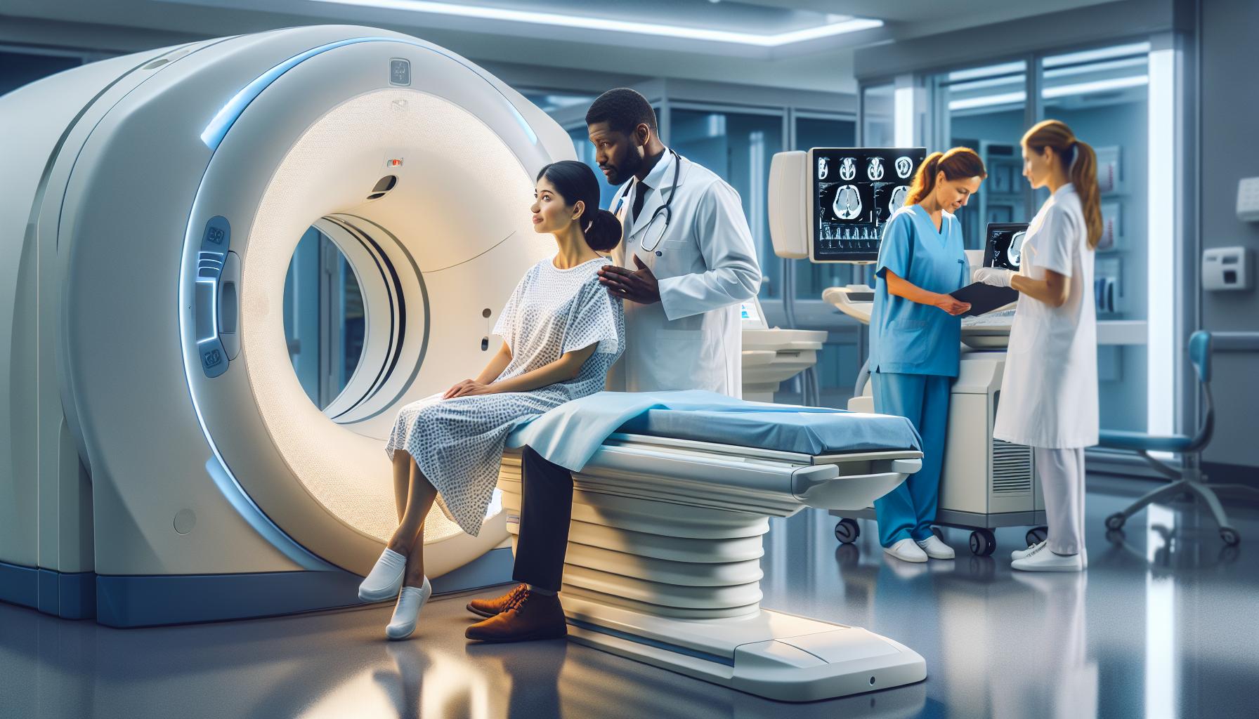
During a CT scan, the patient lies on a motorized table that moves through a circular opening of the CT machine, known as the gantry. As the machine rotates, it emits X-rays and simultaneously captures images from various angles. The images are then processed by a computer, resulting in detailed pictures of the brain’s anatomy. It’s noteworthy that while CT scans are highly effective at detecting certain brain tumors and abnormalities, they may not always capture all possible issues due to factors like tumor size or location, which can affect visibility within the images.
Key Advantages of CT Scans
- Speed: Scans typically take only a few minutes, making them suitable for emergencies.
- Detailed Imaging: Offers clear images of brain structures and helps differentiate types of tissue.
- Accessibility: Widely available in hospitals and imaging centers, enabling faster diagnosis and treatment initiation.
Nevertheless, it’s important for patients to discuss any concerns regarding the scans with their healthcare providers. This dialogue can provide clarity on what to expect before and after the procedure, help alleviate anxiety, and ensure that any specific health considerations are addressed. Engaging with healthcare professionals not only aids in understanding the purpose of the scan but also in navigating the response if a tumor or other abnormalities are detected.
How Accurate Are CT Scans for Detecting Tumors?
CT scans are a powerful tool in the realm of medical imaging, particularly for detecting tumors in the brain. Their ability to create detailed cross-sectional images allows healthcare providers to identify and assess abnormalities effectively. However, the accuracy of CT scans in detecting tumors can vary based on several factors. While these scans have a reasonably high sensitivity-detecting approximately 76.9% of brain tumors-there are instances where they may not capture all abnormalities, especially smaller or less defined lesions or tumors located in hard-to-see positions[[3]].
Several factors can influence the diagnostic accuracy of a CT scan. Tumor size is a significant consideration; smaller tumors may not be visible, or their borders may blend into surrounding tissues. The type of tumor also plays a crucial role; certain tumors, such as low-grade gliomas, may appear less distinct on a CT scan compared to more aggressive types. Additionally, the patient’s specific anatomy and the presence of surrounding conditions, like inflammation, can obscure a tumor’s visibility.
To enhance the accuracy of tumor detection, healthcare providers sometimes use contrast agents during a CT scan. These substances improve the differentiation between various tissues, making it easier to identify abnormalities. If a tumor is suspected based on symptoms or preliminary imaging, further diagnostic measures, including MRI or biopsy, may be warranted to provide a more comprehensive evaluation. Therefore, while CT scans serve as an excellent first-line imaging option, they are not infallible. It’s critical for patients to communicate openly with their healthcare team, discuss their symptoms, and inquire about the best imaging strategy tailored to their individual needs. This proactive approach ensures that any potential issues are accurately identified and managed promptly.
Factors That Can Lead to False Negatives
While CT scans are a valuable tool for detecting brain tumors, they are not without limitations, and certain factors can contribute to false negatives. Understanding these elements can help patients and their families navigate the complexities of brain imaging with greater awareness and assurance.
One primary factor that may lead to a missed diagnosis is the size of the tumor. Smaller tumors, particularly those under a certain threshold, can evade detection as they may not present a clear, distinct image in the scan. Their edges can blend into surrounding healthy tissues, making it challenging for radiologists to identify them. For instance, low-grade gliomas, relatively small and less aggressive tumors, are often more difficult to visualize compared to larger, high-grade tumors, which typically exhibit more pronounced features on a CT scan.
Another important aspect is the type of brain tumor. Some tumors, due to their location or nature, may not elicit significant changes in surrounding brain structures that could easily be detected through imaging. This lack of clear distinction can lead to oversight during the interpretation of the scan. Additionally, patient-specific factors such as anatomy and existing conditions like inflammation or lesions can obscure the visibility of tumors. Inflammation, for example, may mimic the appearance of a tumor or mask its presence, complicating the diagnosic process.
In certain cases, the timing and technique of the scan can also impact results. If a CT scan is performed too early in the development of a tumor, it may not yet be large enough to be detected. Furthermore, the protocols used, such as the angle of imaging and the specific focus of the scan, play a crucial role in the sensitivity and specificity of tumor detection.
Therefore, if a CT scan does not provide conclusive results and symptoms persist or worsen, it is imperative to consult with healthcare professionals. Further evaluations, such as MRI or additional imaging studies, may be necessary to ensure a comprehensive understanding of the patient’s condition, underscoring the importance of ongoing communication with medical providers for the most accurate diagnosis. Empowering patients with knowledge about these factors can lessen anxiety and promote a proactive approach to their health journey.
Common Symptoms That May Indicate a Tumor
Recognizing potential symptoms of a brain tumor is crucial for early detection and treatment. While not all of these symptoms will indicate the presence of a tumor, being aware of them can empower individuals to seek medical attention sooner rather than later. Early intervention can significantly affect outcomes, underscoring the importance of patient awareness.
Some common symptoms to watch for include:
- Headaches: Persistent or severe headaches that differ from typical patterns may be a warning sign. These headaches can be accompanied by nausea or vomiting.
- Seizures: Experiencing new seizures or changes in existing seizure patterns can indicate abnormal brain activity related to a tumor.
- Cognitive Changes: Memory issues, confusion, or changes in reasoning abilities can suggest interference with brain functions.
- Vision or Hearing Problems: Blurred or double vision, loss of peripheral vision, or sudden hearing difficulties can point to tumors affecting areas of the brain responsible for sensory perception.
- Changes in Coordination or Balance: Unexplained problems with coordination, balance, or difficulty walking can indicate potential issues within the brain.
- Personality or Behavioral Changes: Significant mood swings, personality changes, or unusual behavior are potential markers as tumors can affect emotional regulation.
- Weakness or Numbness: Sudden weakness or numbness in limbs, particularly if it affects one side of the body, can be a critical warning sign.
Recognizing these symptoms can create a proactive approach to health. If any of these issues develop, especially if they persist or escalate, it’s essential to consult a healthcare professional for a comprehensive evaluation. Timely discussions with a physician can facilitate the necessary imaging tests, like a CT scan or MRI, to investigate further. Remember, while these symptoms can be alarming, many benign conditions can also produce similar signs. Seeking professional guidance can help clarify the situation and provide peace of mind.
The Role of Contrast Agents in CT Scans
When it comes to enhancing the precision of CT scans, contrast agents play an essential role. These agents, often administered intravenously or orally, provide clarity by highlighting specific areas inside the body, allowing healthcare providers to obtain a more detailed view of potential abnormalities, including brain tumors. By increasing the contrast between different tissues, these agents help distinguish healthy tissue from potentially diseased areas, facilitating more accurate diagnoses.
During a CT scan, the contrast agent enhances the visibility of vascular structures and organs. For brain imaging specifically, using contrast can reveal additional information that might not be evident in standard scans. Tumors, which often have a different blood supply and density compared to surrounding tissues, may absorb the contrast agent differently, making them easier to identify. As a result, healthcare professionals can detect conditions that may otherwise be missed, significantly improving diagnostic accuracy.
While contrast agents are generally safe, it is vital for patients to communicate any allergies or pre-existing medical conditions to their healthcare providers beforehand. Some individuals may experience mild reactions, such as a warm sensation or a metallic taste in the mouth, which are usually harmless. In rare cases, serious allergic reactions can occur, necessitating immediate medical attention. Understanding both the benefits and the considerations associated with contrast use empowers patients to participate actively in their diagnostic process.
If your doctor suggests a CT scan with contrast agent, it is crucial to follow any preparation guidelines, such as fasting before the procedure or staying hydrated. This careful preparation helps ensure the best possible outcomes and minimizes any risks associated with the procedure. Always feel free to ask your healthcare team any questions or express concerns you may have; open communication is key to a positive imaging experience.
CT Scan Limitations: When Is MRI Necessary?
While CT scans are valuable tools for detecting brain tumors, they are not without limitations. Understanding these constraints can help patients and healthcare providers decide when an MRI may be the preferred imaging method. One of the primary challenges with CT scans is their reliance on x-rays, which may not always delineate soft tissues or subtle abnormalities with the clarity that MRIs provide. This limitation is particularly critical in brain imaging where distinguishing between tumor types, grades, and other brain structures is crucial for an accurate diagnosis.
CT scans may miss smaller tumors or those located near complex structures within the brain, which can lead to false negatives. Additionally, certain types of tumors might not present distinct characteristics on a CT scan, necessitating further evaluation with an MRI, which uses magnetic fields and radio waves to produce more detailed images. For example, high-grade gliomas or tumors that infiltrate surrounding brain tissue can often be better visualized using MRI due to its superior soft tissue contrast. When initial CT imaging reveals an abnormality but lacks clarity, an MRI can provide the additional detail needed to confirm or rule out a diagnosis.
Patient experience often drives the decision to use an MRI in conjunction with CT scans. If there are ongoing symptoms-such as headaches, seizures, or cognitive changes-that persist despite CT results, healthcare providers commonly recommend MRI for a more comprehensive view. It’s essential for patients to communicate openly about their symptoms and any concerns with their healthcare team, as these discussions can significantly influence the choice of imaging modality.
In instances where a tumor is suspected but not confirmed through CT findings, the next steps typically involve an MRI to evaluate the extent and type of the lesion. The process begins with a thorough review of the CT results, followed by an MRI if the initial findings suggest further investigation is necessary. By ensuring that patients understand these processes, healthcare professionals can alleviate anxiety and help them feel more engaged in their care decisions, emphasizing that their active participation is vital for achieving the best outcomes.
What Patients Should Expect During a CT Scan
Undergoing a CT scan can be an important step in assessing potential issues within the brain, including tumors. Knowing what to expect during the procedure can help alleviate anxiety and prepare you for the experience. The process is typically straightforward and designed to be as comfortable as possible, but understanding the details can make it much easier.
When you arrive for your CT scan, you will first be greeted by a radiologic technologist who will guide you through the process. You may be asked to change into a gown and remove any metal objects that could interfere with the imaging. The scan itself usually takes about 10 to 30 minutes, during which you will lie on a narrow table that moves through a large, doughnut-shaped machine. It’s important to remain still during the procedure since movement can blur the images and affect the results.
For some scans, a contrast agent may be administered intravenously to enhance the images. This contrast can help highlight specific areas of the brain, making abnormalities more visible. You might feel a slight pinch when the needle is inserted, followed by a warm sensation as the dye enters your bloodstream. This feeling is normal and typically subsides quickly. If you have any concerns or previous allergic reactions to contrast agents, be sure to discuss them with your healthcare provider beforehand.
After the scan, you’ll be able to return to your normal activities, though you might be advised to drink plenty of fluids to help flush the contrast dye from your system. The images will be reviewed by a radiologist, who will send a report to your doctor, detailing any findings. If there is any indication of a tumor or other issue, your doctor will discuss the next steps with you, which may include additional imaging studies or referrals to specialists. Being informed and prepared can help transform a potentially stressful experience into a straightforward part of your health journey.
Evaluating the Risks: Radiation Exposure Insights
Undergoing a CT scan is a crucial step in diagnosing various health conditions, including brain tumors. However, it’s natural to have concerns about radiation exposure associated with this imaging technique. A CT scan delivers a small amount of radiation, which is a key consideration for many patients and their families. Understanding the risks and the measures taken to ensure your safety can significantly alleviate any anxiety.
Radiation exposure from a single CT scan is generally considered low, and for most patients, the benefits of accurate imaging far outweigh the risks involved. For instance, the amount of radiation from a CT scan of the brain is comparable to the background radiation a person is exposed to over a few years. Healthcare professionals always assess the necessity of the scan, ensuring that it’s only performed when clinically warranted. Here’s what you can expect regarding radiation exposure:
- Justification of the Scan: Before a CT scan is approved, your doctor will evaluate whether the advantages of obtaining vital diagnostic information outweigh any potential risks from radiation.
- Minimization of Exposure: Modern CT technology is designed to limit radiation exposure as much as possible without compromising image quality. Techniques such as adjusting the radiation dose based on patient size and the specific area being scanned help to reduce unnecessary exposure.
- Alternatives: In certain cases, alternative imaging methods, like MRI, can be considered, as they do not involve ionizing radiation. Discussing these options with your healthcare provider can provide clarity based on your unique situation.
While radiation exposure is an important concern, it’s essential to keep in mind that the knowledge gained from a CT scan can lead to early detection and improved treatment outcomes for serious conditions like brain tumors. If you have specific worries about radiation or personal medical history that might affect your scan, don’t hesitate to discuss these with your doctor. Being informed and involved in your healthcare decisions can transform concerns into confidence, helping to ensure that your imaging experience is a positive step towards understanding and maintaining your health.
Interpreting CT Scan Results: How Tumors Are Identified
When it comes to diagnosing brain tumors, understanding how CT scan results are interpreted is vital for patients and their families. CT scans provide detailed cross-sectional images of the brain, which can reveal the presence of abnormal growths, including tumors. Medical imaging relies heavily on the experience and expertise of radiologists who analyze the scans for any irregularities or signs of tumors. These professionals look for differences in density, shape, and size of brain tissues in the images that may indicate a tumor, along with other potential issues such as swelling or hemorrhage.
Key Indicators in CT Scan Imaging
During the interpretation of a CT scan, radiologists focus on several important factors to identify tumors. They assess:
- Size and Shape: Tumors often appear as mass lesions that disrupt the normal structure of the brain. A tumor’s irregularity or abnormal size compared to surrounding tissues can be a crucial indicator.
- Location: The specific area of the brain affected by the tumor helps determine its type and severity. Certain tumors have typical locations, influencing both diagnosis and treatment options.
- Enhancement Patterns: If contrast agents are used during the scan, the way the tumor absorbs the contrast can provide additional insights. Tumors may show varying levels of enhancement compared to normal brain tissue.
Challenges to Accuracy
While CT scans are valuable tools, they are not infallible. Several factors can lead to the misinterpretation or oversight of tumors. For instance, small tumors may not be easily visible, particularly when they are located in complex regions of the brain. Additionally, conditions that mimic tumor symptoms, such as inflammation or vascular issues, can complicate interpretations. It is also essential for healthcare providers to consider the patient’s clinical history and symptoms when reviewing results, as this context can significantly impact the evaluation process.
For patients, the key takeaway is the importance of follow-up consultations and potential additional imaging, such as MRI, if there are concerns about a missed diagnosis. Understanding how CT scans work and what radiologists seek can empower patients to ask informed questions about their diagnosis and care pathway. Always discuss any findings or next steps with your healthcare provider to ensure that you are fully informed and supported in your health journey.
Follow-Up Procedures: What Happens If a Tumor Is Suspected?
If concerns arise after a CT scan regarding the possibility of a brain tumor, it’s essential to understand the typical follow-up procedures that may be involved. The healthcare team is dedicated to ensuring that any potential diagnosis is thoroughly investigated, aiming to provide peace of mind and accurate assessments. Following the initial scanning, if a tumor is suspected, your physician will likely discuss the results in detail, explaining any findings from the CT scan and addressing your questions and concerns.
The next step often involves additional imaging studies, with MRI being a common choice. An MRI offers greater detail of soft tissues and can provide clearer images of brain structures, enhancing the ability to identify small or ambiguous lesions that a CT scan might miss. Patients may also undergo neurologic assessments and follow-up scans to monitor any changes over time, which can be critical for making informed decisions about treatment.
In some instances, further diagnostic procedures such as a biopsy may be necessary to conclusively determine the nature of any findings. This involves obtaining a small tissue sample for laboratory analysis, which can confirm whether a growth is benign or malignant. Throughout this process, maintaining open communication with your healthcare professionals is paramount. They can explain each step in a manner that makes sense, ensuring that you feel supported and well-informed. Engaging with other patients going through similar experiences can also be beneficial, providing a sense of community and shared understanding.
Additionally, keep in mind that emotional health is just as important during this time. Reaching out to counselors, support groups, or even trusted friends and family can be invaluable. This type of support can help alleviate anxiety as you navigate the next steps in your care, making the journey a little bit easier amidst uncertainty. Remember, being proactive and well-informed is your best defense when facing any health diagnosis, allowing you to advocate for your needs with confidence.
Patient Stories: Experiences with CT Scans and Tumor Detection
Experiencing a brain imaging process, like a CT scan, can be daunting, especially when the stakes involve the possibility of detecting a tumor. Many individuals have navigated this journey, with a range of experiences that highlight both the emotional impact and the practical aspects of undergoing a CT scan. For example, Sarah, a 34-year-old mother, sought a CT scan after experiencing persistent headaches. The results indicated an abnormal area, and though the initial feeling was one of anxiety, her doctor reassured her that not all abnormalities signify a tumor. This reassurance proved crucial, as it enabled her to focus on the next steps, including follow-up imaging with an MRI, which provided further clarity.
In another story, Mark, a 47-year-old teacher, had faced numerous challenges with vague neurological symptoms. His earlier CT scans returned normal, but he felt that something was amiss. After advocating for further tests, his healthcare team recommended a specialized MRI, revealing a small tumor that the CT scan had missed. Mark’s experience underlines the importance of ongoing communication with healthcare professionals and personal advocacy. His journey to diagnosis involved the support of family and friends, who played an essential role in alleviating his fears during the waiting period.
These personal accounts illuminate common patient questions and fears surrounding CT scans and tumor detection. It’s essential for individuals to approach their healthcare conversations with openness, asking specific questions about their imaging results and the next steps if concerns arise. Having a network of support, whether through friends, family, or patient groups, can make a significant difference. Many find that sharing experiences not only eases anxiety but also provides valuable insights into navigating the medical landscape.
Understanding the variance in individual experiences encourages empathy and reassurance among those facing similar situations. A CT scan, while a helpful tool, sometimes requires further investigation to ensure clarity in diagnosis. Whether through additional imaging, more in-depth discussions with medical professionals, or connecting with others who have faced similar challenges, patients like Sarah and Mark remind us of the importance of active participation in one’s health care journey.
Consulting Healthcare Professionals: Why It Matters
The journey through medical imaging can be confusing and filled with uncertainty, particularly when considering the possibility of brain tumors. It’s crucial to understand that consulting healthcare professionals is not just beneficial-it’s essential for navigating this landscape effectively. Medical experts provide insights and expert knowledge that can significantly impact outcomes, especially when it involves complex conditions like tumors that may not be easily detected via initial scans.
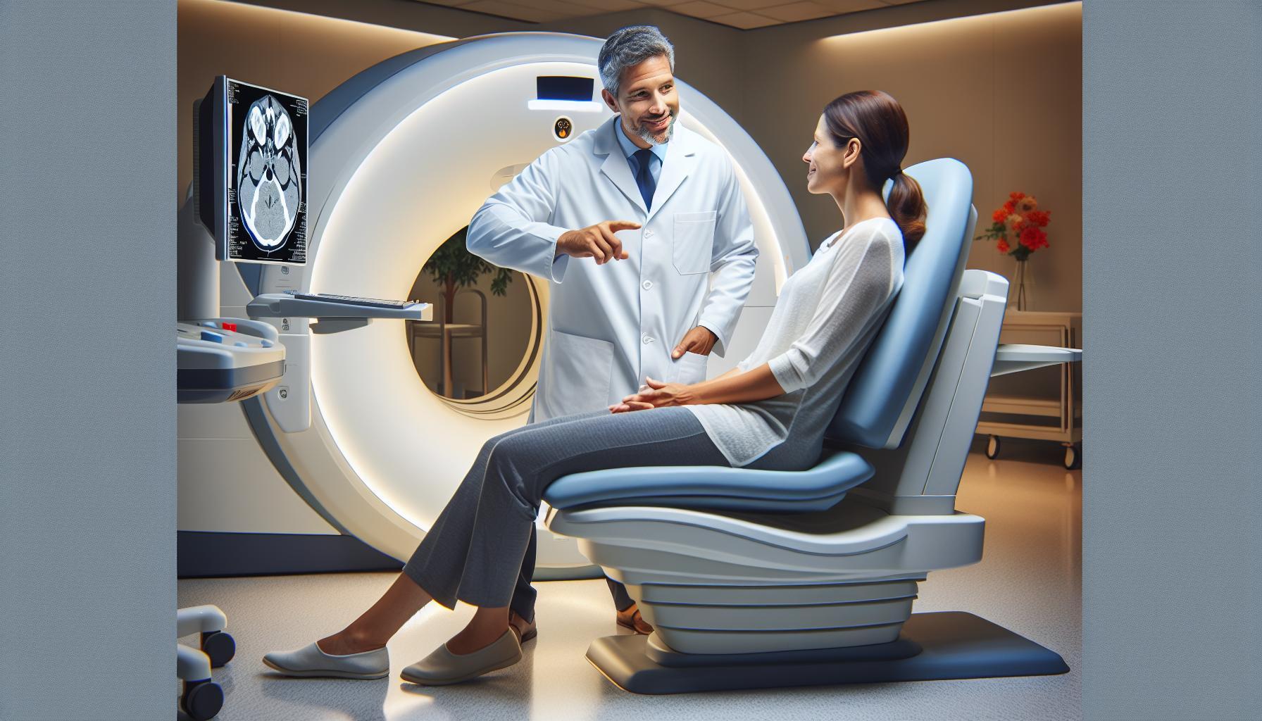
When you receive a CT scan, it’s natural to have questions and concerns, especially if the results show abnormalities or if symptoms persist. Engaging with your healthcare provider offers an opportunity to discuss these findings in detail. They can explain the implications of what was seen on the scan, consider your complete medical history, and suggest further evaluations or necessary follow-up tests, such as MRIs that provide more detailed images of soft tissues. The dialogue between you and your healthcare team is vital, as they can help clarify potential next steps and alleviate anxiety surrounding ambiguous results.
Furthermore, healthcare professionals can enhance your understanding of the limitations of CT scans. For instance, while these scans are excellent for providing a broad overview, they may sometimes miss smaller tumors or specific types of brain lesions. Being aware of this helps in advocating for your health. If you feel that your symptoms are not adequately addressed, trust your instincts. You have the right to seek further opinions or request additional tests.
In addition to the medical expertise, having an ongoing conversation with your healthcare provider can foster a supportive partnership. This relationship can be incredibly empowering, allowing you to ask questions-no matter how small-and gain clarity about the medical processes you’re undergoing. Remember that you don’t have to navigate this alone; your healthcare team is there to support you through understanding your condition, addressing your concerns, and determining the best course of action, ensuring that you receive the most accurate and comprehensive care available.
Frequently Asked Questions
Q: Can a CT scan completely rule out a brain tumor?
A: A CT scan is effective for detecting larger tumors but may miss smaller ones or specific types of tumors. Therefore, while it can identify significant abnormalities, additional imaging like MRI might be necessary for a comprehensive evaluation. Always discuss results with your healthcare provider for appropriate next steps.
Q: What factors can lead to a false negative result in a CT scan for brain tumors?
A: Factors contributing to false negatives in CT scans include the size and location of the tumor, the tumor’s type, and the imaging technique used. Small tumors or those located in complex areas of the brain may not be easily visible. Regular follow-ups and thorough evaluations are crucial if symptoms persist.
Q: How long does it take to receive CT scan results for a brain tumor?
A: Typically, CT scan results are available within a few hours to a day. However, interpretation by a radiologist and subsequent discussion with your doctor may take longer. Don’t hesitate to ask your healthcare provider about the timeline, especially if you’re anxious about your results.
Q: What should I do if my CT scan shows abnormalities but no tumor?
A: If your CT scan reveals abnormalities without a definitive tumor diagnosis, your doctor may recommend further testing, such as an MRI or additional imaging studies, along with monitoring symptoms. This approach helps ensure any potential issues are addressed promptly and accurately.
Q: Can a CT scan miss brain tumors in certain populations?
A: Yes, there are populations that may be at higher risk for missed diagnoses on a CT scan, such as children or patients with specific symptoms that may not correlate with common signs of a tumor. It’s vital to advocate for thorough evaluations if symptoms persist despite normal imaging results.
Q: Is it necessary to use contrast agents during a CT scan for brain tumors?
A: Using contrast agents during CT scans can enhance the visibility of tumors, making it easier for radiologists to identify abnormalities. While not always necessary, your doctor may recommend it based on your individual case and the information needed for an accurate diagnosis.
Q: What are the common symptoms that might indicate a brain tumor?
A: Common symptoms of a potential brain tumor include persistent headaches, vision problems, seizures, difficulty with balance or coordination, and unexplained changes in mood or behavior. If you experience these symptoms, consult your healthcare provider for further evaluation and imaging.
Q: When should I consider a follow-up MRI after a CT scan?
A: Follow-up MRI is advisable when there are abnormalities detected on a CT scan, when symptoms persist, or when initial CT findings are inconclusive. Discuss any concerns you have with your healthcare provider to ensure an appropriate plan for further evaluation is in place.
The Way Forward
In conclusion, understanding the limitations and capabilities of a CT scan in detecting brain tumors is essential for making informed healthcare decisions. While a CT scan can aid in detection, it may not always provide a definitive answer, which is why consulting with your healthcare provider is crucial for personalized guidance. If you’re still concerned about your health or symptoms, consider scheduling a consultation for more comprehensive imaging options like MRI, known for clearer results.
Explore our resources on brain tumor diagnosis and treatment for further insights, or read about the implications of imaging technology on your health. Don’t leave your health to chance; empower yourself with knowledge and take proactive steps today. Remember, your journey to understanding starts with one click-dive deeper into our related articles and equip yourself with critical information. Your health matters, and we’re here to support you every step of the way.


