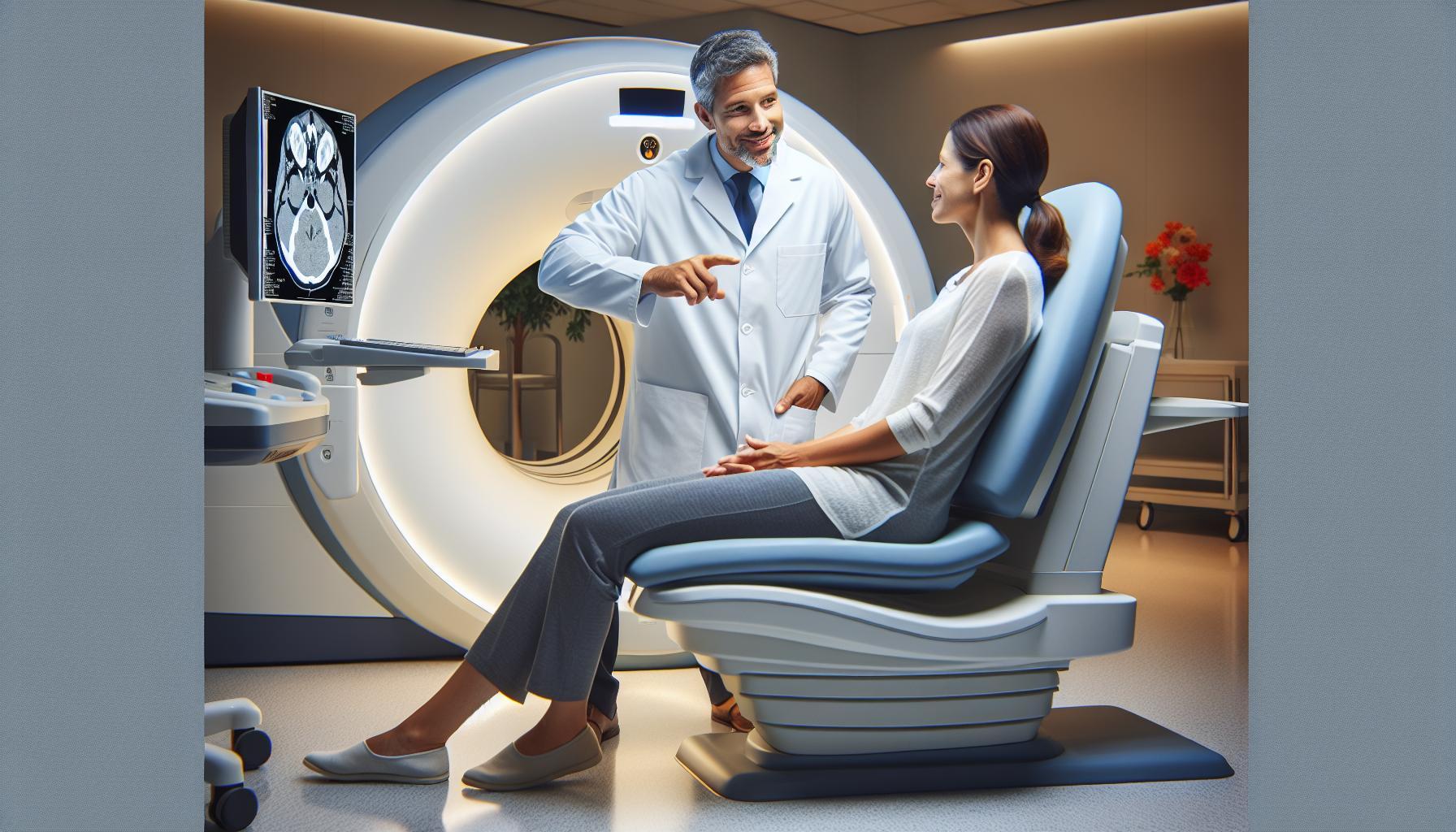
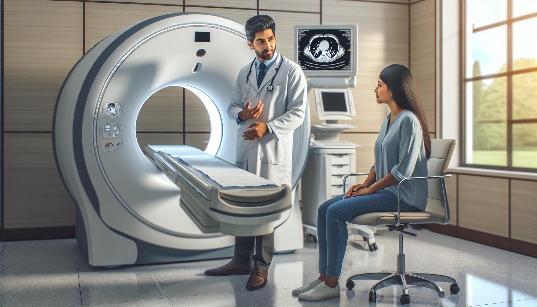
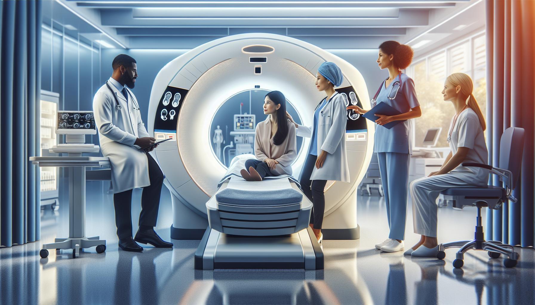
Every year, millions of people experience concussions, yet many wonder how these brain injuries are properly diagnosed. While a CT scan is a powerful imaging tool, it’s crucial to understand its role in detecting concussions. This article will explore whether concussions can be effectively identified through CT scans, offering insights that could ease concerns for those who have suffered head injuries.
Understanding how concussions manifest and the limitations of diagnostic imaging can empower you or your loved ones in seeking the right medical guidance. If you’ve experienced a blow to the head, knowledge is your best ally in navigating the complexities of brain health. Join us as we uncover the facts surrounding concussions and CT scans, helping you make informed decisions about your health and recovery.
Understanding Concussions: Key Facts You Need to Know
Understanding concussions is essential for anyone involved in activities where head injuries can occur, from athletes to parents and coaches. A concussion is a type of traumatic brain injury that results from a blow to the head or body, causing the brain to move rapidly back and forth within the skull. This movement can create chemical changes in the brain and, in some cases, damage brain cells. Symptoms of a concussion can vary widely and may not appear immediately after the injury. These can include headaches, confusion, dizziness, nausea, and difficulty concentrating.
Recognizing the potential seriousness of a concussion is crucial. Even though they are often categorized as mild traumatic brain injuries, concussions can lead to significant long-term effects if not adequately addressed. Immediate evaluation by a healthcare provider is necessary, especially if symptoms worsen. It’s important to remember that management typically includes rest and a gradual return to normal activities, but that process should be guided by a healthcare professional.
Most people are aware that imaging tests can help identify more severe head injuries, like skull fractures or hemorrhages, but many wonder if CT scans can detect concussions. The reality is that concussions do not usually show up on CT scans. Instead, these scans are effective for ruling out more severe injuries, allowing doctors to determine the best course of action for the patient. Understanding what a CT scan does-and does not-reveal can alleviate some anxiety surrounding the examination, making it clear that while CT scans are valuable tools, they primarily serve in managing significant neurological concerns rather than diagnosing subclinical injuries like concussions.
Awareness and education about concussions can empower individuals to act swiftly and effectively when a head injury occurs. If you or someone you know experiences a potential concussion, prioritizing professional medical advice is key to ensuring a safe recovery.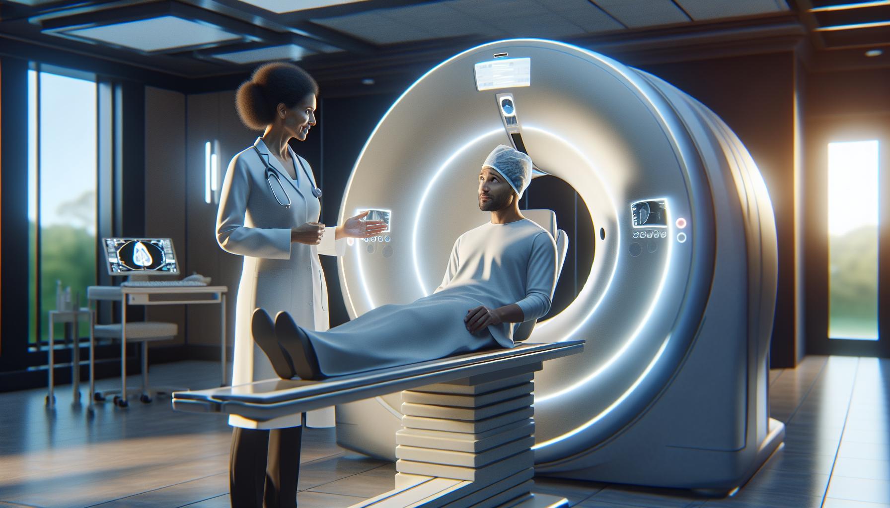
How CT Scans Work: A Step-by-Step Guide
A CT scan, or computed tomography scan, is a powerful imaging tool that allows healthcare professionals to visualize the inside of the body, particularly the brain, with remarkable detail. Understanding how this technology works can alleviate concerns and clarify its role in diagnosing and managing medical conditions, including head injuries.
During a CT scan, a series of X-ray images are taken from different angles around the body. These images are processed by a computer to create cross-sectional views, or slices, of the tissues and organs. Here’s a simple step-by-step breakdown of the CT scan process:
Step-by-Step Process of a CT Scan
- Preparation: Before the scan, patients may be asked to change into a hospital gown and remove any metal objects, such as jewelry, that could interfere with the imaging. If a contrast material is needed to enhance the images, the healthcare provider will explain the procedure and assess any potential allergies.
- Positioning: Patients lie on a motorized table that slides into the CT scanner. It’s essential to remain still during the scan to ensure the images are clear and accurate. Some scans, particularly those of the brain, might require you to be positioned with your head in a specific way.
- Scanning: The CT machine will rotate around the body, capturing multiple images as it does so. You might hear a humming or buzzing sound while the scan is in progress, which typically lasts only a few minutes.
- Completion: After the scan, patients can usually resume normal activities immediately. If contrast material was used, drinking plenty of fluids helps flush it from the body.
Unlike regular X-rays, CT scans are particularly useful in identifying more severe injuries like fractures or internal bleeding, but they are not designed to detect concussions or subtle imaging findings associated with them. This distinction is crucial to understand, as it helps manage expectations regarding what a CT scan can reveal.
As you approach a CT scan, it can be reassuring to remember that you are in the hands of experienced healthcare professionals who are committed to your safety and comfort. Always feel free to ask questions or express concerns; clarity can greatly reduce anxiety surrounding medical procedures. If you’re ever in doubt about your symptoms or the necessity of a CT scan, consulting a healthcare provider is the best course of action to ensure your health is prioritized.
Are Concussions Visible on CT Scans? Exploring the Science
While a CT scan is a powerful diagnostic tool, it’s essential to understand that it has limitations when it comes to identifying concussions. Unlike more severe brain injuries, concussions often do not result in detectable structural changes in the brain that a CT scan can visualize. This means that even after experiencing a concussion, a CT scan might appear normal, which can be confusing and concerning for patients and their families.
Concussions are classified as a type of mild traumatic brain injury (mTBI) and typically don’t show up on imaging tests like CT scans or MRIs because the damage is often functional rather than structural. Concussions may involve disruptions in brain activity and temporary alterations in consciousness without clear physical evidence that imaging can capture. This functional nature of concussions means they can sometimes lead to symptoms such as headaches, confusion, dizziness, and nausea, even when no visible injuries are present on a scan.
The distinction is critical because patients may seek a CT scan for reassurance after sustaining a head injury. While a normal CT scan can rule out other severe issues such as fractures or bleeding, it does not confirm the absence of a concussion. Therefore, it’s paramount to focus on the symptoms and clinical assessments by healthcare professionals to gauge brain health following an injury. If symptoms persist or worsen, consulting with a medical professional about your condition is vital for appropriate management and care.
Understanding this can help in managing expectations and navigating the recovery process more effectively. While imaging plays a role in evaluating head injuries, it is essential to complement it with thorough assessments and patient history to develop a comprehensive treatment plan. This holistic approach ensures that those suffering from a concussion receive the best care tailored to their specific needs.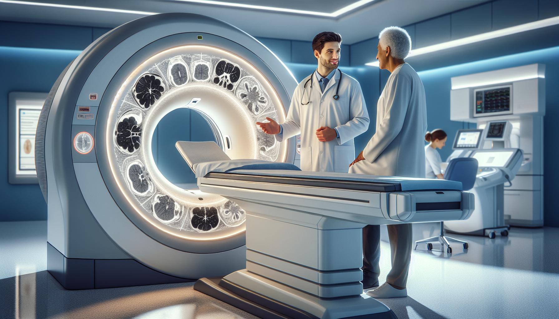
Common Misconceptions About CT Scans and Concussions
Many individuals believe that undergoing a CT scan after a head injury will provide definitive answers regarding the presence of a concussion. However, this is a significant misconception. CT scans are designed to identify structural injuries such as fractures or bleeding in the brain but are not equipped to detect concussions, which are primarily functional brain injuries. Even a normal CT scan following a head injury does not guarantee that a concussion is absent; the brain’s functional disruptions may not manifest as visible changes on imaging.
Understanding the limitations of CT scans is essential for patients and their families. For instance, concussions can result in a range of symptoms-including headaches, dizziness, and cognitive difficulties-that may persist even when imaging results appear normal. This lack of visible injury can lead to unnecessary anxiety, as patients might expect a clear diagnosis or reassurance from imaging. It’s crucial to remember that concussions can significantly impact daily life, and clinical assessment and symptom management remain paramount.
Another common belief is that CT scans can rule out all forms of brain injury. In reality, while CT technology serves as an invaluable tool in emergency settings to exclude severe conditions, it shifts focus away from the subtle nature of concussions. Clinicians will often rely on patient history, reported symptoms, and neurological examinations to assess the situation effectively. Patients should be encouraged to communicate openly with healthcare providers about their experiences post-injury, allowing for a more comprehensive evaluation and tailored care plan.
In navigating the complexities surrounding concussions and CT scans, an empathetic approach is key. Providing education about the purpose of the scan, combined with reassurance regarding the recovery process, can mitigate concerns. For those who have sustained a head injury, emphasizing the importance of follow-up assessments and the role of healthcare professionals in managing symptoms can cultivate a supportive environment where informed decisions are made collaboratively.
Signs and Symptoms: When to Seek a CT Scan
Experiencing a head injury can be alarming, especially when symptoms start to appear. Recognizing when to seek a CT scan is crucial in ensuring proper evaluation and care. While most concussions do not show visible signs on a CT scan, certain symptoms may warrant immediate medical attention to rule out severe injuries such as fractures, bleeding, or swelling in the brain. If you, a loved one, or an athlete has sustained a head injury, staying vigilant about specific signs can guide your decision-making when seeking medical help.
If symptoms such as persistent headaches, nausea, vomiting, dizziness, or confusion develop following a head injury, it’s essential to consult with a healthcare professional. Other critical signs to monitor include:
- Loss of consciousness: Even a brief loss of consciousness can be significant.
- Severe or worsening headache: A headache that intensifies rather than subsides should be evaluated.
- Changes in vision: Blurry or double vision, or any sudden vision changes can be concerning.
- Difficulty with balance or coordination: Feeling unsteady can indicate underlying issues.
- Unusual behavior or confusion: If someone seems dazed or acts differently than usual, it’s a sign to seek help.
It’s vital to approach this with calmness and clarity. If any of these symptoms arise, especially in the first few hours or days post-injury, consider visiting an emergency room or urgent care facility. The primary purpose of a CT scan in this context is to identify acute injuries that may require immediate intervention. Remember that while CT scans are beneficial for detecting structural injuries, they cannot confirm the presence of a concussion itself; therefore, discussing symptoms and medical history with a healthcare provider remains essential for a comprehensive evaluation. Empowering yourself with knowledge not only reduces anxiety but ensures that you are well-prepared to seek the necessary care when it matters most.
Patient Preparation: What to Expect Before Your CT Scan
Undergoing a CT scan can be a crucial step in diagnosing potential brain injuries, particularly after a concussion. Understanding what to anticipate before your appointment can alleviate anxiety and help you prepare effectively for the procedure. Firstly, it’s essential to arrive with a clear mind about the process. A CT scan is a painless imaging technique that uses X-rays to create detailed pictures of the brain, allowing healthcare providers to evaluate any structural damage or bleeding that may have resulted from a head injury.
Before the scan, you will typically be asked to remove any metallic items such as jewelry, glasses, or hairpins, as these can interfere with imaging quality. Depending on your specific case, your doctor may provide instructions regarding food and drink intake beforehand. In many instances, you can continue to eat and drink as normal; however, if contrast material is required for the scan, you might be advised to fast for a few hours prior to your procedure.
It’s also beneficial to wear comfortable, loose-fitting clothing, and some facilities may provide patient gowns for you to change into. If you have any concerns about the scan or if you are sensitive to contrast materials or radiation, don’t hesitate to communicate this with your healthcare team. They are there to reassure you, answer any questions, and ensure that you feel as comfortable as possible during the process.
Finally, consider discussing any recent medications or health conditions with your doctor, as this information helps tailor the procedure to your specific needs. By preparing adequately and understanding what to expect, you can navigate your CT scan experience with confidence, knowing it’s a vital tool for assessing and ensuring your health following a concussion.
Safety and Risks of CT Scans in Concussion Detection
A CT scan can be a critical tool in evaluating concussions, as it allows doctors to see any structural changes or bleeding in the brain. However, understanding the safety and risks associated with this imaging technique can help alleviate apprehensions and ensure informed decisions about your health.
One of the primary concerns regarding CT scans is the exposure to radiation. While CT scans do involve a level of radiation-more than standard X-rays-the benefits often outweigh the risks, particularly in acute settings like concussion evaluation. Modern CT technology aims to minimize radiation exposure. For example, in many cases, low-radiation protocols are used without compromising image quality. It’s important to discuss your specific situation with your healthcare provider, who can assess the necessity of the scan versus alternative options.
Another aspect to consider is the potential for contrast material, which may be used during some CT scans to enhance image clarity. While this substance can help identify problems more effectively, some individuals may experience allergic reactions or adverse effects. If you have a history of allergies or kidney issues, it’s crucial to inform your healthcare team beforehand. They can take necessary precautions, such as prescribing alternative imaging methods if needed.
As with any medical procedure, open communication with your healthcare provider is key to mitigating risks and enhancing safety. They can provide personalized insights based on your medical history, current health condition, and specific symptoms you’re experiencing. Empowering yourself with knowledge will not only help prepare you for the scan but also foster a sense of confidence in the healthcare journey you’re navigating. Remember, the goal of the CT scan is to provide valuable information to ensure your well-being after a concussion.
The Role of MRI vs. CT in Identifying Brain Injuries
Detecting brain injuries such as concussions often involves advanced imaging techniques, and among the most commonly used are CT (Computed Tomography) scans and MRI (Magnetic Resonance Imaging). Each of these technologies offers unique advantages and specific use cases, making it essential for patients to understand their roles in diagnosing brain conditions.
CT scans are particularly effective in assessing acute situations due to their speed and efficiency. They are widely used in emergency settings to quickly identify hemorrhages, skull fractures, or other structural abnormalities that could be life-threatening. The imaging process generates detailed cross-sectional images of the brain, allowing physicians to make swift, informed decisions about treatment. However, CT scans primarily focus on identifying major injuries and may miss subtle damage associated with concussions, such as small contusions or changes in brain tissue.
On the other hand, MRI scans excel in providing detailed images of soft tissues, making them more sensitive to minor brain injuries, including those that may not be immediately visible on a CT scan. MRI can detect microbleeds or diffuse axonal injuries, which are common in concussions. Although MRIs take longer and are usually not performed in emergency situations, they can be invaluable for ongoing assessment after an initial CT scan has been completed.
Both imaging methods have their place, and the choice between a CT scan and an MRI often depends on the specific circumstances surrounding the patient’s condition. For instance, while a CT scan may be the first step in acute concussion assessment, an MRI might be recommended during follow-up evaluations to monitor recovery and address any lingering symptoms. Engaging in a thorough conversation with your healthcare provider can help clarify which scan is appropriate for your situation and ensure tailored care to your needs. Always remember that your healthcare team is there to provide guidance based on personal medical history and current health conditions, empowering you on your journey to recovery.
Interpreting CT Scan Results: A Patient’s Perspective
Interpreting CT scan results can often feel like deciphering a code, especially for those who are grappling with the immediate and overwhelming reality of a concussion. Understanding the outcome of your CT scan is crucial in guiding your next steps, whether that means reassurance, further investigation, or treatment. Keep in mind that while CT scans are an essential tool in identifying acute brain injuries, they have limitations in detecting subtle damage caused by concussions. This makes the communication between you and your healthcare provider vital.
When you receive the results of your CT scan, your doctor will explain the findings in relation to your specific symptoms and injury history. They may use terms like “hemorrhage” or “contusion” to describe injuries that are clearly visible on the scan. It’s natural to feel anxious or unsure during this process, but remember that understanding the context of these findings is key. If anything is unclear, don’t hesitate to ask questions. For example, if a “normal” result is reported but you still experience symptoms, inquire about the possibility of follow-up imaging, like an MRI, which can provide more detailed insights into soft tissue injuries often associated with concussions.
It’s important to set expectations for what your CT scan can show. While it may identify significant issues-such as fractures or bleeding-it might overlook milder concussions, which can lead to confusion about your overall health. This is why ongoing discussions with your healthcare provider are essential. Be proactive; share your symptoms and any changes in your condition, as well as any concerns you have after receiving the scan results. This collaboration can enhance your understanding of your injury and support you in making informed decisions about treatment and recovery strategies.
In summary, interpreting CT scan results should not be a solitary journey. Engaging with your healthcare provider and seeking clarity will empower you during your recovery process. Remember, every step taken in understanding your brain health is significant for your wellbeing. Your healthcare team is there to help you navigate this complex landscape with compassion and expertise, ensuring that you are informed and cared for throughout your recovery journey.
Advancements in Imaging Technology: What Lies Ahead
As advancements in medical imaging technology continue to evolve, they promise to revolutionize the identification and management of concussions. Traditional CT scans have served a crucial role in diagnosing acute brain injuries, yet they can sometimes miss subtle changes associated with mild traumatic brain injuries. Looking ahead, innovative imaging modalities are being developed to enhance our understanding and detection of these complex conditions.
One exciting advance is the integration of functional imaging techniques, such as functional MRI (fMRI) and diffusion tensor imaging (DTI). These methods allow medical professionals to visualize brain function and the integrity of neural pathways, respectively. For example, fMRI assesses brain activity by measuring blood flow changes, providing insights into how the brain responds to stimuli post-injury. DTI, on the other hand, can help identify microstructural changes in brain tissue that indicate injury, even when CT scans appear normal.
Moreover, researchers are exploring the use of biomarkers in combination with advanced imaging. By developing blood tests that can detect specific proteins associated with brain injury, healthcare providers may have an additional layer of information to guide treatment decisions alongside imaging results. This holistic approach could lead to earlier detection of concussive impacts, better tracking of recovery, and ultimately more personalized treatment plans.
Despite the promise of these innovations, patient comfort and safety remain paramount. It’s essential that individuals understand these new technologies will be introduced gradually and integrated with existing practices. As always, consulting with healthcare professionals is crucial when discussing imaging options, as they can provide guidance tailored to individual circumstances, ensuring both safety and accuracy in the diagnosis of concussions. As these advancements unfold, they hold the potential to greatly enhance our ability to identify, monitor, and treat concussions more effectively.
Consulting a Healthcare Professional: Your Next Steps
When faced with a potential concussion, understanding the next steps can significantly ease anxiety and provide a clear path forward. Consulting a healthcare professional is essential for receiving personalized medical advice tailored to your individual needs. With concussions being complex injuries, a thorough assessment by a knowledgeable expert will help determine the appropriate course of action, including whether a CT scan is warranted.
During your consultation, be prepared to discuss your symptoms in detail. Common indicators of a concussion include headaches, dizziness, confusion, and difficulty concentrating. It’s helpful to relay the circumstances of the injury, including any loss of consciousness or subsequent symptoms. This information allows healthcare providers to evaluate the severity of your condition and decide on the necessity of imaging studies like CT scans. If a CT scan is deemed appropriate, your clinician will explain what to expect, reassuring you about the safety and effectiveness of the procedure.
Before your appointment, consider keeping a symptom diary. Documenting when symptoms occur, their intensity, and any triggers can provide valuable insights for your healthcare provider. Additionally, prepare a list of medications and supplements you are currently taking, as this information is crucial for determining any potential interactions or contraindications for imaging procedures.
After your CT scan, the medical team will interpret the results and discuss them with you, outlining both your current condition and the next steps in your care plan. It’s natural to have questions; don’t hesitate to ask for clarification on the results or about further testing, treatment options, and follow-up appointments. Remaining engaged in your recovery process is vital, and your healthcare provider is there to guide you toward a safe return to daily activities. By taking your health seriously and seeking professional advice, you empower yourself to take proactive steps toward recovery.
Frequently Asked Questions
Q: How accurate are CT scans in detecting concussions?
A: CT scans are not specifically designed to detect concussions, as concussions typically involve functional brain changes rather than structural damage. While CT can reveal serious brain injuries, concussions may not show apparent abnormalities on these scans. Consult a healthcare professional for proper assessment of concussion symptoms.
Q: What are the limitations of using CT scans for concussion diagnosis?
A: The primary limitation of CT scans in diagnosing concussions is their inability to detect subtle brain changes associated with mild traumatic brain injury. CT scans excel in identifying structural damage like bleeding but may miss functional issues. For comprehensive evaluation, an MRI or specialized tests may be better.
Q: When should someone get a CT scan after a concussion?
A: A CT scan should be considered if a person experiences severe headaches, repeated vomiting, confusion, or loss of consciousness after a head injury. It’s crucial to seek medical advice to determine if a CT is necessary based on the presence of these symptoms.
Q: Can concussions lead to permanent brain damage detectable by CT scans?
A: While most concussions are reversible, repeated head injuries can lead to permanent damage, which might be visible on a CT scan. Long-term effects such as chronic traumatic encephalopathy (CTE) require further investigation and monitoring by healthcare professionals over time.
Q: Are there alternative imaging techniques to CT scans for concussion assessment?
A: Yes, MRI (Magnetic Resonance Imaging) is an alternative that provides detailed images of brain structures and can detect changes associated with concussions better than CT scans. Functional imaging techniques may also help assess brain activity and identify potential issues related to concussions.
Q: How can I prepare for a CT scan if I suspect a concussion?
A: Preparation for a CT scan typically involves fasting for a few hours before the procedure and informing the technician of any allergies or existing medical conditions. It’s also important to remove any metal objects that may interfere with imaging. Follow your healthcare provider’s specific instructions for a smooth process.
Q: What should I do if my CT scan results are normal but I still have concussion symptoms?
A: If CT scan results are normal but symptoms persist, follow up with your healthcare provider for further evaluation. They may recommend additional tests, such as an MRI or cognitive assessments, to explore underlying issues and ensure appropriate management of your symptoms.
Q: How does a CT scan help in the context of sports-related concussions?
A: In sports, CT scans assist in ruling out serious injuries like fractures or internal bleeding after a player sustains a head impact. However, it’s essential to remember that a normal CT does not imply the absence of a concussion, and ongoing monitoring of symptoms is necessary.
To Wrap It Up
Understanding the nuances of concussion detection is crucial for your health. While CT scans are essential in ruling out severe brain injuries, they may not always reveal concussions, which can have serious implications for recovery. For further insights on managing concussions, explore our comprehensive guide on what constitutes a concussion or check our article on baseline concussion testing for preventative strategies.
If you have concerns about potential symptoms or the next steps after an injury, don’t hesitate to reach out for personalized advice from a healthcare professional. Stay proactive about your cognitive health and, importantly, never dismiss the impact of even mild head injuries. Remember, your well-being is worth the attention! Share your thoughts and experiences in the comments, and explore our site for more valuable resources on understanding and recovering from concussions.


