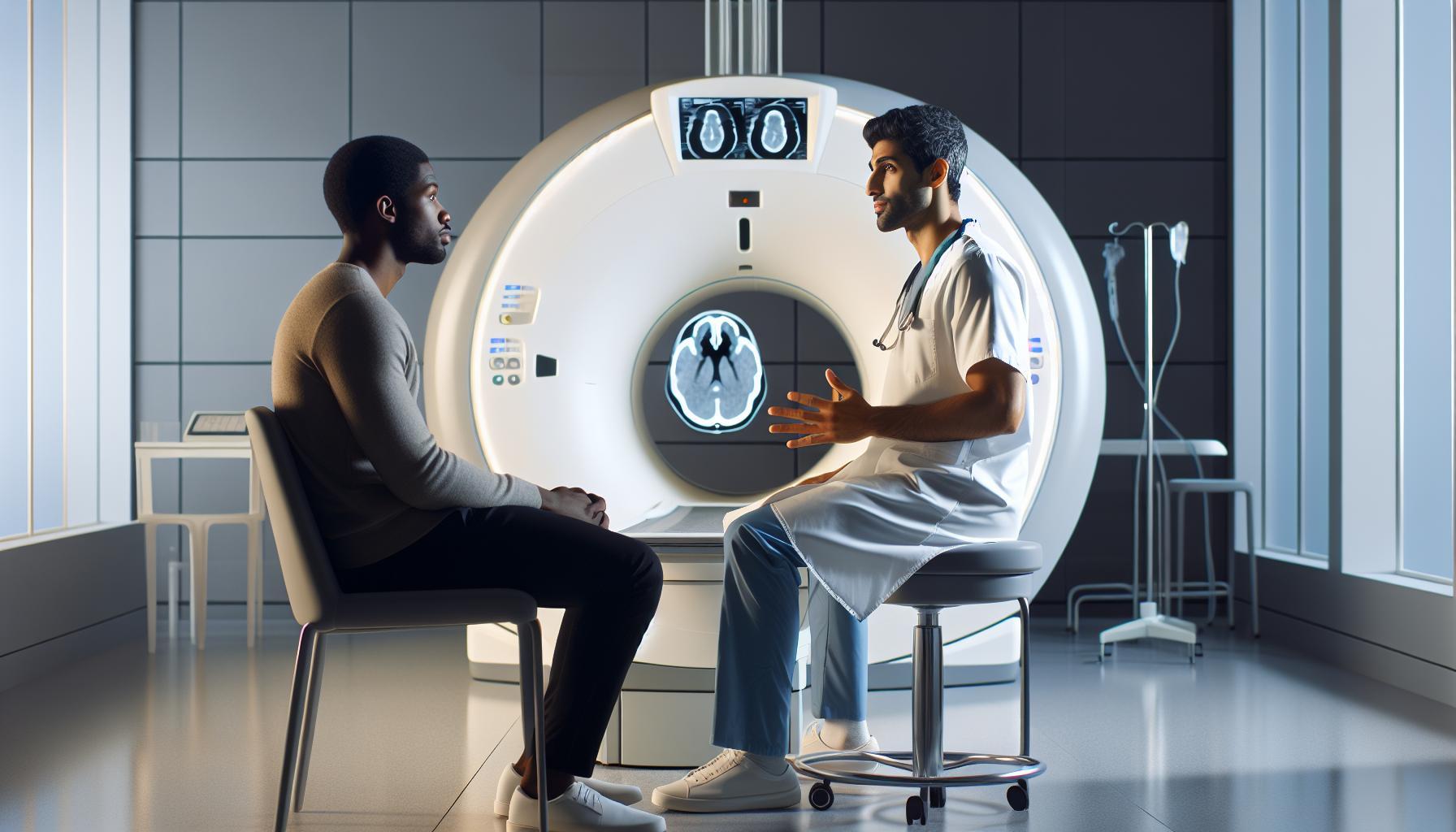
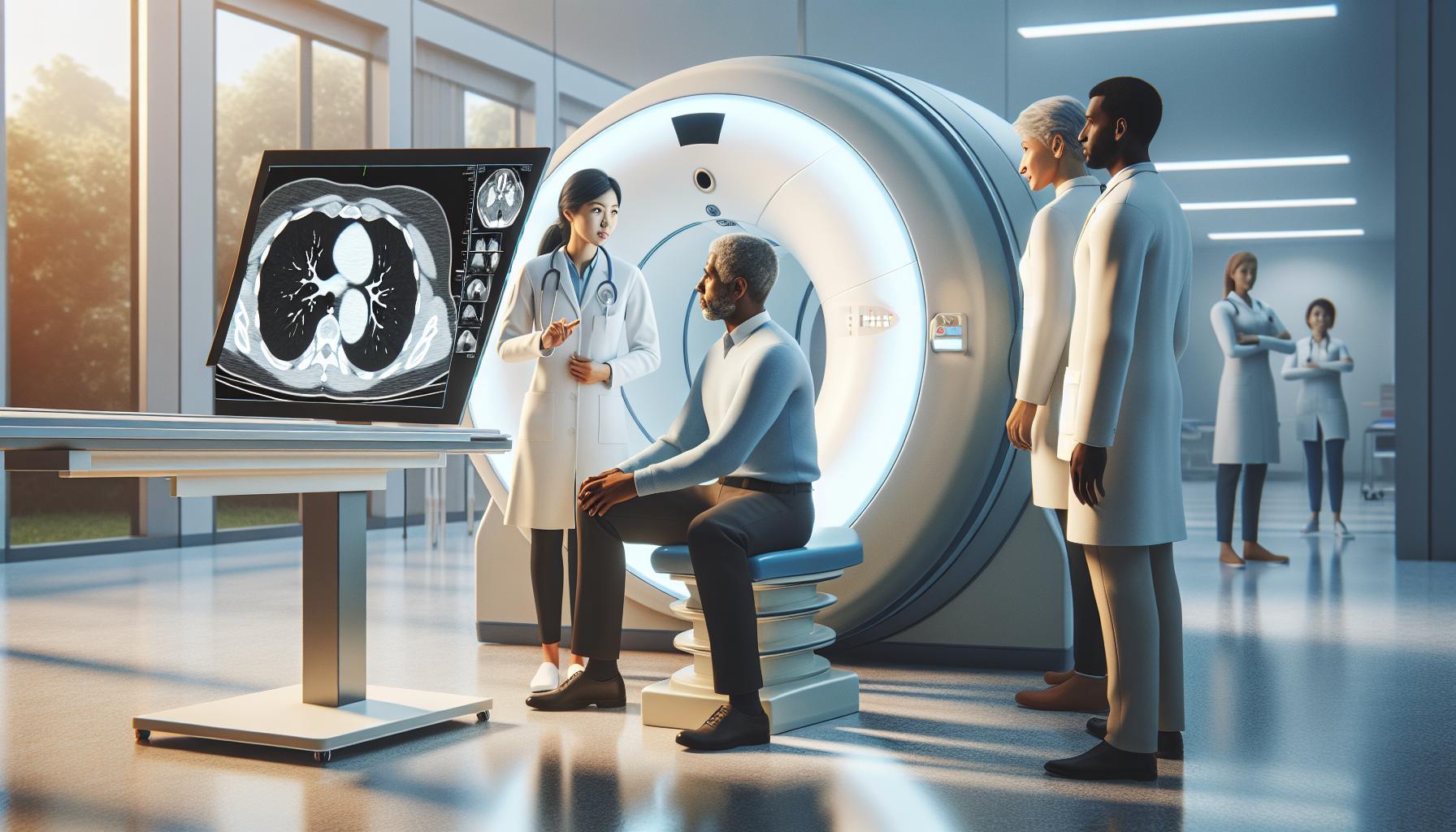
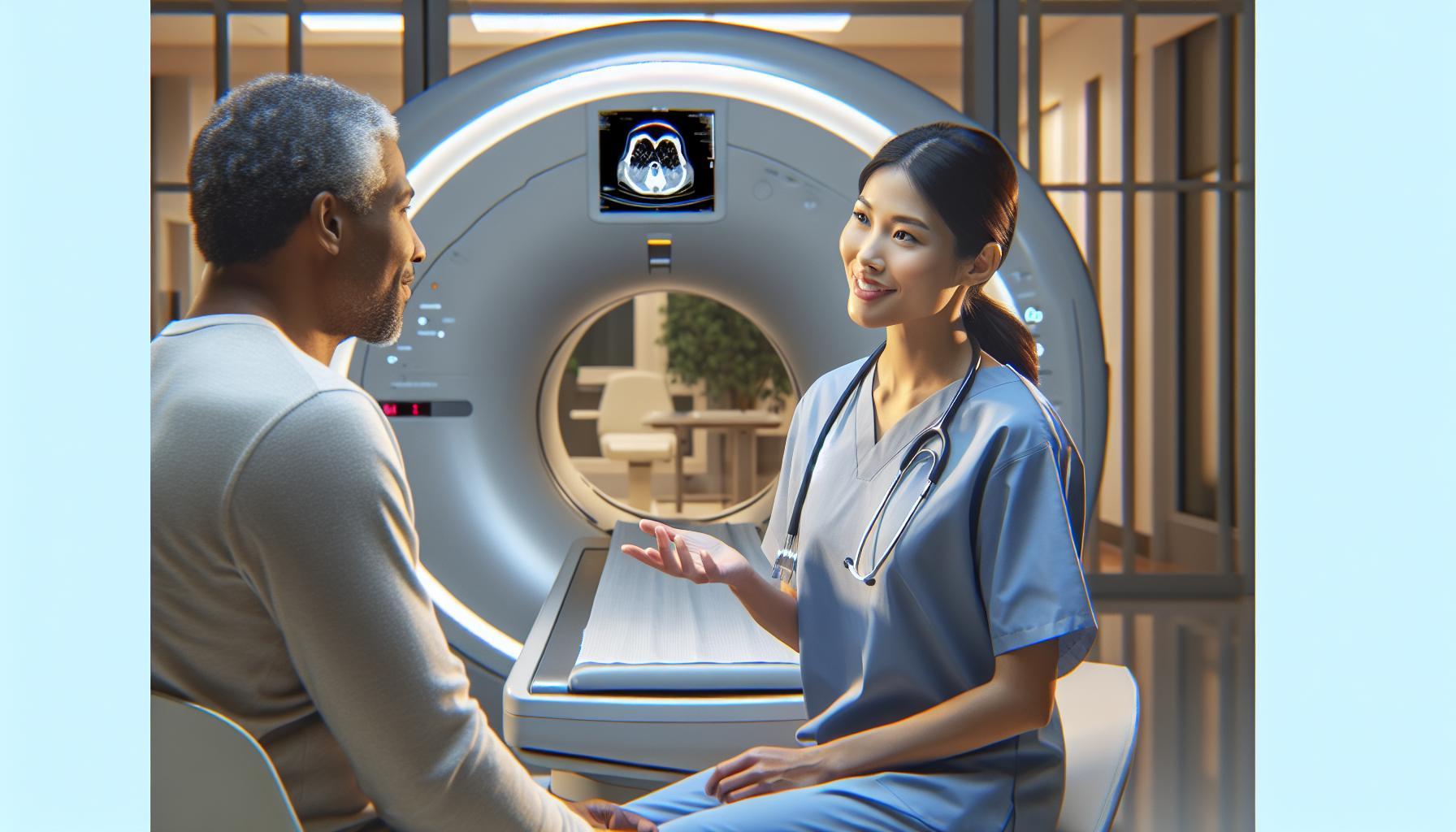
Concussions are common yet often overlooked injuries that can have lasting effects on health and well-being. Understanding whether a CT scan can effectively detect a concussion is crucial for those concerned about brain injuries. While traditional imaging may not reveal all nuances of a concussion, the role of CT scans in identifying potential complications like bleeding is vital.
As awareness of concussion-related risks grows, many find themselves asking, “How can I be sure my health is safeguarded?” This article delves into the capabilities and limitations of CT imaging in concussion detection, empowering you with knowledge to guide discussions with healthcare professionals. With clarity on this topic, you can make informed decisions about your health and that of your loved ones.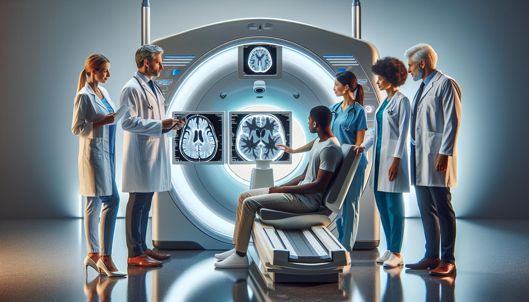
Understanding Concussions: What You Need to Know
In recent years, concussions have gained attention due to their prevalence in sports and the potential long-term effects on brain health. Defined as a type of traumatic brain injury, concussions can occur even with mild impact. Recognizing the signs-such as headaches, confusion, dizziness, and memory disturbances-is crucial for timely treatment. However, diagnosing a concussion can be challenging as conventional imaging techniques, including CT scans, often reveal no visible damage.
Contrary to more severe brain injuries that may show fractures or bleeding on a CT scan, concussions typically do not present detectable structural changes. This limitation highlights the necessity of thorough clinical assessments. Medical professionals often rely on a combination of patient history, symptom evaluation, and cognitive tests alongside imaging to form a complete picture of an individual’s condition.
It’s equally important for individuals who experience a head injury to seek medical guidance, even in the absence of obvious symptoms. Early intervention can significantly enhance recovery and prevent complications. To empower patients, healthcare providers should engage in open discussions about the utility of imaging options, including CT scans, while emphasizing the significance of ongoing monitoring and personalized management plans.
Through understanding the complexities of concussions and their diagnosis, patients can feel more supported in navigating their treatment journey, fostering a collaborative relationship with healthcare professionals focused on recovery and health optimization.
How CT Scans Work for Brain Injury Detection
Understanding how CT scans function in the context of brain injuries can be crucial for patients and families grappling with the aftermath of a head injury. A CT (computed tomography) scan utilizes a series of X-ray images taken from different angles, coupled with computer processing, to create cross-sectional views of the brain. This imaging technique helps in identifying various types of brain injuries that may result from trauma, including hemorrhages, fractures, and more severe conditions that may necessitate immediate medical intervention.
When a patient presents with a suspected concussion, CT scans can be instrumental in ruling out more serious injuries. For instance, while a concussion may not manifest any visible changes on the scan, such as swelling or bleeding, it is essential to ensure there are no other complications, like an intracranial hematoma. The scan will highlight any abnormal accumulation of blood or fluid that could compromise brain function, offering a safety net for quick decision-making regarding further treatment.
Preparation for a CT scan is straightforward and designed to minimize anxiety. Patients may be asked to remove jewelry and any metal objects that could interfere with the imaging. In most cases, no special preparation-like fasting-is necessary. During the procedure, the patient lies on a table that moves through the CT scanner. The scan usually lasts just a few minutes, and it’s painless, although the machine may produce loud noises. Having a supportive family member or friend can provide comfort and reassurance before and after the scan.
It is important to understand that while CT scans are a useful tool, they have limitations when it comes to concussions. They do not always reveal the extent of brain function impairment or detect subtle brain injuries. Therefore, healthcare professionals often complement CT results with comprehensive assessments like neurological examinations and cognitive testing to achieve a holistic understanding of the patient’s condition. This combination of technological and clinical evaluation is vital for establishing proper care plans tailored to the individual’s recovery needs.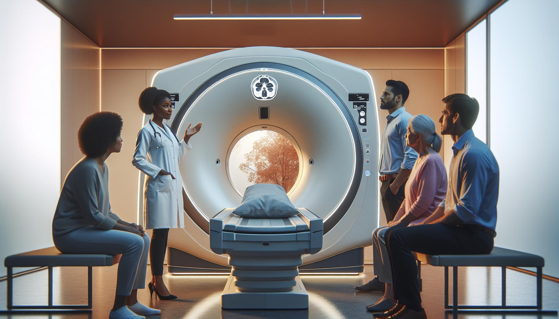
Limitations of CT in Concussion Diagnosis
Many people may not realize that a CT scan, while a powerful tool for assessing brain injuries, has significant limitations when it comes to diagnosing concussions specifically. Concussions are complex injuries that often do not produce overt structural damage visible on imaging tests. This means that even though a CT scan can effectively identify severe brain injuries, such as fractures or bleeding, it may not show any abnormalities in cases of concussion, which primarily involve functional disturbances in the brain rather than physical injuries.
One of the main challenges with using CT scans for concussion diagnosis is their inability to detect subtle changes in brain function. For instance, a person with a concussion might experience symptoms like headaches, dizziness, or cognitive difficulties without any visible signs of trauma on the CT images. Consequently, healthcare providers often need to rely on detailed neurological examinations and patient-reported symptoms alongside imaging results to assess the full extent of a concussion. Understanding this distinction is crucial for patients and their families so they do not underestimate the effects of a concussion merely because a CT scan appears normal.
Moreover, CT scans are typically not sensitive enough to identify microstructural brain injury that could occur in mild traumatic brain injury situations. Advanced imaging techniques, such as MRI, might offer a better alternative in some cases, especially when ongoing symptoms persist despite a normal CT scan. However, MRIs are less commonly employed in acute settings due to their longer duration and increased costs. Thus, many patients may leave the hospital with a false sense of security when they receive normal CT results, inadvertently undermining the importance of monitoring symptoms and seeking further evaluation or rest.
In summary, while CT scans can provide valuable information about potential complications following a head injury, they are not comprehensive tools for diagnosing concussions. The lack of detectable abnormalities often leads to a gap in understanding the injury’s impact on the brain. This makes it essential for patients to remain vigilant about their symptoms and maintain open communication with healthcare providers about any ongoing issues they experience after a concussion.
Comparing CT Scans to Other Imaging Techniques
While CT scans have become a go-to imaging technique for assessing various types of brain injuries, they are often compared to other methods like MRI and PET scans, each with its own strengths and limitations when it comes to detecting conditions like concussions. Understanding these differences can help patients make informed decisions about their care and the evaluation process following a suspected brain injury.
CT scans use X-rays to create detailed cross-sectional images of the brain, which can effectively reveal major injuries such as skull fractures or significant bleeding. However, when it comes to concussions-subtle brain injuries that may not cause visible damage on imaging-CT scans fall short. They lack the ability to detect functional disruptions within the brain that occur during a concussion. In contrast, MRI (Magnetic Resonance Imaging) offers a more nuanced view of soft tissue and can identify microstructural changes that CT scans may miss. This makes MRI an appealing choice for ongoing symptoms or when patients have persistent complaints despite normal CT results, although MRIs typically require more time and can be more expensive.
Another alternative is PET (Positron Emission Tomography) scans, which work by injecting a small amount of radioactive material to measure metabolic activity in the brain. These scans can highlight areas of the brain that are functioning abnormally, providing insights into mild traumatic brain injuries where standard imaging techniques might not reveal structural issues. While PET scans may not be as widely available as CT or MRI, they can be invaluable in research or complex cases of brain injury.
Ultimately, it’s crucial for patients to have open discussions with their healthcare providers about what imaging options are best suited for their specific situation. Each technique contributes uniquely to the overall understanding of brain health, and the choice of which to use often depends on the specific symptoms presented, the urgency of the situation, and the resources available. This multi-faceted approach to imaging ensures that patients receive comprehensive care tailored to their needs.
Patient Experience: Preparing for a CT Scan
When faced with the prospect of a CT scan, especially after a head injury, it’s common to feel anxious or uncertain about the process. Understanding what to expect can significantly ease these concerns. A CT scan, or computed tomography scan, involves multiple X-ray images taken around your head to create cross-sectional views. This powerful imaging tool is used to detect significant issues such as skull fractures or bleeding, but it’s essential to be prepared to ensure a smooth experience.
Before Your Appointment
Prior to your scan, your healthcare provider may give you specific instructions tailored to your situation. Generally, you might be advised to refrain from eating or drinking for a few hours before the procedure, particularly if a contrast dye is required. If you’re taking any medication, don’t forget to discuss this with your healthcare provider; they can guide you on whether to take it as usual or hold off until after the scan.
What to Expect During the Scan
Upon arrival, you’ll likely be asked to change into a hospital gown and remove any metal accessories like jewelry or eyeglasses, as these can interfere with the images. The procedure itself is quick, often taking only about 10 to 15 minutes. You will lie on a table that slides into a large, doughnut-shaped machine. During the scan, the machine will make a series of clicking and buzzing sounds. It’s crucial to remain still and follow instructions, such as holding your breath briefly when prompted; this helps ensure clear images.
After the Scan
Once the scan is complete, you can typically resume normal activities immediately, unless you were given a sedative or specific instructions otherwise. If contrast dye was used, drinking extra fluids afterwards can help flush it out of your system. You can expect to receive your results within a few days, at which point your healthcare provider will discuss the findings and the next steps in your care.
Having a CT scan can be a key step in assessing brain injuries like concussions, even if its effectiveness in detecting subtle changes is limited. Knowing what to expect during preparation and the procedure itself can help ease your worries and empower you for this important diagnostic test. Always feel free to ask your healthcare staff any questions you might have, as they can provide reassurance and clarity tailored to your individual circumstances.
Interpreting CT Scan Results for Brain Injuries
Interpreting the results of a CT scan can often feel like deciphering a complex puzzle, especially when the focus is on whether a concussion has occurred. While CT scans are a vital diagnostic tool for detecting serious brain injuries, they often fall short in visualizing the subtle brain changes associated with a concussion. It’s essential to understand what these scans can reveal and how to interpret the information that radiologists and healthcare providers present.
The primary purpose of a CT scan following a head injury is to identify acute intracranial changes, such as hematomas, skull fractures, or signs of brain swelling. A normal CT scan can be reassuring as it suggests that there are no major structural injuries. However, it is crucial to remember that a normal scan does not definitively rule out a concussion or other functional brain issues. Concussions may result in symptoms like dizziness, confusion, or headaches that are not visible on a CT despite significant impact.
Understanding the Report
Your healthcare provider will receive a radiology report detailing the findings from the scan. Key aspects found in this report could include:
- Presence of bleeding or hematomas: These indicate that there might be significant trauma or damage requiring urgent intervention.
- Skull fractures: A description of any fractures can help assess the severity of the injury.
- Brain swelling or edema: Any signs of expanding brain tissues can be critical in managing potential complications.
It’s important to discuss these findings with your healthcare provider to gain a full understanding of what they mean for your specific situation. They can provide context on how the results correlate with your symptoms and guide you on the next steps in your care plan.
When to Seek Further Evaluation
If the CT scan indicates a normal result but symptoms persist, it may be necessary to explore other diagnostic tools. Functional MRI (fMRI) or neuropsychological assessments can help uncover underlying issues associated with concussion that a CT scan cannot detect. These assessments focus on the brain’s operations and cognitive function-important for developing a comprehensive treatment plan.
By fostering an open dialogue with your healthcare team about your CT scan results and any ongoing symptoms, you can better understand the implications for your health and recovery. Always feel empowered to ask questions and clarify any uncertainties, as this collaborative approach will provide the confidence you need in your concussion care journey.
Recent Advances in CT Technology for Concussions
Recent advancements in CT technology are transforming how concussions and brain injuries are detected and diagnosed. Traditionally, CT scans have primarily focused on identifying structural damage such as fractures and hematomas, often overlooking the subtle changes associated with concussions. However, innovations like high-resolution imaging and iterative reconstruction techniques have significantly improved the diagnostic capabilities of CT scans.
The development of low-dose CT protocols has been a major breakthrough, allowing for clearer images without increasing radiation exposure. These protocols are crucial, especially for children and young adults who are more sensitive to radiation. Enhanced imaging algorithms help to delineate minor brain alterations that may indicate a concussion, providing healthcare professionals with a more comprehensive view of the brain’s condition post-injury.
Moreover, the integration of artificial intelligence (AI) into CT imaging is paving the way for more accurate and efficient evaluations. AI systems can analyze CT scans against vast datasets to identify patterns and anomalies that may not be readily visible to the human eye. This can lead to quicker diagnoses and tailored treatment plans, ensuring that those who have experienced concussions receive the necessary care promptly.
As technology continues to evolve, the ongoing research into diffusion tensor imaging (DTI) is worth noting. This advanced imaging technique focuses on the movement of water molecules in the brain, offering insights into the microstructural integrity of brain tissues. Coupled with traditional CT scans, DTI could enhance understanding of concussion-related injuries, helping to identify not only the presence of concussion but also its potential impact on brain function.
With these advancements, it is essential for patients to stay informed and work closely with healthcare professionals to understand the significance of their imaging results. The evolving landscape of CT technology serves as a beacon of hope for improving concussion detection, ultimately leading to better outcomes and recovery strategies for those affected.
Common Questions About CT and Concussion
When it comes to understanding concussions and the role of CT scans, many patients have a myriad of questions rooted in both curiosity and concern. One common inquiry is whether a CT scan can actually show a concussion. It’s essential to clarify that, while CT scans are incredibly useful for identifying structural brain injuries, such as skull fractures or bleeding, they may not always detect the subtle functional changes that occur during a concussion.
Patients often wonder about the symptoms that might warrant a CT scan. If you experience persistent headaches, confusion, dizziness, or any unusual behavior following an impact to the head, these could be signs that a CT scan is necessary. The decision is ultimately guided by healthcare providers, who will assess the severity of symptoms and any potential risk factors. It’s also natural to worry about the safety of undergoing a CT scan, especially regarding radiation exposure. Fortunately, advancements in CT technology, such as low-dose protocols, have significantly reduced radiation risks while still providing high-quality images.
Another frequent question is how to prepare for a CT scan and what the experience will be like. Preparing for the scan typically involves removing any metal objects, such as jewelry, and wearing comfortable clothing. Most scans take only a few minutes, but it’s important to arrive with a calm mindset. If you feel anxious, communicating this to the staff can help them accommodate your needs, ensuring that you’re relaxed throughout the process.
Afterward, patients often want to know when they will receive results and what to expect. Understanding CT scan images can be complex; therefore, healthcare providers will review the results and explain them in the context of your symptoms and medical history. If a concussion is suspected but not visible on the CT scan, additional testing or observation may be recommended to ensure proper management of your condition. Always feel empowered to ask your healthcare team any questions regarding your care-they’re there to support you.
Cost Considerations: Are CT Scans Worth It?
The decision to undergo a CT scan, particularly in the context of suspected concussions, often comes with concerns about costs and the overall value of the procedure. A CT scan can range significantly in price, typically between $300 and $3,000 depending on factors such as location, the facility performing the scan, and whether or not you have health insurance. It’s essential to note that while the upfront costs may seem daunting, the long-term benefits of obtaining a clear diagnosis and ensuring appropriate treatment can far outweigh these expenses.
When contemplating the worth of a CT scan for concussion assessment, it’s important to consider the potential consequences of not getting one. Untreated or misdiagnosed head injuries can lead to ongoing symptoms, chronic issues, and even long-term brain damage. For instance, the ability of a CT scan to detect serious complications such as bleeds or fractures can be invaluable and serve as a critical gatekeeper to further treatment. In many cases, the early detection of these serious conditions may not only save lives but also reduce the need for more extensive and expensive interventions in the future.
Additionally, many insurance plans cover CT scans, especially when deemed medically necessary. Always consult with your insurance provider to understand your coverage details, including any out-of-pocket costs you might incur. Ask your healthcare provider about alternatives or financial assistance programs if cost is a major concern. Understanding payment options can ease some anxiety surrounding the decision, allowing you to focus on your health and recovery.
Ultimately, while the financial considerations of a CT scan are important, prioritizing your health and well-being should be the primary concern. Engaging in conversations with your healthcare team about the necessity and benefits of potential imaging can empower you to make informed choices. Being proactive about your health can lead to better outcomes and potentially lower costs associated with complications down the road.
Safety of CT Scans: What Patients Should Know
It’s natural to feel anxious about undergoing a CT scan, especially when it involves concerns over a concussion. Understanding the safety aspects can alleviate some of that concern. CT scans utilize X-rays to create detailed images of the brain, providing crucial insights into potential injuries. While they are considered safe for most patients, it is important to be informed and prepared.
One of the primary safety concerns associated with CT scans is exposure to radiation. However, the amount of radiation you receive during a CT scan is relatively low compared to the possible risks of untreated head injuries. Modern CT technology has significantly reduced radiation exposure, and your healthcare provider will only recommend a CT scan when the potential benefits outweigh the risks. Ensuring that the procedure is medically necessary can give you peace of mind.
Patients typically do not need much preparation for a CT scan, but it’s always advisable to follow specific instructions provided by your healthcare team. This may include guidelines on eating or taking medications before the scan. If the scan involves contrast dye, your healthcare provider will discuss any allergies or medical conditions that may need consideration, enhancing the overall safety of the procedure. Additionally, it’s always helpful to communicate any concerns or questions you may have to the medical staff, who are there to support you.
After the scan, the results will be analyzed by a radiologist, who will provide a detailed report to your healthcare provider. You’re encouraged to discuss these results with your doctor to understand their implications fully. This collaborative approach not only empowers you with knowledge but also ensures that you make informed decisions about your health and subsequent treatment. Remember, prioritizing your health and staying informed about the safety protocols can help you feel more comfortable and confident as you navigate the process of brain injury assessment.
The Role of Healthcare Professionals in Concussion Care
The process of managing a concussion extends far beyond the immediate event of injury; it involves a comprehensive team of healthcare professionals dedicated to patient recovery and ongoing care. Individuals experiencing concussion symptoms should ideally consult with a multidisciplinary team that may include primary care physicians, neurologists, sports medicine specialists, and physical therapists. Each of these professionals plays a critical role in assessing the situation, diagnosing the severity of the injury, and formulating a tailored treatment plan.
Understanding the Healthcare Team’s Role
The primary care physician often serves as the first point of contact. They conduct an initial assessment, looking for visible signs of distress and taking a detailed medical history. Based on this evaluation, they might refer patients to specialists for further testing, such as CT scans, to rule out complications like intracranial hematomas, which require immediate attention. Neurologists further specialize in understanding the intricate workings of the brain, providing in-depth diagnostic tests and overseeing recovery protocols. They help translate CT scan results into actionable insights regarding brain health and can guide necessary lifestyle changes.
Coordinated Care for Recovery
In cases where additional rehabilitation is necessary, a sports medicine specialist or physical therapist can play a crucial role. These professionals focus on safe return-to-activity protocols and may offer rehabilitation exercises tailored to strengthen cognitive and physical function gradually. Conversations about activity restrictions are vital to avoid re-injury, and these specialists help integrate the necessary training for athletes with gradual exposure to physical stress.
Healthcare professionals also prioritize patient education. Understanding the symptoms of a concussion, such as headaches, dizziness, or confusion, can empower patients and their families to identify when to seek further medical help. The emphasis is on comprehensive recovery, meaning that healthcare providers will evaluate both physical symptoms and emotional well-being, recognizing that concussions can impact mental health as well.
Maintaining open lines of communication with healthcare providers is essential throughout the recovery process. Regular follow-ups are crucial for adjusting treatment plans and ensuring that any emerging symptoms are addressed promptly. This collaborative effort leads not only to effective management of the injury but also to a supportive environment that fosters healing, ensuring that patients feel cared for and informed every step of the way.
Future Perspectives: Enhancing Concussion Detection with CT
As the understanding of concussions evolves, advancements in CT technology are enhancing the detection and management of brain injuries. While traditional CT scans provide valuable insights into structural damage, ongoing research is focusing on improving the sensitivity of these scans for subtle brain changes often associated with concussions. These efforts could lead to better diagnostics, potentially enabling healthcare professionals to identify injuries that might not have been visible in earlier imaging techniques.
One promising direction is the integration of advanced imaging methods, such as machine learning algorithms, to analyze CT scan data. This technology may help differentiate between normal anatomical variations and pathological changes, allowing for a more nuanced understanding of concussion impacts on brain health. Furthermore, research into the use of CT scans in combination with other imaging techniques, like MRI, could provide a more comprehensive view of both structural and functional brain changes after a concussion.
In addition to refining imaging techniques, education for healthcare providers is crucial. By equipping medical professionals with the latest knowledge on reading and interpreting CT scans in the context of concussions, they can make more informed treatment decisions. Increased awareness of how to recognize subtle signs of concussion-related brain changes will empower clinicians to provide better care and potentially identify patients who need intensive rehabilitation sooner.
The future of concussion detection with CT also hinges on collaboration across disciplines, including neurology, radiology, and sports medicine. Such interdisciplinary teamwork fosters innovation, ensuring that the latest scientific findings translate into improved diagnostic practices. This collaborative approach not only enhances the accuracy of concussion evaluations but also paves the way for developing tailored management plans that address each patient’s unique needs, promoting safer recovery pathways in the process.
FAQ
Q: Can a CT scan detect a concussion?
A: A CT scan can show structural brain damage resulting from a concussion, such as bleeding or swelling, but it may not detect the concussion itself, which often involves functional changes rather than visible injury. For more on this, see the section on limitations of CT in concussion diagnosis.
Q: What are the symptoms of a concussion?
A: Common symptoms of a concussion include confusion, headaches, dizziness, nausea, balance issues, and sensitivity to light or noise. Monitoring these symptoms is crucial for determining the necessity of imaging tests like CT scans.
Q: How long does it take to get CT scan results for a concussion?
A: CT scan results are typically available within a few hours after the procedure. However, the time may vary depending on the hospital or imaging center’s workload.
Q: Are there alternatives to CT scans for diagnosing concussions?
A: Yes, alternatives include MRI scans, which provide detailed images of brain tissues, and neurocognitive tests that measure mental functioning. These methods can offer insights into brain health without radiation exposure.
Q: What should you do if you suspect a concussion?
A: If a concussion is suspected, seek medical attention immediately. A healthcare provider can assess symptoms, recommend appropriate imaging, and develop a treatment plan.
Q: Can CT scans show long-term effects of a concussion?
A: CT scans may not show long-term effects directly but can reveal changes in brain structure over time, such as atrophy or changes due to repeated concussions. Continual monitoring is essential for a comprehensive assessment.
Q: Why might a CT scan be recommended after a concussion?
A: A CT scan may be recommended to rule out serious complications, such as bleeding or skull fractures, especially if the patient exhibits severe symptoms or has experienced a significant impact.
Q: Is it safe to have multiple CT scans for concussion assessment?
A: While CT scans are useful for diagnosing injuries, repeated use should be carefully considered due to radiation exposure. Patients should discuss risks and benefits with their healthcare provider to ensure informed decision-making.
To Wrap It Up
As we conclude our discussion on whether CT scans can detect concussions, it’s essential to highlight that while they play a crucial role in identifying structural brain injuries, the subtle changes often associated with concussions may not be visible. Understanding the limitations of CT imaging empowers you to seek further evaluation and guidance from healthcare professionals if you’re experiencing symptoms.
For those looking to deepen their knowledge, explore our articles on “Signs of Concussion” and “Advanced Imaging Techniques” for a comprehensive understanding of brain injury detection. Don’t hesitate to reach out for a consultation or join our newsletter for more insights on brain health and injury prevention. Your well-being is our priority, and we’re here to provide you with the knowledge and support you need on this crucial topic. Remember, staying informed is the first step towards better health-keep engaging with us for more valuable resources!

