Every year, millions rely on brain CT scans to diagnose conditions ranging from tumors to strokes. Understanding how to interpret these images can seem daunting, yet it’s a vital skill for both healthcare professionals and interested patients. This guide demystifies brain CT interpretation, equipping you with the knowledge to discern critical details that can influence treatment decisions and improve outcomes.
With this understanding, you’ll gain confidence in discussing results with your healthcare team, empowering you to ask informed questions and seek clarity where needed. The ability to read brain CT scans not only enhances your understanding of the diagnosis process but also fosters a sense of partnership in your healthcare journey. Join us as we explore the fundamentals of neuroimaging, unlocking the valuable insights these scans provide for brain health.
Understanding Brain CT Scans: A Comprehensive Guide
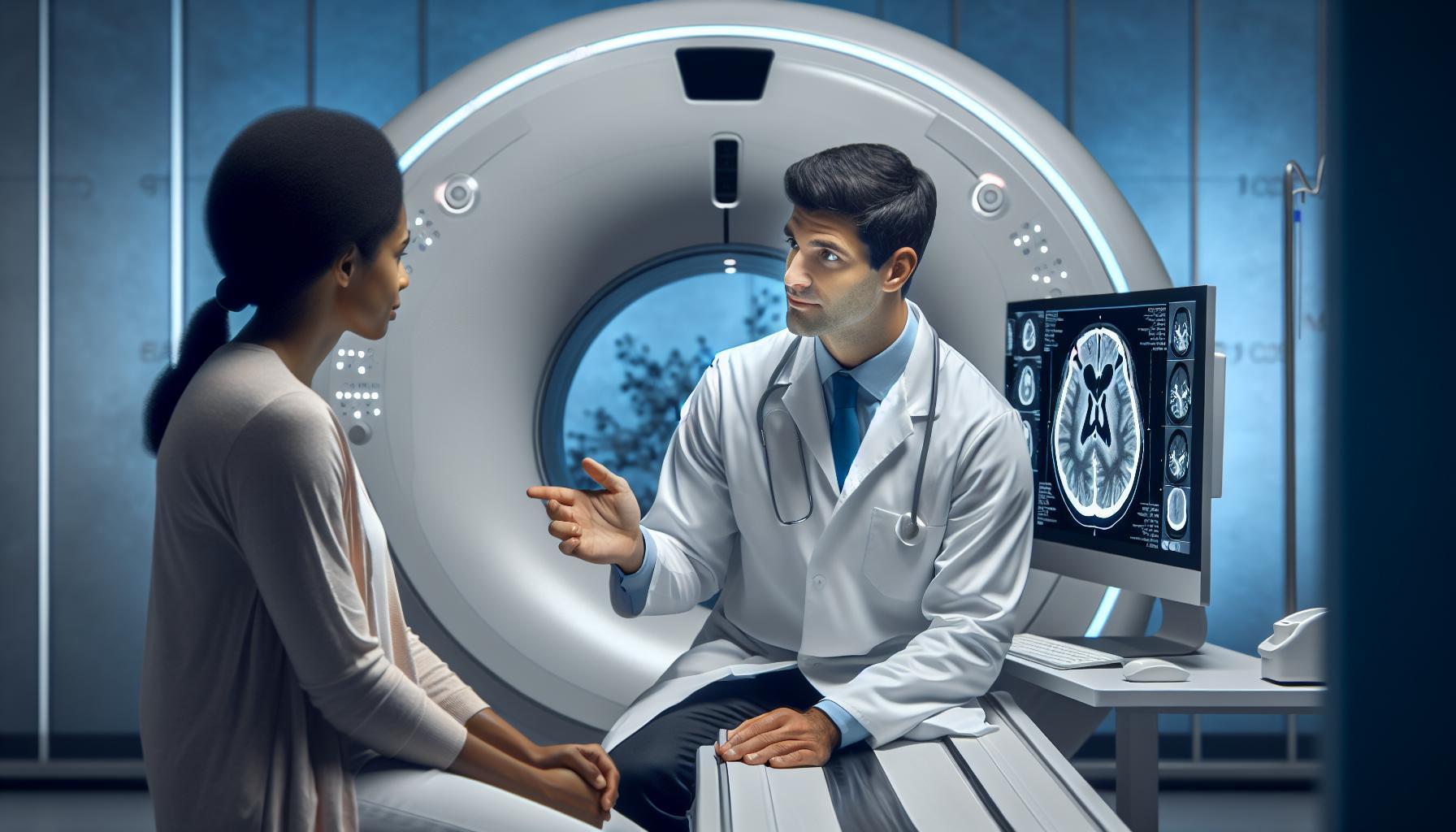
A brain CT scan is a vital diagnostic tool that provides detailed images of the brain, helping physicians to identify potential issues swiftly. These scans use X-ray technology to take multiple images of the brain from various angles, which are then processed by a computer to create cross-sectional images. This technique is particularly useful in emergency situations, such as assessing traumatic brain injuries, strokes, or tumors, where immediate diagnosis can be crucial for effective treatment.
Before undergoing a brain CT scan, patients often have various questions and concerns. It’s important to prepare mentally and physically for the procedure to enhance the experience. Patients should inform their healthcare provider about any allergies, especially to contrast dyes, previous reactions to imaging procedures, and any other relevant medical history. In most cases, no special preparation is required; however, patients may need to avoid food or drink for a few hours before the scan if contrast materials will be used.
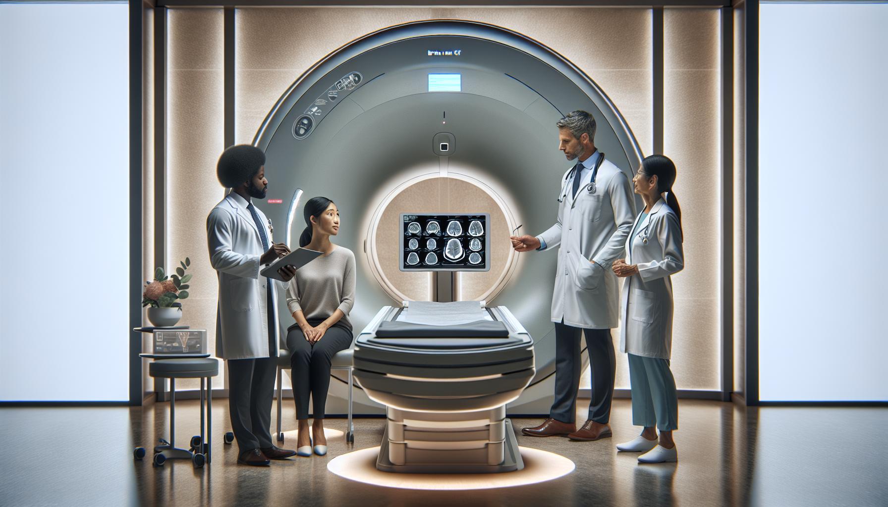
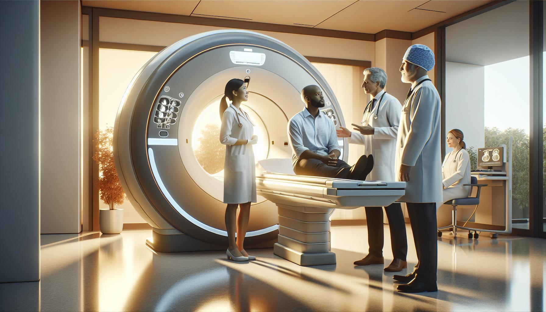
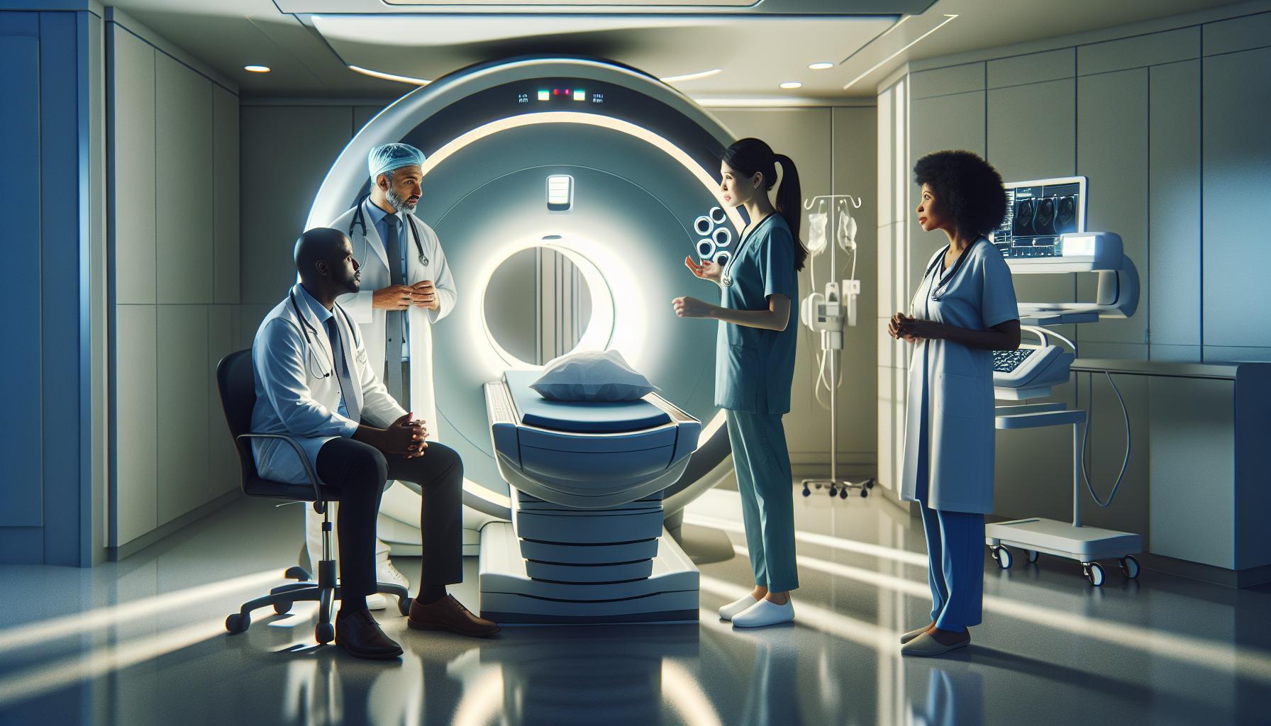
During the scan, patients lie on a table that slides into the CT machine, which resembles a large doughnut. The test is painless, though some may experience a feeling of warmth or flushing if contrast dye is administered. This imaging technique efficiently captures data about brain structures, making it easier for doctors to analyze any irregularities or signs of disease. Understanding these foundational aspects of brain CT scans can empower patients, alleviating anxiety and equipping them with knowledge about the essential role this imaging plays in neurodiagnostics.
Lastly, it’s essential to underscore the importance of consulting with healthcare professionals for personalized advice and guidance related to specific health concerns, as they can provide tailored information based on individual circumstances.
Key Indicators and Abnormalities in Brain CT
A brain CT scan is a remarkable tool that provides crucial insights into the internal structures of the brain, making it indispensable for diagnosing conditions that require immediate attention. Key indicators and abnormalities seen on brain CT scans can help healthcare professionals quickly assess a patient’s condition and determine the best course of action. Understanding these indicators not only aids in diagnosis but also empowers patients with the knowledge of what to expect from their imaging results.
One of the most significant indicators observed in a brain CT scan is the presence of hemorrhage, which can manifest as hyperdense areas due to pooling blood. This finding is often associated with trauma, aneurysms, or stroke. Edema, which appears as hypodense regions, signals swelling and can indicate issues such as infections or tumors. Additionally, the identification of mass effect-a shift in the midline structures of the brain-can suggest a tumor or significant swelling that requires urgent intervention.
Other abnormalities may include lesions, which can indicate various underlying conditions ranging from tumors to abscesses. Radiologists also look for signs of calcifications, which can signify prior injuries or specific diseases like tuberous sclerosis. Furthermore, the evaluation of the ventricular system is essential; any enlargement could indicate hydrocephalus, while a narrowing may point towards congenital abnormalities.
For patients and their families, understanding these indicators and abnormalities is vital. If a CT scan reveals any concerning signs, such as bleeding or significant swelling, your healthcare provider may discuss immediate treatment options. As patients navigate their healthcare journey, it’s essential to maintain open communication with medical professionals, always seeking clarity on findings and implications. Remember, every patient’s situation is unique, and personalized guidance from a qualified healthcare provider can ensure the best outcomes.
Step-by-Step Approach to Analyzing Brain CT Images
Analyzing brain CT images requires a careful and systematic approach to ensure accurate interpretation. With a detailed understanding of brain CT imaging, healthcare professionals can identify crucial abnormalities effectively. To start, forming a mental checklist for the analysis can be immensely helpful.
Systematic Review Process
Begin the interpretation by assessing the overall image quality. Ensure that the images are clear and that proper techniques were employed during the scan. Once established, follow these key steps:
- Identification of Structures: Familiarize yourself with the major brain structures, including the cortex, white matter, ventricles, and cerebellum. Know what normal anatomy looks like to recognize deviations.
- Evaluation of Symmetry: Check for symmetry between the left and right hemispheres, as asymmetries can suggest pathologies. For instance, a shift in midline structures may indicate the presence of a mass or edema.
- Assessment of Density: Analyze the density of different areas on the scan. Look for hyperdense regions indicating blood products, or hypodense areas suggesting edema. It’s essential to compare these densities against expected normal values.
- Search for Abnormalities: Use your checklist to guide the search for specific abnormalities, such as lesions, calcifications, or ventricle enlargement. Document these findings accurately for further assessment.
Contextual Considerations
Context is critical when analyzing CT scans. Consider the patient’s clinical history, presenting symptoms, and relevant laboratory findings. For example, in cases of trauma, look closely for hemorrhagic changes or fractures. If the patient presents with neurological deficits, focus on potential infarcts or areas of ischemia.
Additionally, be aware of common pitfalls during analysis. Misinterpreting normal anatomical variations as pathologies can lead to unnecessary anxiety for patients and their families. Always cross-reference findings with clinical data and follow up with more advanced imaging modalities if necessary.
By taking a thorough and methodical approach, healthcare professionals can enhance their confidence in reading brain CT scans. This skill not only aids in timely diagnosis but also supports the emotional well-being of their patients by ensuring that concerns are addressed promptly and effectively.
Common Brain Pathologies Detected by CT Scans
Detecting brain pathologies through CT scans can provide critical insights into a patient’s health, guiding timely and appropriate treatment. CT imaging is particularly adept at visualizing various conditions that affect brain structures, including hemorrhages, tumors, and signs of stroke. Understanding these common pathologies not only aids radiologists and physicians in making accurate diagnoses but also reassures patients about the information gleaned from their scans.
One notable condition that CT scans can effectively diagnose is intracerebral hemorrhage, which appears as a hyperdense region within the brain tissue. This is often a result of trauma, hypertension, or vascular anomalies, and immediate action may be necessary to prevent further complications. Another important pathology is subdural hematoma, which typically presents as a crescent-shaped area of increased density on the CT image. It often arises from a head injury, especially in older adults where brain atrophy may predispose them to such injuries.
Moreover, ischemic strokes are frequently visible on CT scans, particularly within the first 24 hours after onset. In these cases, clinicians look for areas of hypodensity that indicate regions of brain tissue affected by reduced blood flow. Time is critical in these cases, as swift intervention can significantly improve patient outcomes. Finally, brain tumors, whether primary or metastatic, can also be delineated via CT scans. Tumors typically show as irregularly shaped, contrast-enhancing lesions, depending on their composition and relation to surrounding brain structures.
Understanding these common pathologies helps patients feel more informed as they undergo imaging tests, equipping them to engage in conversations with their healthcare providers about their conditions. It’s important to remember that while CT scans can reveal significant abnormalities, the interpretation of the results must be done by a qualified medical professional who can integrate clinical data and additional imaging studies for accurate diagnosis and management.
Preparing Patients for a Brain CT: What to Expect
Undergoing a brain CT scan can be an important step in diagnosing various medical conditions, and knowing what to expect can significantly ease any anxiety. The process usually begins with a simple check-in at the imaging center, where you will be asked about your medical history and any medications you are currently taking. It’s crucial to communicate any health concerns, such as allergies to contrast materials if applicable, as this information helps healthcare professionals determine the safest approach for your scan.
As you prepare for the actual CT scan, you may be instructed to change into a gown and remove any metal objects, such as jewelry or hairpins, which could interfere with the imaging. You’ll then be positioned on a movable table that slides into the CT machine, which resembles a large donut, designed to provide a 360-degree view of your brain. It’s important to remain as still as possible during the procedure, as movement can blur the images and complicate interpretation. The scan itself is painless and usually lasts only a few minutes, although the preparation might take longer.
If contrast material is needed, you may receive an injection through an intravenous (IV) line. This substance enhances the clarity of the images by highlighting certain areas of the brain. While most people tolerate contrast well, some may experience a warm sensation or mild discomfort during the injection. After the scan, you can typically resume normal activities immediately. The results will be reviewed by a radiologist, who will provide your healthcare provider with a detailed interpretation, leading to informed discussions about your health moving forward. Always remember, consulting with your healthcare team is vital for understanding your specific situation and the implications of the scan results.
Understanding Radiation Safety in CT Imaging
Radiation exposure is often a source of concern for patients undergoing a CT scan, especially for brain imaging where precision is crucial. Understanding the principles of radiation safety can significantly alleviate anxiety and help patients make informed decisions regarding their health. A CT scan utilizes X-rays to produce detailed images of the brain, offering critical insights into conditions that may affect brain function. However, the key lies in ensuring that the benefits of the scan thoroughly outweigh any potential risks associated with radiation exposure.
It’s essential to realize that modern CT equipment is designed with advanced technology to minimize radiation doses while maximizing diagnostic quality. Controlled by well-trained radiologists and technicians, protocols are in place to adjust the dose based on factors like patient size, age, and the specific area being scanned. Patients can contribute to this safety by actively communicating their medical history, particularly any previous imaging that could have resulted in cumulative exposure.
When preparing for a brain CT, it is beneficial to follow some practical safety tips:
- Discuss Concerns: Have an open dialogue with your healthcare provider about your concerns regarding radiation.
- Accurate Medical History: Always inform your provider about your previous imaging tests to prevent unnecessary repeat scans.
- Child Safety: If the patient is a child, inquire whether the facility uses pediatric protocols for radiation dose.
- Follow Pre-Scan Instructions: Adhering strictly to pre-scan guidelines can lead to more efficient imaging and reduce the need for repeat scans.
Ultimately, the goal of a brain CT scan is to provide your care team with vital information that can lead to effective treatment decisions, outweighing the minimal risks posed by radiation exposure. Understanding these safety measures empowers patients to feel more secure in their decision to undergo this invaluable diagnostic tool. Always consult with your healthcare team for personalized advice on radiation safety and imaging options tailored to your specific needs.
Interpreting Contrast Agents in Neuroimaging
When it comes to neuroimaging, contrast agents play a pivotal role in enhancing the visualization of the brain’s intricate structures. These substances, often iodine-based for CT scans, help improve the contrast of images, allowing healthcare professionals to differentiate between normal and abnormal tissues more effectively. Their use can be essential in diagnosing a variety of conditions, from tumors to vascular issues, by highlighting areas that require closer examination.
Understanding how contrast agents work begins with recognizing their purpose. When injected into the bloodstream, these agents distribute throughout the vascular system, filling blood vessels and helping to delineate them against the surrounding tissues. This process is especially beneficial in identifying abnormalities like aneurysms or lesions that may otherwise blend into the background. However, patients may have questions or concerns regarding the use of contrast agents, particularly about potential allergic reactions or kidney function implications. It’s crucial to consult with a healthcare provider beforehand to address these concerns and ensure that the contrast agent used is appropriate for the individual’s health status.
Before undergoing a CT scan with contrast, there are a few preparatory steps that can facilitate a smooth experience. Patients should inform their healthcare team about any allergies, especially to iodine, as well as pre-existing kidney conditions, as these factors influence the decision to use contrast agents. Staying well-hydrated before the scan can also help support kidney function and aid in the elimination of the contrast from the body post-procedure.
Having an open and informed dialogue with your healthcare provider can alleviate anxiety and enhance the overall experience of neuroimaging procedures. By understanding how contrast agents function and their critical role in enhancing diagnostic capabilities, patients can feel more empowered and reassured as they navigate their imaging journey. Consulting with professionals throughout the process ensures that individual needs are addressed, making the experience as smooth as possible.
Advanced Techniques in Brain CT Analysis
Advancements in brain CT analysis have revolutionized diagnostic capabilities, allowing healthcare professionals to assess neurological conditions with remarkable precision. These sophisticated techniques often leverage high-resolution imaging, advanced algorithms, and refined software tools to enhance the quality and interpretability of CT scans. One such method is iterative reconstruction, which improves image quality while reducing radiation exposure. This technique uses sophisticated mathematical models to reconstruct images from raw data more effectively, resulting in clearer images that can reveal even subtle abnormalities.
Furthermore, the integration of machine learning and artificial intelligence (AI) is transforming the landscape of neuroimaging. AI algorithms can analyze vast datasets quickly, identifying patterns that might be missed by the human eye. For instance, they can assist radiologists in detecting early signs of conditions such as tumors, hemorrhages, or ischemic strokes by highlighting key areas of concern in the scans. These technologies not only enhance diagnostic accuracy but also save valuable time in urgent situations, enabling faster treatment decisions.
Another crucial development is the use of dual-energy CT, which utilizes two different energy levels to provide richer information regarding tissue composition. This technique can help differentiate between types of tissues, such as calcium and soft tissue, thus improving the detection of pathologies like acute hemorrhages or calcifications. Additionally, post-processing techniques such as volume rendering and multiplanar reconstruction enable healthcare providers to visualize complex anatomical relationships, which are especially beneficial for surgical planning or evaluating trauma cases.
Finally, as neuroimaging methodologies evolve, so does the importance of enhancing radiologist training in these advanced techniques. Regular workshops and training sessions can help practitioners stay updated on the latest scans, tools, and interpretative methodologies. Engaging in collaborative learning and interdisciplinary discussions within healthcare teams fosters a comprehensive approach to patient care, ultimately leading to more confident diagnoses and improved patient outcomes. By staying informed and utilizing these advanced techniques, healthcare professionals can empower their patients with more accurate and timely information, generating reassurance during potentially stressful medical situations.
Comparing CT Scans with Other Imaging Modalities
The choices in imaging modalities available today can be overwhelming, yet understanding the differences can empower patients and caregivers to make informed decisions. CT scans, while invaluable for assessing the brain, are just one tool in a comprehensive diagnostic toolbox that includes MRI (Magnetic Resonance Imaging), PET (Positron Emission Tomography), and ultrasound, each with its unique strengths and applications.
CT scans are particularly effective for quickly evaluating acute conditions, such as hemorrhages or skull fractures, due to their speed and the detailed images they provide. They utilize ionizing radiation, which can be a concern for some patients, particularly if multiple scans are needed. In contrast, MRI, which employs magnetic fields and radio waves, excels in providing detailed images of soft tissue. This makes it especially helpful in diagnosing conditions such as multiple sclerosis, brain tumors, and other abnormalities that may not be as visible on a CT scan. Moreover, MRI does not use radiation, making it a safer option for certain patient populations, such as children and pregnant women.
- CT Scan: Quick imaging, ideal for acute situations, excellent for detecting bleeding and trauma.
- MRI: Offers high-resolution images of soft tissue, preferred for chronic conditions and detailed brain studies.
- PET Scan: Useful for assessing metabolic processes and conditions like Alzheimer’s or tumors; highlights areas of high activity.
- Ultrasound: Non-invasive and radiation-free, often used in neonatal imaging, but limited for adult brain assessments.
When considering which imaging modality best suits a patient’s needs, it’s essential to consult with healthcare professionals who can provide personalized guidance based on individual health circumstances. Discussions may involve the specific goals of imaging-whether for screening, diagnosis, or treatment planning-and may explore concerns regarding safety, contrast agents, and overall comfort during the procedure. Understanding the advantages and limitations of each imaging technique can alleviate anxiety and foster a collaborative approach to diagnostic care. Remember, the best imaging strategy often involves a comprehensive evaluation that employs the strengths of multiple modalities to provide the most accurate and timely diagnosis.
Common Mistakes in Brain CT Interpretation to Avoid
Mistakes in interpreting brain CT scans can lead to significant consequences, affecting diagnosis and treatment plans. One common error involves overlooking subtle signs of injury or pathology. For instance, small hemorrhages may be easily missed, especially if they are located in complex regions of the brain. Radiologists must be meticulous in their review, utilizing high-quality images and advanced software to enhance detection accuracy.
Another frequent pitfall is not correlating imaging findings with clinical history. A brain CT scan might reveal an anomaly that seems benign in isolation but is critical when viewed alongside a patient’s symptoms and medical background. For example, finding a cyst may raise concerns that require further investigation only if the patient reports headaches or neurological deficits, emphasizing the importance of a comprehensive approach.
Additionally, reliance on previous scans without recognizing changes can lead to misinterpretation. If a radiologist interprets a scan based solely on established norms from past images, they may miss an evolving condition. Staying updated with new imaging guidelines and remaining aware of the patient’s previous medical history is essential for accurate assessment.
Lastly, the use of non-standard protocols or inadequate patient preparation can also compromise image quality, leading to diagnostic errors. Proper positioning and adherence to scan protocols are crucial; motion artifact can obscure critical details, resulting in misdiagnosis. Radiologists should always ensure that the images are of sufficient quality to evaluate potential issues thoroughly.
To avoid these common mistakes, continuous education and collaboration among healthcare professionals are vital. Engaging in multidisciplinary discussions, attending workshops, and utilizing peer reviews can enhance the interpretative process and ultimately improve patient outcomes.
Future Trends in Neuroimaging: Enhanced CT Techniques
The landscape of neuroimaging is rapidly evolving, driven by advances in technology and a deeper understanding of the brain. As researchers and healthcare providers strive for earlier detection and more accurate diagnosis of neurological conditions, enhanced CT techniques are emerging as pivotal tools in this journey. With innovations that improve image quality, reduce radiation exposure, and enhance diagnostic capabilities, the future of brain CT imaging holds promising developments for both patients and clinicians.
One significant trend is the integration of artificial intelligence (AI) in CT image analysis. AI algorithms are increasingly being employed to assist radiologists in detecting abnormalities-such as tumors or hemorrhages-at earlier stages. These algorithms are trained on vast datasets to recognize patterns that might elude the human eye. For instance, studies have shown that AI can help identify subtle changes in brain scans over time, supporting radiologists in their conclusions and reducing the likelihood of misdiagnosis.
Advancements in scan technology are also noteworthy. High-definition and dual-energy CT scans offer improved resolution and contrast, allowing for better differentiation between normal and abnormal tissue. These technologies enable the visualization of microstructural changes within the brain, which is crucial for diagnosing conditions such as multiple sclerosis or early-stage Alzheimer’s disease. Moreover, advancements in iterative reconstruction techniques, which enhance image quality while reducing noise, ensure that clinicians have clearer visuals to guide their interpretations.
Radiation safety remains a top priority in neuroimaging, and new techniques are emerging to minimize exposure. Innovations such as low-dose CT protocols aim to achieve high-quality images while exposing patients to less radiation. These methods are especially beneficial for vulnerable populations, including children, who may require imaging studies for various conditions.
Ultimately, as these enhanced CT techniques continue to evolve, they offer the potential to improve patient outcomes significantly. By empowering healthcare professionals with advanced tools and technologies, neuroimaging is set to become more accurate and accessible. Nevertheless, it’s essential for patients to understand that while these advancements are exciting, they should always consult with their healthcare providers to interpret findings appropriately and discuss any concerns related to their imaging procedures.
Resources for Continued Learning in Neuroimaging Interpretation
Staying informed about neuroimaging interpretation is essential, particularly as technology advances and the understanding of brain conditions evolves. If you’re keen to grasp the intricacies of reading brain CT scans, a wealth of resources is available to deepen your knowledge, enhance your skills, and ensure that you are well-prepared to engage in discussions with your healthcare providers about imaging procedures and results.
Educational Websites and Online Courses
There are numerous online platforms offering comprehensive courses and learning materials focused on neuroimaging and brain CT scan interpretation. Websites such as Coursera and edX provide access to courses taught by industry experts, covering topics from basic imaging principles to complex case studies. Additionally, resources like Radiopaedia.org present a plethora of free case studies, quizzes, and articles that can help you practice reading CT scans and familiarize yourself with common pathologies.
Books and Journals
A selection of textbooks tailored to neuroimaging can serve as invaluable references. Titles such as “Neuroimaging: The Essentials” or “Computed Tomography for Technologists: Exam Study Guide” offer a great blend of theory and practical insights. Furthermore, professional journals like the “American Journal of Neuroradiology” frequently publish peer-reviewed articles that delve into the latest research and advancements in neuroimaging techniques.
Professional Organizations and Workshops
Joining professional bodies such as the Radiological Society of North America (RSNA) or the American Society of Neuroradiology (ASNR) can also provide access to webinars, workshops, and conferences focused on imaging techniques, including CT. Networking opportunities within these organizations allow you to connect with experts and peers for shared knowledge and insights.
Real-World Applications
Engage with case studies presented in medical literature or online forums where professionals discuss real-world applications and interpretations of brain CT scans. Such examples provide context to theoretical knowledge and present opportunities to reflect on and discuss various interpretations of CT findings.
Ultimately, continuing education in neuroimaging not only enhances your understanding but also equips you with the ability to engage more confidently with healthcare professionals regarding your or a loved one’s imaging procedures. Remember, while these resources are instrumental in grasping concepts and interpretations, consultation with qualified healthcare providers remains crucial for receiving personalized and accurate medical advice.
Faq
Q: What common signs indicate the need for a brain CT scan?
A: Common signs include persistent headaches, sudden neurological symptoms (like weakness or numbness), confusion, seizures, or after head trauma. If these symptoms arise, consult a healthcare professional to determine if a brain CT scan is necessary for further evaluation.
Q: How can I prepare for a brain CT scan?
A: To prepare for a brain CT scan, follow your physician’s instructions, which may include fasting for a few hours and discussing current medications. Wear comfortable clothing and remove any metal objects. This ensures optimal imaging results and safety during the procedure.
Q: What variations might be seen in normal vs. abnormal brain CT scans?
A: Normal brain CT scans will show clear and symmetrical structures without lesions or unusual density. In contrast, abnormal scans may reveal tumors, strokes, hemorrhages, or brain swelling, each showing distinct changes in texture and brightness that aid in diagnosis.
Q: What does the term “contrast agents” mean in brain CT imaging?
A: Contrast agents are substances used to enhance the visibility of internal structures during a CT scan. They help delineate blood vessels and highlight abnormalities, improving diagnostic accuracy. Your healthcare provider will explain the risks and benefits before use.
Q: How are brain CT scans compared to MRI scans?
A: Brain CT scans use X-rays to create images and are faster and often used for emergency cases, while MRI provides more detailed images of soft tissues using magnetic fields. Each modality has specific indications, with CT often used for acute conditions.
Q: What should I expect after a brain CT scan?
A: After a brain CT scan, you can typically resume normal activities unless instructed otherwise. If a contrast agent was used, you might be monitored temporarily. Results usually require a follow-up visit to discuss findings and next steps in treatment.
Q: How long does it take to get results from a brain CT scan?
A: Results from a brain CT scan can usually be available within a few hours to a couple of days, depending on the facility and the urgency of the case. Always ask your doctor when you can expect to receive the results.
Q: What are the possible risks associated with undergoing a brain CT scan?
A: The primary risks include exposure to radiation and allergic reactions to contrast agents, though such reactions are rare. Discuss any concerns with your healthcare provider, who can explain how benefits outweigh risks in usual diagnostic scenarios.
Key Takeaways
As you explore the intricacies of reading brain CT scans, remember that understanding neuroimaging is a valuable skill that can enhance your clinical practice or personal knowledge. Start applying the techniques you’ve learned today, and consider diving deeper into our related guides on CT scan technology and patient preparation for imaging. These resources will provide additional insights and practical tips to further bolster your expertise.
Don’t hesitate to share your thoughts or questions in the comments below; your engagement helps us create a thriving community of learners. Also, for ongoing updates and expert insights, subscribe to our newsletter. We’re here to support your journey in mastering neuroimaging interpretation, reminding you that while knowledge is power, consulting healthcare professionals for personalized guidance is essential in any medical context. Thank you for joining us, and we look forward to your continued exploration!


