Did you know that a CT scan is one of the most effective tools for detecting brain tumors? As concerns about unexplained headaches or neurological symptoms arise, understanding the role of CT imaging can be incredibly reassuring.
CT scans create detailed cross-sectional images of the brain, allowing healthcare professionals to identify abnormalities such as tumors with precision. If you’re worried about your health or facing the uncertainty of potential symptoms, knowing how this imaging technique works can empower you.
In this article, we will explore the capabilities of CT scans in detecting brain tumors, shedding light on the process, its effectiveness, and why consulting with a healthcare professional is essential for your peace of mind and health journey.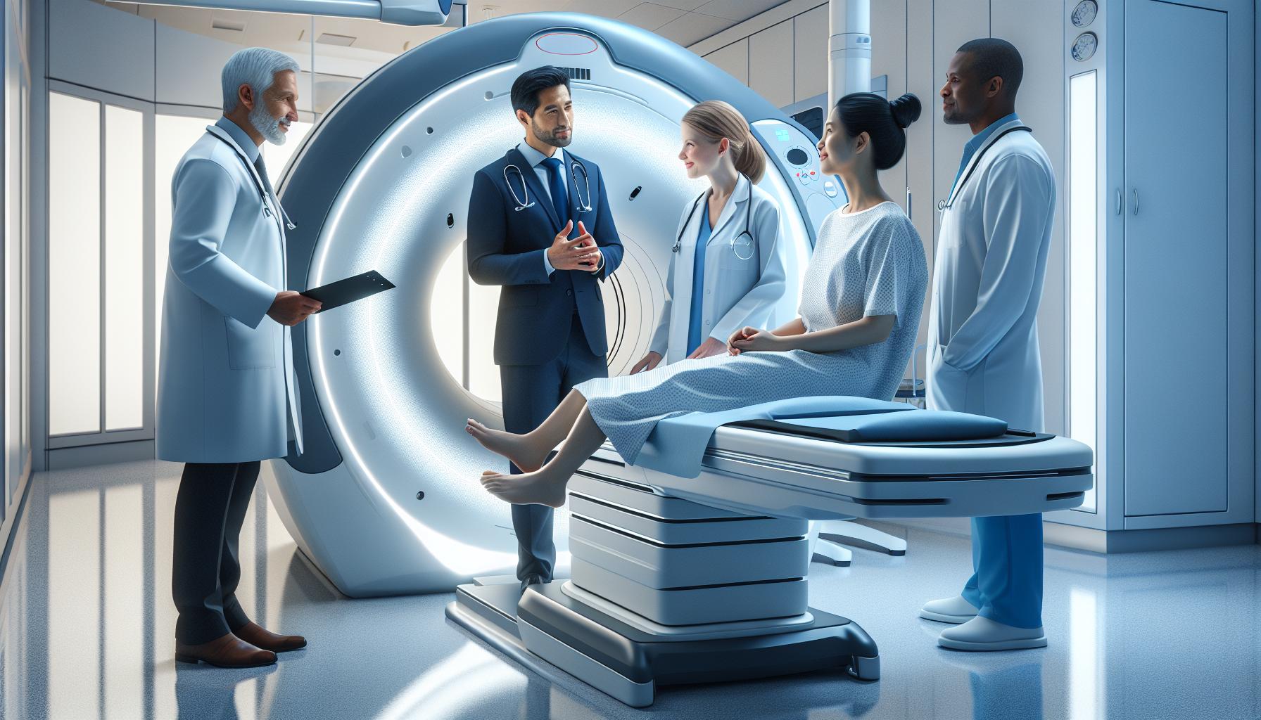
Understanding How CT Scans Detect Brain Tumors
CT scans have revolutionized the way medical professionals detect and diagnose brain tumors, providing high-resolution images that offer critical insights into the structures of the brain. Utilizing a series of X-rays taken from different angles, a CT scan creates cross-sectional images, allowing healthcare providers to visualize tumors much more effectively than traditional X-rays. One of the primary advantages of CT scans is their ability to highlight the density and mass of different tissues, which can reveal the presence of abnormalities such as brain tumors.
When a patient undergoes a CT scan for suspected brain issues, a contrast dye may be administered, enhancing the clarity of the images. This contrasts with surrounding tissues, making tumors more distinguished on the scan. It is essential to understand that while CT scans are highly effective at identifying many types of tumors, they might not always detect very small tumors or those located in challenging areas of the brain. In such cases, additional imaging techniques, like MRI, may be recommended to provide a more comprehensive view.
Patients can prepare for a CT scan by ensuring they follow any specific instructions provided by their healthcare team, which may include fasting for a few hours prior to the procedure if a contrast agent is used. During the scan, which typically takes only about 10 to 30 minutes, individuals lie still on a table that moves through the CT scanner. This simplicity means many patients experience minimal discomfort, though some may feel anxiety or concern about the findings. It’s important to communicate these feelings with healthcare providers, who can offer support and information to ease concerns.
Understanding the implications of a CT scan should extend beyond the procedure itself. After the scan, results are typically reviewed by a radiologist, who will analyze the images for any signs of abnormalities, including tumors. Patients should feel empowered to discuss the results with their doctors, ensuring they grasp the information and next steps clearly. This collaborative dialogue not only helps demystify the process but also reinforces a patient-centered approach to care, fostering a sense of agency in the face of medical challenges.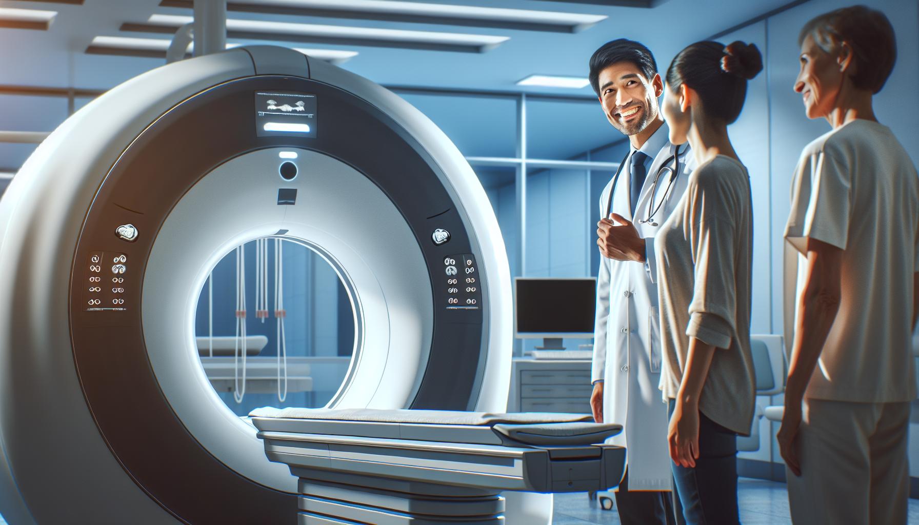
The Technology Behind CT Scans Explained
The journey of a CT scan begins long before the procedure itself, as it employs advanced technology that has transformed diagnostic imaging. At the core of the process is the utilization of X-rays, which are emitted in a circular pattern around the patient from multiple directions. This technique enables the machine to generate intricate cross-sectional images of the brain, revealing not only the structure of various tissues but also the density variations that can indicate the presence of abnormalities, including tumors. Each image captured during the scan is then processed by a sophisticated computer system that reconstructs these slices into detailed three-dimensional visuals, providing highly accurate representations of brain anatomy.
To enhance the visibility of the brain during the scan, physicians may opt to use a contrast dye. This innovative approach highlights areas of interest more distinctly, allowing for a clearer differentiation between normal tissue and potential tumors. By altering the way X-rays pass through different materials, the contrast agent improves diagnostic accuracy dramatically. This is particularly helpful in revealing tumors that might be obscured due to their size or location, showcasing the advantages of CT technology in the detection process.
Understanding the mechanics of CT scans can help alleviate concerns about the procedure itself. Typically completed within 10 to 30 minutes, patients are required to lie still on a table that moves through a donut-shaped scanner. Many find this process comfortable, although pre-scan preparation may involve fasting, especially if a contrast agent is used. Minimal discomfort is expected, though anxiety about the results can arise. Healthcare providers are there to support patients through this journey, offering reassurance and guidance to foster a more positive experience.
Ultimately, the technology behind CT scans not only aids in detecting brain tumors but also empowers patients with critical information about their health. Armed with knowledge about the process and its capabilities, individuals can approach their upcoming scans with greater confidence, knowing they have taken a proactive step toward understanding their health. Collaboration with healthcare professionals before, during, and after the scan enhances this experience, transforming a potentially daunting process into an opportunity for informed decision-making and assurance in their care.
How Accurate Are CT Scans for Tumor Detection?
A CT scan is one of the most effective tools in diagnosing brain tumors, stemming from its ability to produce detailed images of the brain’s internal structures. Although no diagnostic method is infallible, studies indicate that CT scans can identify brain tumors with a significant degree of accuracy. Typically, the sensitivity of CT imaging for detecting tumors ranges around 70% to 80%, meaning it can successfully identify the presence of a tumor in a majority of cases, particularly when used alongside contrast agents that enhance image clarity.
The precision of a CT scan in detecting brain tumors, however, can depend on various factors, including the tumor’s size, type, and location. Larger tumors are generally more detectable, with imaging quality improving as the tumor grows because it causes greater disruption in normal brain architecture. Smaller tumors or those located in complex areas of the brain may not be as easily visualized, which is why healthcare professionals may recommend complementary imaging techniques, such as MRI. MRI scans offer superior detail of soft tissues and can better depict abnormalities that may not clearly show up on a CT scan.
Prior to your scan, it is vital to communicate openly with your healthcare team about your symptoms, as this information will aid in achieving the most accurate diagnosis possible. Early intervention is crucial; therefore, mentioning specific issues such as headaches, vision changes, or other neurological signs can prompt your physician to fine-tune imaging strategies. If needed, a follow-up with additional imaging or testing may be warranted to ensure comprehensive examination and accurate results.
In summary, while CT scans are a highly reliable option for detecting brain tumors, understanding their limitations and the circumstances under which they excel is vital. Engaging in a conversation with healthcare providers about scan results and next steps can empower you, reducing uncertainty and fostering a proactive approach to your health care.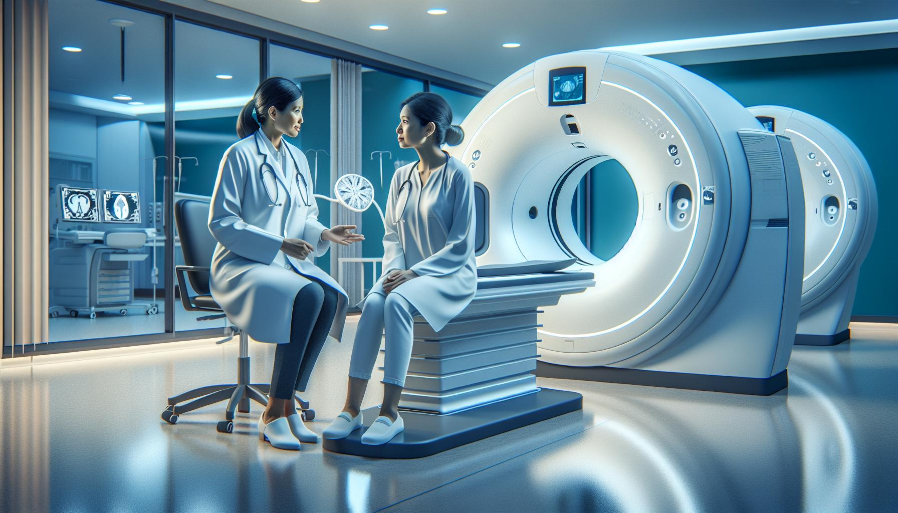
Preparing for a CT Scan: What Patients Should Know
Preparing for a CT scan can be an important step in understanding your health, especially when it concerns potential brain tumors. Knowing what to expect and how to prepare can significantly ease the anxiety often associated with medical procedures. One key point to remember is that a CT scan is a non-invasive procedure that utilizes advanced imaging technology to provide detailed pictures of your brain, helping doctors diagnose any abnormalities.
To ensure the best possible imaging results, follow these practical steps in your preparation:
Communicate Openly with Your Healthcare Team
Before the procedure, speak candidly with your doctor about any medications you are currently taking, allergies, and previous reactions to contrast dyes, if applicable. This communication is crucial, especially if a contrast agent is to be used during your scan, as it enhances visibility within your brain images.
Follow Dietary and Activity Guidelines
Depending on your specific CT scan directives, you may be instructed to refrain from eating or drinking for several hours before the test. Typically, a fasting period of about 4 to 6 hours is common if contrast material is being used, as it helps prevent complications and ensures the best imaging outcomes.
- Wear Comfortable Clothing: It’s advisable to wear loose-fitting clothing without metal fasteners, as metal can interfere with imaging quality. You may be asked to change into a hospital gown.
- Arrive Early: Arriving early allows time to complete any necessary paperwork and to discuss final preparations with the staff.
Emotional Preparedness
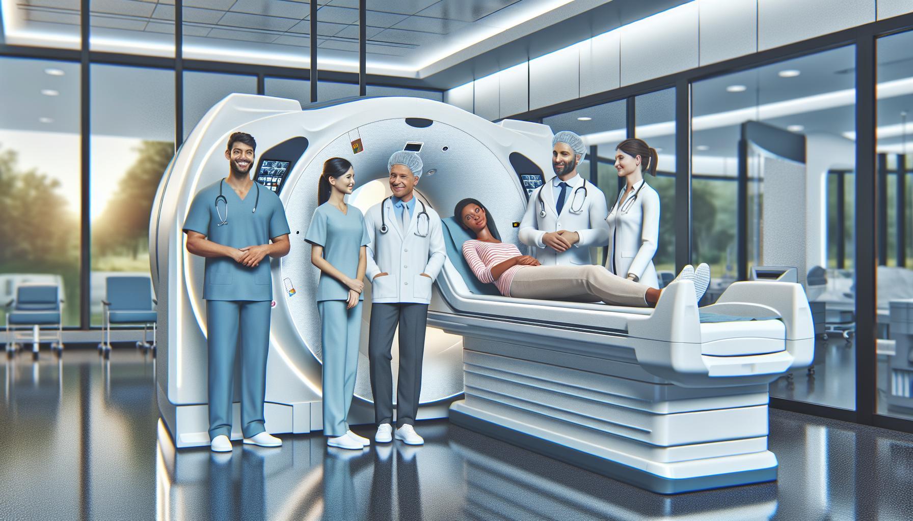
Feeling anxious about medical procedures is completely normal. Understanding that the process is quick-usually taking between 10 to 30 minutes-can help mitigate some of that anxiety. During the CT scan, you’ll lie on a padded table that slides into the scanning machine. You’ll need to remain still while images are taken, but the staff will be there to guide and reassure you throughout.
After the Scan
Post-scan, there are generally no restrictions, allowing you to resume daily activities immediately, unless directed otherwise by your doctor. If contrast dye is used, you may be encouraged to drink plenty of fluids to help flush it out of your system.
By equipping yourself with the right information and guidance, you can approach your CT scan with confidence. The knowledge gained from this procedure can be empowering, as it plays a vital role in understanding your health and guiding subsequent medical decisions. Always consult your healthcare provider for personalized advice tailored to your needs.
What to Expect During a CT Scan for Brain Tumors
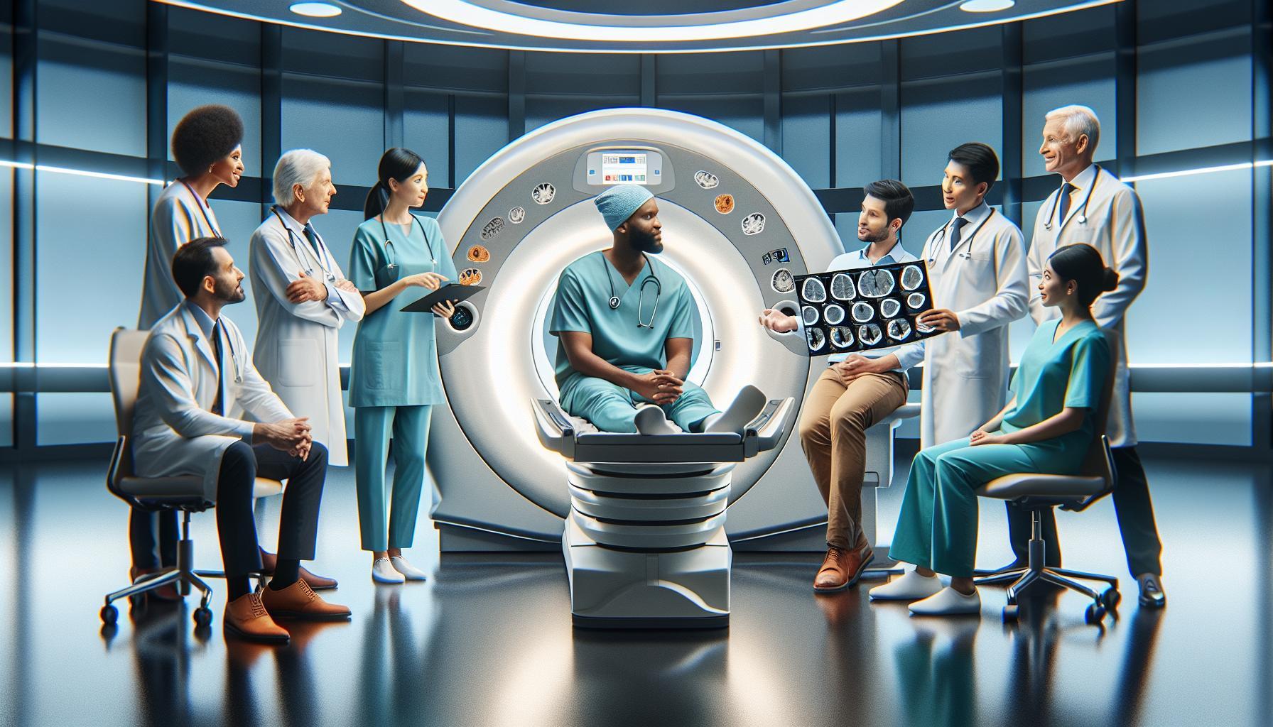
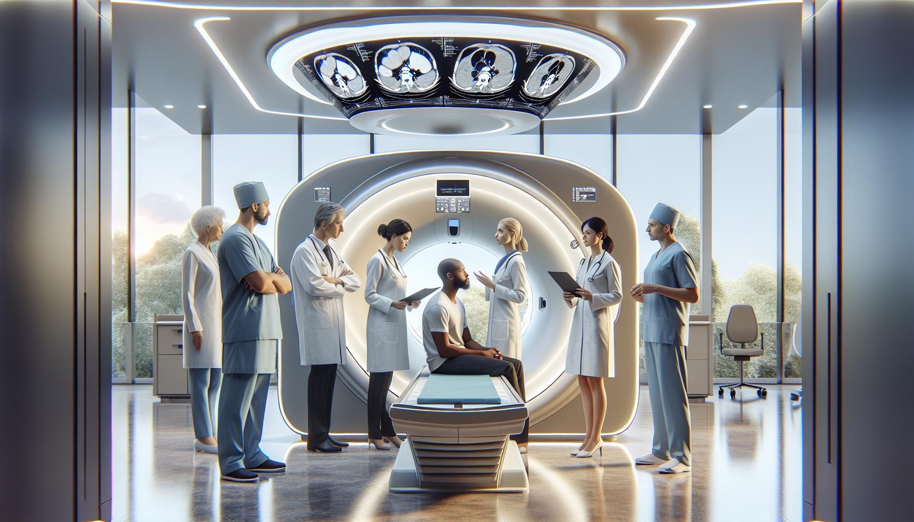
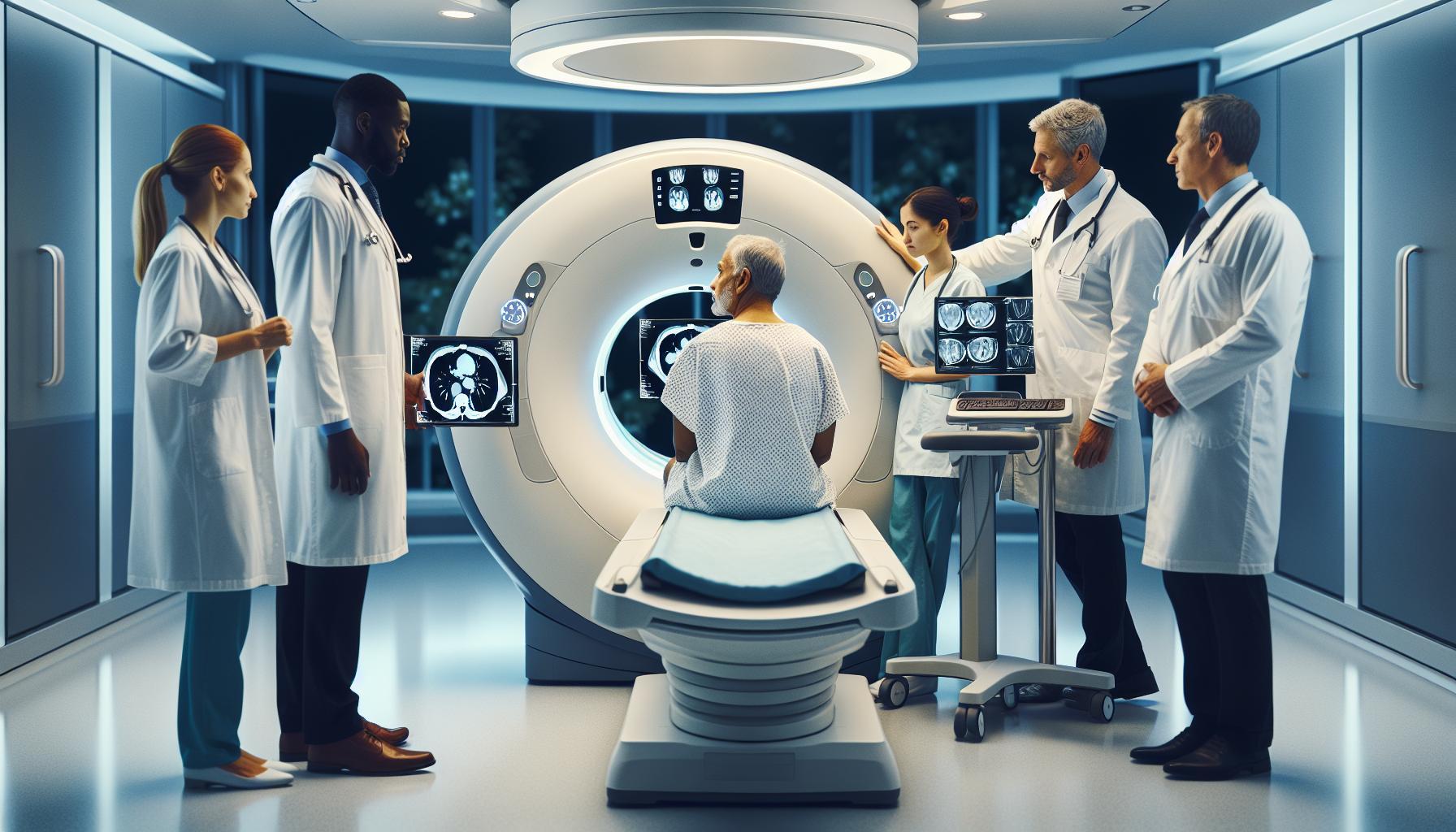
A CT scan is a powerful tool in the detection of brain tumors, and understanding what happens during the procedure can help alleviate any concerns you may have. The process typically takes between 10 to 30 minutes, and during that time, you’ll be closely monitored by a skilled team of healthcare professionals. Imaging is performed with you lying on a padded table that slides into a large, doughnut-shaped machine known as the CT scanner. This machine uses X-ray technology to capture detailed cross-sectional images of your brain, enabling doctors to identify any potential abnormalities, such as tumors.
Before the scan begins, you might be asked to remove any metal objects, including jewelry and eyeglasses, since they can interfere with the imaging quality. If a contrast material is being used to enhance image clarity, the medical staff will explain how it will be administered, often through an intravenous (IV) line. While the process of getting the contrast may cause a brief sensation-sometimes described as warmth or flushing-this usually subsides quickly.
During the scanning process, it’s crucial to remain still to ensure clear images are captured. You may hear a series of whirring and clicking sounds as the machine operates, which is entirely normal. The staff will be present to guide you, reassuring you throughout the procedure. A common concern for many is feeling claustrophobic inside the scanner. If this is something you worry about, don’t hesitate to express your concerns to the medical team beforehand; they can provide helpful strategies or adjust the setup to accommodate you better.
Once the CT scan is complete, you can typically resume normal activities immediately, unless specific instructions have been given. If contrast dye is used, drinking fluids afterward can help flush it from your system. Understanding these steps and what to expect can significantly enhance your comfort level during this essential diagnostic procedure, empowering you to take control of your health journey. Always remember to consult with your healthcare provider for personalized advice tailored to your individual needs.
Risks and Safety Considerations of CT Scans
While CT scans are invaluable in detecting brain tumors and providing detailed insights into brain health, it’s essential to be aware of various risks and safety considerations associated with the procedure. One of the prominent concerns is the exposure to radiation. CT scans utilize X-rays, and while the dose of radiation is generally low and considered safe for most individuals, repeated scans can accumulate radiation exposure over time, which may raise the risk of cancer later on. It’s vital to discuss any previous scans with your healthcare provider, especially if you’re being advised to undergo multiple imaging sessions.
Moreover, if a contrast material is used to enhance the clarity of the images, there may be additional risks. Some patients may experience allergic reactions to the contrast dye, including mild symptoms like itching or rash, and in rare cases, more severe reactions such as anaphylaxis. Your medical team will assess your history of allergies and kidney function prior to administering contrast to mitigate these risks. Staying hydrated before and after the scan can also assist your kidneys in processing the contrast material more effectively.
Another safety consideration involves the environment of the scanning facility. Patients who are anxious about enclosed spaces might feel uneasy during the procedure. It’s important to communicate any claustrophobia or anxiety to the medical staff prior to the scan, as they can offer solutions such as sedation or adjusting the CT scanner settings to reduce discomfort. The presence of sounds like whirring and clicking is normal during the scan, but understanding these noises can help diminish anxiety.
Lastly, ensure that you inform the healthcare provider about any medical conditions or other imaging procedures you have had, as these can influence the interpretation of the scan findings and ensure your overall safety. Empower yourself with knowledge and open dialogue with your healthcare provider to navigate the CT scan process with confidence, prioritizing both your health and peace of mind.
Comparing CT Scans to Other Imaging Techniques
When it comes to imaging techniques for detecting brain tumors, a variety of options exist, each with its unique strengths and limitations. CT scans, or computed tomography scans, are often favored for their speed and ability to capture high-resolution images of the brain. However, understanding how they compare to other imaging modalities can assist patients in making informed decisions regarding their diagnostic journey.
One of the primary alternatives to CT scanning is magnetic resonance imaging (MRI). MRIs use magnetic fields and radio waves to produce detailed images without radiation exposure, making them particularly attractive for patients needing frequent imaging. MRIs excel in differentiating between types of soft tissues, which can be crucial when evaluating complex structures in the brain. For instance, while CT scans are excellent at identifying bleeding or large tumors, MRIs can provide better differentiation between tumor types and can visualize changes in brain tissue, making them a preferred choice for many neurologists.
Another imaging option is positron emission tomography (PET). PET scans are particularly useful in assessing metabolic activity and can help distinguish between benign and malignant tumors. Often, PET scans are used in conjunction with CT or MRI to provide a comprehensive view of the tumor’s nature and behavior, revealing not just the structure but also functional aspects. This synergy can provide a more complete diagnosis, allowing for better treatment planning.
As patients navigate imaging options, it’s also worth mentioning the role of ultrasound in specific cases, especially in pediatrics. Though not commonly used for brain tumors in adults, ultrasound can help evaluate brain anomalies in infants and young children due to its safety and non-invasive nature.
Ultimately, the choice of imaging technique should be guided by a healthcare professional who can assess individual circumstances, medical history, and specific symptoms. Engaging in an open dialogue about the benefits and limitations of each of these methods can empower patients, ensuring they are comfortable and informed about their diagnostic path.
Recognizing Symptoms That May Require a CT Scan
Many people are unaware of the subtle signs that may indicate a need for a CT scan, especially when it comes to brain health. Early recognition of symptoms can be pivotal in identifying issues like brain tumors, which might otherwise go undetected until they have progressed significantly. If you experience any of the following symptoms, it is crucial to consult with a healthcare professional who can determine if a CT scan is warranted.
Common Symptoms
- Persistent Headaches: If you experience frequent, severe headaches that differ from your usual pattern or worsen over time, it’s important to seek medical advice.
- Vision Changes: Blurred or double vision, difficulty seeing in one or both eyes, or sudden changes in vision can be red flags.
- Cognitive Disturbances: Changes in memory, difficulty concentrating, or sudden personality changes should be discussed with a doctor.
- Nausea and Vomiting: Unexplained and persistent nausea or vomiting, particularly if accompanied by headaches, may indicate increased intracranial pressure.
- Seizures: New-onset seizures in adults, especially if they have no previous history, can be a serious symptom.
- Balance and Coordination Issues: Difficulty walking or maintaining balance and coordination may signal neurological processes that need evaluation.
Recognizing these symptoms early on can lead to timely interventions. For instance, a sudden, severe headache described as a “thunderclap” may warrant immediate medical evaluation, as it can indicate a serious condition. Similarly, experiencing a seizure can prompt a rapid assessment of brain function and potential abnormalities.
Consulting Healthcare Professionals
While these symptoms can be concerning, it’s essential to approach them calmly. Various conditions, not just brain tumors, can manifest through similar symptoms. Consulting with a healthcare provider is the first step to understanding the underlying issues and determining whether a CT scan is necessary. Your physician may also recommend additional tests or imaging modalities based on your specific symptoms and medical history.
Being proactive in addressing health concerns can significantly impact outcomes. Always prioritize open communication with your healthcare provider about your symptoms, as this can lead to timely and effective treatment options tailored to your needs.
Post-Scan Process: Understanding Your Results
Receiving the results of a CT scan can be an anxiety-inducing experience, especially when the purpose of the scan is to investigate the possibility of a brain tumor. Understanding the post-scan process is crucial not only for alleviating concerns but also for ensuring you are equipped to discuss your health comprehensively with your healthcare provider. Typically, your physician will receive the images and a radiologist’s report within a few hours to a few days after the scan. This timeline may vary based on the facilities involved and the urgency of your symptoms.
Once your provider has reviewed the results, they will schedule an appointment with you to discuss what the images reveal. During this discussion, it’s essential to ask questions and express any concerns you may have. If a tumor is detected, your healthcare professional will explain the findings in detail, including the type of tumor, its size, and its location. They may also discuss potential next steps, ranging from further imaging tests, biopsy options, or referrals to specialists such as oncologists or neurosurgeons.
If the results indicate no signs of a tumor or other serious conditions, it still may be beneficial to follow up with your physician regarding any ongoing symptoms. They may suggest alternative evaluations or management strategies to address your health concerns. In addition, it’s important to prioritize a path forward-whether that involves regular monitoring or lifestyle adjustments based on your physician’s recommendations.
Ultimately, the key to navigating this process smoothly is open communication. Be proactive in discussing your results and treatment options, and don’t hesitate to seek a second opinion if you feel uncertain about the findings. Remember that understanding your health is a shared journey between you and your healthcare team, and being well-informed is your best tool for taking an active role in your healthcare decisions.
Next Steps After a Positive CT Scan Finding
Receiving a positive result from a CT scan raises many important questions and can understandably create feelings of anxiety and uncertainty. However, it is essential to remember that a positive finding is not an immediate indication of severe illness; rather, it prompts further evaluation and understanding of the situation. Navigating the next steps after your scan is crucial, as it lays the foundation for your ongoing health journey.
Understanding Communication with Your Healthcare Provider
At this stage, communication with your healthcare provider is vital. Schedule a follow-up appointment where you can discuss the findings in detail. Come prepared with a list of questions, such as:
- What exactly did the scan reveal?
- What are the possible next steps?
- Will I require further tests or imaging?
- What specialists might I need to see?
Being open and proactive can help clarify the situation and ensure you thoroughly understand your options moving forward. Your healthcare provider will likely explain the nature of any potential tumors, including their type, size, and possible implications based on your health history.
Exploring Further Diagnostics and Treatments
If a tumor is detected, your next steps may involve additional imaging tests, such as MRI or PET scans, which provide more detailed information. Depending on the characteristics of the tumor, further evaluations like biopsies may be recommended to determine if it is benign or malignant.
Discussing treatment options is also essential. Various possibilities exist, ranging from surgical interventions to radiation therapy or chemotherapy, depending on the tumor type and your overall health. Each option will come with its own benefits and risks, and your healthcare team will help guide you through the best course of action tailored to your specific case.
Emotional Support and Education
Don’t forget the importance of emotional support during this time. It may be beneficial to speak with a counselor or join a support group for individuals facing similar health challenges. Engaging with others who understand your concerns can provide comfort and reduce feelings of isolation.
Furthermore, educate yourself about your condition. Understanding brain tumors and the treatment landscape can empower you, making you feel more in control of your health. Reputable medical websites, books, and resources provided by your healthcare team can be excellent starting points.
Navigating the aftermath of a positive CT scan finding may feel daunting, but taking these structured steps can help manage anxiety and set a clear path forward. Always remember that knowledge is empowering, and working closely with your healthcare professionals will ensure you receive the most appropriate and personalized care possible.
Patient Experiences: Real Stories of CT Scans
When faced with the uncertainty of a potential brain issue, many patients share their experiences surrounding CT scans. These shared stories often highlight the anxiety and emotional challenges of waiting for results. For example, one patient recounted how they felt a whirlwind of emotions leading up to their scan. They noted that understanding the procedure-how a CT scan uses X-ray technology to create detailed images of the brain-helped alleviate some of their fears. Knowledge about why the scan was necessary often empowers the patient, serving as a comforting reminder that these procedures are essential tools for diagnosis.
Patients have also shared their experiences on what it feels like during the scan itself. Many describe the process as quick and non-invasive. A woman who underwent a CT scan vividly recalled the moment she entered the machine. Initially apprehensive, she was soon reassured by the technician’s calming presence and clear instructions. Understanding that the scan would only take a few minutes gave her a sense of control amidst the uncertainty.
The waiting period for results can be daunting. It’s common for patients to experience anxiety while waiting for their doctor to provide feedback. Someone once shared that during this phase, they found comfort in talking to others who had gone through a similar experience. The insights they gained about managing anxiety, such as deep breathing exercises and journaling, proved beneficial during this stressful time. Engaging in support groups or forums can be a constructive way to process emotions and gather valuable information.
Ultimately, each patient’s journey with a CT scan is unique, but shared experiences often reveal common threads of fear, empowerment through knowledge, and the importance of support systems. Those preparing for a CT scan can benefit from connecting with others who’ve walked in their shoes. Building a network of support-whether through friends, family, or online communities-can provide significant solace during this challenging time.
Financial Considerations: Cost of CT Scans Explained
While the prospect of undergoing a CT scan may understandably invoke anxiety, it is essential to also consider the financial implications of this diagnostic tool. A CT scan can often be a necessary step in detecting potential brain tumors, but the costs associated with it can vary widely based on several factors, including location, facility type, and insurance coverage.
Typically, the price of a CT scan can range from $300 to $3,000. This wide range depends on whether the scan is performed in a hospital, outpatient center, or private practice. In metropolitan areas, for example, costs tend to be on the higher end because of increased operational costs. Furthermore, a CT scan that includes contrast material-often necessary for clearer imaging-may also increase the expense by an additional $100 to $500.
Insurance Coverage and Out-of-Pocket Costs
For many patients, health insurance can significantly alleviate the financial burden associated with medical imaging. However, the extent of coverage can vary greatly between policies. Patients should contact their insurance providers to inquire whether CT scans are covered under their plan and what out-of-pocket costs to expect. Coverage may vary based on whether the scan is deemed medically necessary and where the scan is performed.
In some cases, patients might face co-pays or deductibles that further impact their financial responsibilities. Understanding these details upfront can prevent unforeseen expenses and provide peace of mind. Some facilities offer payment plans or financial assistance programs, which can be helpful for those facing high costs or who are uninsured.
Financial Assistance and Preparing for Costs
If mounting costs become a concern, patients should not hesitate to explore financial assistance options. Here are a few strategies to consider:
- Discuss with your doctor: Your healthcare provider may have knowledge of lower-cost facilities or imaging centers.
- Shop around: Prices can vary widely, so it’s beneficial to compare costs across different facilities.
- Negotiate prices: Some facilities may offer discounts for self-pay patients or negotiate lower rates based on financial hardship.
- Seek community resources: Non-profit organizations and local health departments may offer resources or assistance with medical imaging costs.
Taking the time to understand the costs associated with a CT scan not only prepares patients financially but also reduces anxiety about the entire process. Ultimately, being informed and proactive can help empower individuals to focus on their health and the next steps in their diagnostic journey.
Faq
Q: Can a CT scan detect all types of brain tumors?
A: No, a CT scan may not detect all types of brain tumors. While it is effective for many tumors, some smaller or less dense tumors may not be visible. For unclear results, further imaging like MRI may be recommended. Refer to the section on accuracy in your article for more details.
Q: How does a CT scan differentiate between a tumor and other brain conditions?
A: A CT scan uses cross-sectional imaging to identify abnormalities in brain tissue density. Tumors usually appear as distinct masses compared to surrounding brain tissue. However, definitive diagnosis often requires additional imaging or biopsy. Please see your article’s section on technology behind CT scans for further clarification.
Q: What symptoms indicate the need for a CT scan?
A: Symptoms that may necessitate a CT scan include persistent headaches, vision changes, unexplained nausea, or neurological deficits like weakness or seizures. Recognizing these symptoms early is key for timely diagnosis and treatment. Check the symptoms section in your article for more information.
Q: Are there any alternatives to CT scans for brain tumor detection?
A: Yes, alternatives to CT scans include MRI and PET scans, which may provide clearer images for some types of tumors or conditions. Each method has its own advantages and is chosen based on the specific situation. More on this can be found in the comparison section of your article.
Q: How long does it take to get results from a CT scan?
A: Typically, results from a CT scan can be available within a few hours to a couple of days, depending on the facility and urgency. Patients should discuss expected timelines with their healthcare provider. Refer to the post-scan process section for further details.
Q: What precautions should be taken before undergoing a CT scan?
A: Before a CT scan, inform your doctor of any allergies, especially to contrast material, and any medications you are taking. It’s also important to follow preparatory instructions, such as fasting if required. More tips can be found in the preparation section of your article.
Q: Can a brain tumor be missed on a CT scan?
A: Yes, a brain tumor can be missed on a CT scan due to its size or type. False negatives are possible, especially for smaller tumors. A follow-up with additional imaging may be necessary if symptoms persist. Please see your article for discussions on accuracy and next steps after scanning.
Q: What should patients do if they have concerns about their CT scan results?
A: Patients should discuss any concerns regarding CT scan results with their healthcare provider, requesting further tests or second opinions if necessary. Understanding results is critical for informed decision-making. Your article’s section on next steps after positive findings provides useful insights on this.
Final Thoughts
Understanding the role of CT scans in detecting brain tumors is crucial for anyone concerned about their health or the health of a loved one. While these scans provide critical insights, it’s essential to consult with your healthcare provider for a comprehensive evaluation. If you’re feeling anxious about your health or would like to learn more about CT scan technology, explore our articles on “Understanding Your Imaging Procedure” and “Navigating the Path to Diagnosis.”
Don’t hesitate to take the next step in your health journey. Whether that means scheduling a consultation or signing up for our newsletter for the latest updates in medical imaging, every action counts. Your well-being is paramount, and being informed is a powerful tool. Share your thoughts or any questions you have in the comments below-your experience could help others find clarity too.

