Did you know that chest CT scans are primarily designed to examine the lungs and other structures in the chest rather than breast tissue? This raises a crucial question: can a chest CT effectively detect breast cancer? As the importance of early detection continues to resonate with many, understanding the capabilities and limitations of imaging procedures is vital.
While a chest CT scan can incidentally reveal abnormalities in the breast, it is not the gold standard for breast cancer screening. Readers may wonder what this means for their health and how they should proceed if they have concerns about breast cancer. By delving into the accuracy of chest CTs for detecting breast cancer, we hope to address your concerns, empower you with knowledge, and guide you on the best practices for diagnostic imaging. Continue reading to uncover essential insights that could shape your approach to health and wellness.
Will a Chest CT Scan Detect Breast Cancer?
While a chest CT scan is primarily designed for assessing the lungs and the structures within the thoracic cavity, it can occasionally incidentally reveal abnormalities in breast tissue. However, it is crucial to understand that chest CT scans are not standard for breast cancer detection. They lack the specificity and sensitivity required for identifying breast cancers compared to dedicated breast imaging techniques like mammograms or breast ultrasounds.
Breast cancers typically present as distinct masses or microcalcifications that are more effectively visualized in imaging specifically tailored for breast evaluation. For instance, mammography, which employs low-energy X-rays, is the gold standard for early breast cancer detection due to its ability to highlight subtle changes in breast tissue. On the other hand, CT scans utilize higher-energy radiation and offer a broader view of the body, making them less equipped for detecting the finer details relevant to breast tissue.
If there is a suspicion of breast cancer based on initial imaging or clinical findings, healthcare providers will generally recommend mammograms or MRIs designed for breast assessment rather than relying on a chest CT scan. It’s essential to discuss with your healthcare provider which imaging method is best suited for your individual circumstances, especially if you have risk factors or symptoms indicating potential breast issues. Understanding these distinctions can empower you to make informed decisions about your breast health and screening options.
Understanding Chest CT Scans and Their Role
A chest CT scan is a powerful imaging tool that provides detailed cross-sectional images of the thoracic area, primarily focusing on the lungs, heart, and other structures within the chest cavity. While it is designed for diagnosing conditions related to these organs, patients often find themselves wondering about its role in detecting diseases outside of its primary purpose, particularly breast cancer. Understanding the scope and limitations of a chest CT scan can help alleviate concerns and contribute to informed decisions about health screenings.
Chest CT scans utilize advanced X-ray technology to create comprehensive images, revealing abnormalities that may not be visible through conventional X-rays. For instance, a CT scan can help identify lung nodules, tumors, or infections, but its utility in breast cancer detection is limited. This imaging modality does not target breast tissue specifically, which can result in missed subtle signs of malignancy that specialized breast imaging techniques are adept at identifying. Rather than providing a definitive answer for breast cancer, CT scans may sometimes reveal unexpected findings during evaluations for other conditions, leading to potential follow-up investigations.
It’s important to recognize that imaging for breast cancer screening is best performed using methods designed specifically for breast tissue evaluation, such as mammograms or breast ultrasounds. These techniques are more effective in detecting the types of changes, such as microcalcifications or dense masses, that signal the possibility of breast cancer. As a patient, it is crucial to communicate openly with your healthcare provider about your concerns and risk factors; they can guide you to the most appropriate imaging strategy tailored to your specific health needs.
While a chest CT scan may have limited efficacy in breast cancer screening, it can serve as a valuable part of a comprehensive diagnostic approach when conducted for other medical reasons. Keeping abreast of the advances in imaging technology and discussing them with your healthcare professionals can empower you to navigate your health journey with confidence and clarity.
Accuracy of CT Scans in Breast Cancer Detection
The use of chest CT scans in breast cancer detection presents a nuanced landscape filled with both potential benefits and limitations. While a chest CT scan is a highly detailed imaging technique known for revealing abnormalities in the chest, such as lung nodules or tumors, its effectiveness in identifying breast cancer specifically is not optimal. Studies have shown that chest CT scans do not adequately visualize breast tissue, which means that the subtle indicators of breast cancer, like microcalcifications or dense masses, may be overlooked.
Understanding the accuracy of CT scans in detecting breast cancer involves recognizing the conditions under which these scans provide imaging insights. On their own, chest CT scans are not designed for breast cancer screening and can yield a false sense of security. They may occasionally pick up incidental findings in breast tissue, but these findings often necessitate follow-up imaging with targeted breast diagnostics, such as mammograms or breast ultrasounds, which are tailored specifically for this purpose. Routine screening methods remain the gold standard for breast cancer detection because they are optimized for the unique structures and characteristics of breast tissue.
What’s more, it is essential for patients to be aware that relying on a chest CT scan for breast cancer screening can lead to both false positives and false negatives. In a scenario where an atypical area in the breast is inadvertently captured in a chest scan, this may stir unnecessary concern and subsequent invasive diagnostics. Conversely, if a breast tumor is small or not clearly visible on the CT images, it might not be detected at all. Consequently, an informed approach next to your healthcare provider can help ensure the right screening strategies are employed according to individual risk factors and medical history.
Ultimately, while chest CT scans are invaluable in providing broad insights into thoracic health, they should not replace dedicated breast imaging techniques when it comes to breast cancer screening. Engaging in a dialogue with your healthcare team about your options can demystify the screening process and lead to a more personalized and effective approach, keeping both your physical health and peace of mind in focus.
When is a Chest CT Scan Recommended?
A chest CT scan plays a crucial role in the diagnostic landscape, especially when more specific imaging techniques are not sufficient. There are various scenarios where a physician may recommend a chest CT scan, largely based on the patient’s medical history or presenting symptoms. For instance, if you experience persistent chest pain, unexplained cough, or shortness of breath, a CT scan may provide essential insights. It is particularly valuable in evaluating lung diseases, identifying tumors, or investigating abnormalities detected on a previous X-ray.
In addition, physicians often rely on chest CT scans when monitoring patients with a history of lung cancer or other respiratory conditions. If a patient is undergoing treatment or is at risk for complications, a CT scan can be an indispensable tool for assessing progress and adjusting care as necessary. The detailed imaging offered by a CT scan can reveal changes in lung tissue that may not be visible through other imaging methods, like standard X-rays or ultrasounds.
Contrary to common misconceptions, a chest CT scan is not typically used for breast cancer screening. Due to its inability to provide a clear view of breast tissue, healthcare providers usually recommend alternative imaging options, such as mammograms or breast ultrasounds, which are specifically designed to detect breast anomalies. Consequently, engaging in a thoughtful discussion with your healthcare provider about your symptoms and risks is vital for determining the appropriate imaging tests for your needs.
Remember, while a chest CT scan can unveil critical information about chest and lung conditions, it is essential to adhere to screening guidelines tailored for breast cancer detection to ensure comprehensive care. Take this opportunity to ask your healthcare team any questions or express concerns you may have about the procedure or its implications for your health. Being well-informed is an empowering step towards managing your health effectively.
Differences Between CT Scans and Mammograms
While many people associate imaging studies with their ability to detect various diseases, it’s crucial to understand that not all imaging modalities are created equal. Chest CT scans and mammograms serve different purposes and are designed with distinct technologies, which directly impacts their effectiveness in diagnosing different conditions.
Chest CT scans are high-resolution imaging techniques primarily used to visualize the lungs, heart, and chest structures. They capture detailed cross-sectional images, making them exceptional for observing lung diseases, tumors, and abnormalities. However, due to the nature of their design, CT scans do not provide the specificity required to effectively visualize breast tissue. This limits their utility in screening for breast cancer, as the breast is not the primary focus for these scans.
In contrast, mammograms are specialized X-ray examinations specifically designed to detect breast cancer. They use low-dose radiation to create images of the breast tissue, highlighting subtle changes that may indicate the presence of tumors long before they can be felt during a physical examination. The compression of breast tissue during a mammogram can reveal masses or calcifications indicative of cancer, making it a preferred method for breast cancer screening.
Key
- Purpose: CT scans primarily assess lung and chest conditions, whereas mammograms focus specifically on breast tissue to detect cancer.
- Imaging Technique: CT scans use multiple X-ray images to create 3D cross-sections, while mammograms produce 2D images of breast tissue.
- Detection Capability: Mammograms are tailored to identify breast anomalies, making them more effective for screening breast cancer than chest CT scans.
- Radiation Exposure: Both involve radiation, but mammograms are designed with lower doses specifically for breast imaging.
Knowing these differences is essential for patients. When considering screenings or diagnostic imaging, consult healthcare providers to discuss which imaging method is the most appropriate based on individual risk factors and symptoms. This collaborative approach can alleviate anxiety and ensure that patients receive the most effective care tailored to their specific needs.
Potential Risks and Safety of Chest CT Scans
Undergoing a chest CT scan can understandably create feelings of anxiety, especially when considering its potential risks and safety. While CT scans are non-invasive and provide crucial diagnostic information, it is essential to be aware of some of the concerns associated with this imaging technique, primarily regarding radiation exposure and contrast materials used during the procedure.
Radiation exposure is the most significant risk associated with chest CT scans. Unlike standard X-rays, a CT scan utilizes a higher dose of radiation to obtain detailed images. While the amount of radiation is generally considered safe and the risk of developing cancer from a single scan is very low, it is crucial to limit exposure whenever possible. For patients with multiple health needs that require frequent imaging, discussing alternative imaging methods, such as MRI or ultrasound, could be beneficial. Working closely with healthcare providers can help assess the necessity of each scan and monitor cumulative radiation exposure.
Moreover, some CT scans require the use of contrast dye to enhance image clarity. While generally safe, the contrast agent can cause allergic reactions in some individuals, ranging from mild to severe. It’s essential for patients to inform their healthcare providers about any known allergies or pre-existing conditions, such as kidney issues, which might influence the choice of contrast material. In instances where there is concern about kidney function, blood tests may be performed prior to the scan.
In addition to these concerns, patients may experience discomfort during the scan itself, such as feeling confined within the machine or mild anxiety from the procedure’s technical environment. Communicating openly about any fears with medical staff can help alleviate discomfort. They can provide reassurance and explanation of each step, making the experience more comfortable. Overall, understanding these potential risks-and knowing how to prepare and what to expect-can empower patients to make informed decisions about their health care and contribute to a more positive imaging experience.
Patient Experience: What to Expect During the Procedure
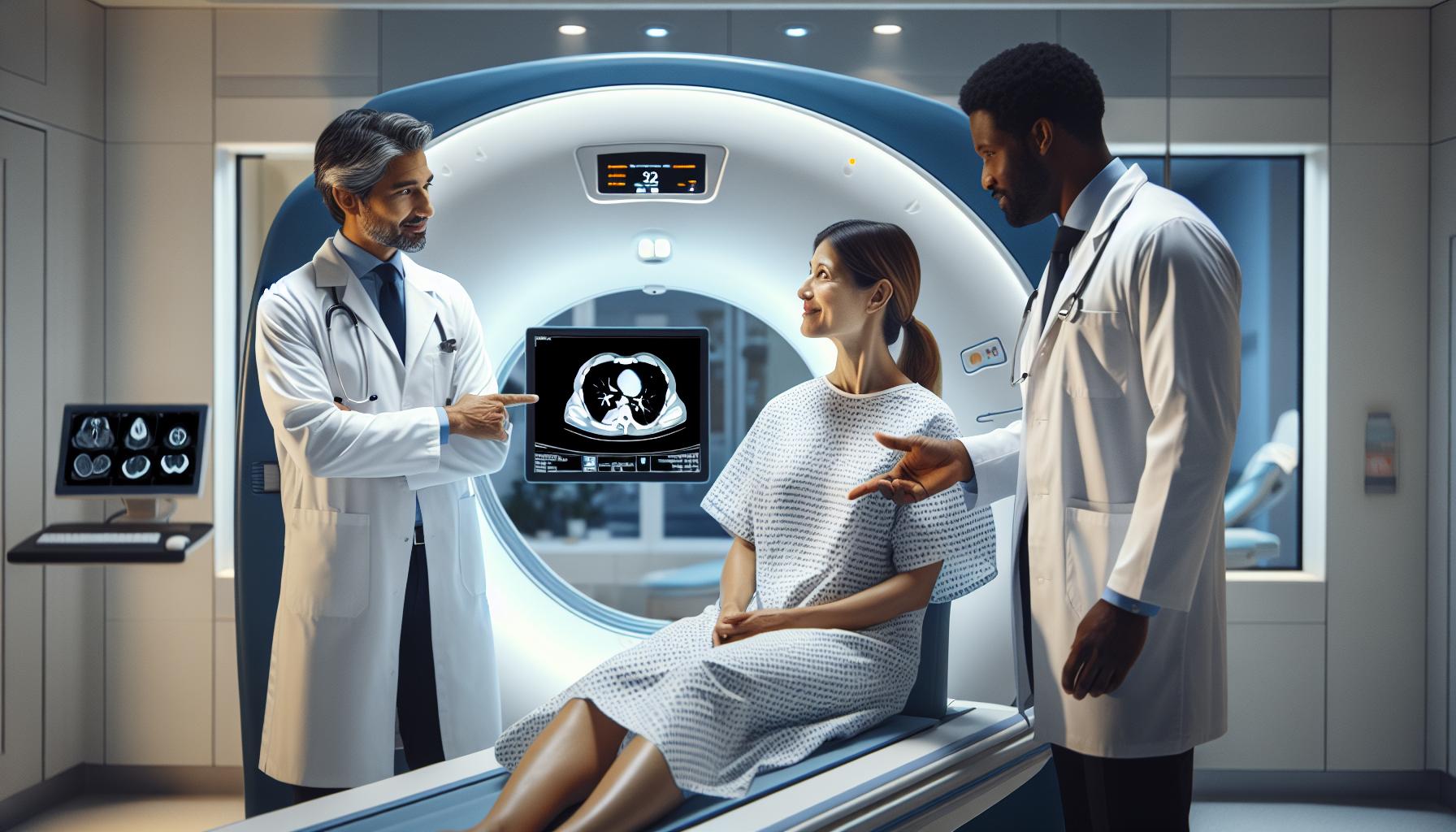
Undergoing a chest CT scan can be an overwhelming experience, especially for those concerned about the potential for breast cancer detection. Understanding what to expect during the procedure can alleviate some anxiety and empower individuals to approach it with confidence. The scan itself is a quick, non-invasive imaging technique that produces detailed cross-sectional images of the chest, including the lungs and surrounding structures.
Upon arrival at the imaging facility, patients will be greeted by a technologist who will guide them through the entire process. After completing a brief pre-scan questionnaire regarding any allergies or medical conditions, the patient will change into a gown and will be asked to lie down on the CT scanner’s table. This table will slide into the machine, which may feel a bit confining. However, it’s reassuring to know that the overall procedure only takes about 10 to 30 minutes. Throughout the scan, patients are often instructed to hold their breath briefly while images are captured, allowing for clearer results without motion blur.
During the scan, it’s important to remain as still as possible to ensure high-quality images. The machine may make buzzing or whirring sounds, which can be unsettling, but hearing this is perfectly normal. If the scan uses a contrast dye for enhancement, a needle will be placed in a vein, typically in the arm; this may cause some mild discomfort or warmth as the dye is injected. Open communication with the medical staff can help manage any discomfort and provide reassurance throughout the procedure.
Post-scan, patients can resume their normal activities immediately, which is another comforting aspect of this imaging technique. Results will be interpreted by a radiologist, and patients are generally contacted by their healthcare provider to discuss findings and any next steps. Being proactive by asking questions about the need for follow-up or further testing can aid in understanding individual health needs.
Preparing for Your Chest CT Scan: Step-by-Step Guide
Preparing for a chest CT scan can feel daunting, but understanding the process can ease your anxiety. This imaging technique plays a significant role in diagnosing various conditions, and knowing how to get ready can ensure a smoother experience. Here’s a step-by-step guide to help you prepare effectively.
Firstly, it’s crucial to consult with your healthcare provider prior to the scan. They will inform you about any specific preparations based on your medical history and the purpose of the imaging. Generally, you may be advised to avoid eating or drinking for several hours before your appointment, especially if contrast dye will be used.
When you arrive at the imaging center, take a moment to relax. Bring any necessary paperwork or identification. You will be greeted by a technologist who will guide you through the next steps. They will likely ask you to complete a questionnaire to gather information about your health history, allergies, and any previous imaging.
Once this is done, you will change into a gown to ensure that clothing does not interfere with the imaging. It’s advisable to wear loose, comfortable clothing on your upper body, as you may be required to remove any metallic items like jewelry, hairpins, or glasses. If contrast dye is used, a small IV will be placed in your arm, which may cause slight discomfort or a warm sensation when injected.
During the scan, you’ll need to lie still on a padded table that slides into the CT scanner. Your healthcare team will explain the process and may provide you with breathing instructions. Although the machine does make noise as it operates, you can expect the scan to be quick, typically lasting 10 to 30 minutes. Once completed, you can resume normal activities without any restrictions.
By following these straightforward steps and maintaining open communication with your healthcare team, you can approach your chest CT scan with confidence, knowing you’re well-prepared for the procedure.
Interpreting Chest CT Scan Results for Breast Cancer
While a chest CT scan is a powerful imaging tool primarily used to investigate lung and chest conditions, its ability to effectively detect breast cancer is limited. The scanning technology excels at identifying abnormalities in soft tissues but struggles with breast tissue due to its density and structure. Hence, interpreting the results of a chest CT scan for potential breast cancer takes a careful and nuanced approach.
When the radiologist evaluates the images from a chest CT scan, he or she will look for any suspicious lesions or masses that might raise concerns for malignancy. However, it’s important to understand that chest CT scans are not designed as a primary screening method for breast cancer. Instead, the results must be interpreted in conjunction with any prior imaging studies, such as mammograms or breast ultrasounds, which are tailored to assess breast tissue more accurately. A finding in a chest CT scan may warrant further investigation through additional imaging, such as a targeted breast MRI or biopsy, based on the characteristics of the observed abnormality.
After the scan is performed, results are typically reviewed within a few days. Patients will receive a detailed report outlining findings, and it is crucial to schedule a follow-up appointment with their healthcare provider to discuss the implications of these findings. During this consultation, patients can seek clarification on whether any abnormalities found in the CT scan indicate a need for additional tests or an alteration in the management plan. The anxiety of waiting for results can be overwhelming, so keeping open communication with the healthcare team is essential in navigating this process.
Ultimately, while a chest CT scan may contribute valuable information about a patient’s overall health and identify other thoracic issues, it should not replace more effective breast cancer screening strategies. In each case, the interpretation of chest CT scan results requires collaboration among healthcare professionals to ensure comprehensive care. Patients should feel empowered to ask their providers any questions they have about their imaging results, follow-up plans, and the best approach to cancer screening tailored to their individual health profiles.
Alternative Imaging Methods for Breast Cancer Detection
When it comes to detecting breast cancer, there are several imaging modalities available that are more effective than a chest CT scan. While CT scans provide a fantastic overview of chest anatomy and can help identify other thoracic diseases, they fall short in specifically targeting breast tissue due to its unique density and structure. This makes it vital to explore alternative imaging methods better suited for breast cancer detection.
Mammography
Mammography remains the gold standard for breast cancer screening and is essential for early detection. Utilizing low-energy X-rays, mammograms are designed to visualize breast tissue in detail, enabling radiologists to identify potential abnormalities such as lumps or calcifications that may indicate cancer. Women are generally encouraged to begin yearly mammograms at the age of 40, or earlier if they have a family history of breast cancer or other risk factors.
Breast Ultrasound
Breast ultrasound is another valuable tool, particularly for women with dense breast tissue where mammograms may be less effective. This technique uses sound waves to create images of the breast, allowing healthcare providers to assess lumps detected during a physical examination or a mammogram. It’s non-invasive, painless, and can often differentiate between solid masses and cystic formations, providing additional context in the evaluation process.
Magnetic Resonance Imaging (MRI)
MRI of the breast is highly sensitive and can detect cancers that mammograms might miss, particularly in women with a high risk for breast cancer or those with dense breast tissue. MRI uses powerful magnets and radio waves to produce detailed images of the breast’s internal structures. This imaging method may be recommended as a supplementary screening tool, particularly for those with family histories or genetic predispositions.
Emerging Techniques
Recent advancements are leading to innovative imaging methods, such as 3D mammography (tomosynthesis), which offers a clearer, layered view of breast tissue compared to traditional mammography. Additionally, contrast-enhanced mammography and novel techniques like photoacoustic imaging are being explored for their potential to improve detection rates and reduce false positives.
Patients should feel empowered to discuss these options with their healthcare providers, as every individual’s case is unique. Consulting with a specialist can help determine the most appropriate imaging strategy based on personal risk factors, breast density, and family history. Always prioritize open communication to navigate this essential aspect of breast health.
Understanding False Positives and Negatives in CT Scans
For individuals undergoing a chest CT scan, understanding the potential for false positives and negatives is crucial in managing expectations and making informed healthcare decisions. A chest CT scan, while a powerful diagnostic tool for assessing various conditions, is not specifically designed for breast cancer detection. This can lead to outcomes that may confuse both patients and providers.
False positives occur when a CT scan suggests there may be an abnormality in the breast that could indicate cancer, but further testing reveals no actual cancer present. These can arise from overlapping densities within the breast tissue or artifacts from surrounding structures, leading to unnecessary stress and additional tests. On the other hand, false negatives happen when a CT scan fails to detect breast cancer that is present. This is especially concerning given that a CT scan is not an ideal method for evaluating breast tissue, as dense or abnormal areas may not be adequately visualized in the absence of dedicated breast imaging technologies.
Patients should be aware of these limitations and the inherent uncertainties in the imaging process. Clear communication with healthcare providers about the purpose of a chest CT scan and its implications for breast cancer screening is vital. Discussing the appropriateness of other imaging modalities, such as mammograms or MRIs, tailored to individual risk factors can help provide clarity and peace of mind. Remember, the goal of these screenings is to ensure that any potential concerns are addressed as early as possible, while also minimizing unnecessary anxiety linked to inconclusive results.
Consulting Your Doctor: Questions to Ask About Screening
When considering breast cancer screening options, having an open and informative dialogue with your doctor is essential. Given the varying effectiveness of different imaging methods, such as chest CT scans and mammograms, it’s important to come prepared with relevant questions. This engagement not only provides clarity but also empowers you to make informed decisions about your health.
Start by asking your doctor about the purpose of the chest CT scan for your specific situation. A helpful question could be, “How does a chest CT scan compare to a mammogram in terms of breast cancer detection?” This will prompt your doctor to explain the limitations of CT scans for breast evaluation and the contexts in which they may be recommended, such as for assessing lung conditions or other thoracic issues unrelated to breast cancer.
Additionally, discussing your personal risk factors is crucial. You might ask, “Given my family history or previous results, what screenings do you recommend?” This allows your healthcare provider to tailor their recommendations to your unique health profile. Don’t hesitate to inquire about alternative imaging methods, such as MRIs or breast ultrasounds, which may provide better visualization of breast tissue.
It’s equally important to address your concerns about potential outcomes from the CT scan. Phrasing questions like, “What should I know about the possibility of false positives or negatives with this scan?” can help you understand the implications of the results and the next steps to consider. Understanding the emotional and physical aspects of these screenings can aid in alleviating anxiety.
Moreover, discussing the logistics surrounding the procedure can equip you with practical insights. For example, ask, “What can I expect during the chest CT scan, and how should I prepare for it?” Knowing what to anticipate helps reduce apprehension and makes the process smoother. Remember, maintaining an open line of communication with your doctor not only enhances your understanding but also fosters a collaborative approach to your health care.
Frequently Asked Questions
Q: Can a chest CT scan detect breast cancer early?
A: A chest CT scan is not primarily designed for early detection of breast cancer. Mammograms are the gold standard for breast cancer screening. However, a CT scan may identify abnormalities indirectly by showing changes in the lungs or surrounding tissues.
Q: What is the accuracy of a chest CT scan for breast cancer detection?
A: The accuracy of a chest CT scan for detecting breast cancer is not well-defined, as it is not the recommended method for this purpose. Its sensitivity can be lower compared to mammograms, and findings may require further investigation.
Q: When should I consider a chest CT scan if I have breast cancer?
A: A chest CT scan may be recommended if breast cancer has been diagnosed and your doctor needs to assess potential spread to the lungs or other chest structures. It is not used as a primary screening tool but for staging or monitoring.
Q: Are there any alternative imaging methods for breast cancer detection?
A: Yes, alternatives include mammograms, breast ultrasounds, and MRIs. Each method has its indications, with mammograms being most effective for routine screening, while MRIs are useful for detailed examinations in high-risk patients.
Q: What are the risks associated with a chest CT scan?
A: Risks of a chest CT scan include exposure to radiation and potential allergic reactions to intravenous contrast material. It’s essential to discuss these risks with your physician, especially in the context of breast cancer evaluation.
Q: How should I prepare for a chest CT scan if breast cancer is suspected?
A: Preparation for a chest CT scan typically involves fasting for a few hours before the procedure and informing your physician about any allergies or medications. Following specific instructions can help ensure accurate results.
Q: What should I do if my chest CT scan shows suspicious findings?
A: If suspicious findings are noted in your chest CT scan, consult your healthcare provider immediately. They may recommend follow-up tests, such as a biopsy or further imaging, to clarify the results and determine the next steps.
Q: How long does it take to receive chest CT scan results?
A: Results from a chest CT scan are usually available within a few days, depending on the facility. Your healthcare provider will discuss the findings with you and outline any necessary follow-up actions or additional testing.
To Wrap It Up
When considering whether a chest CT scan can detect breast cancer, it’s essential to understand that while these scans can provide valuable insights, they are not the definitive tool for breast cancer detection. If you have further questions about imaging procedures or wish to learn about specific breast cancer screenings, explore our articles on breast health and diagnostic imaging.
Act now by speaking with your healthcare provider about your concerns or scheduling a consultation to discuss personalized screening options. Your health and peace of mind are paramount, so don’t hesitate to reach out for more information. As you navigate your health journey, remember to access our resources on understanding CT scans and preparing for imaging tests, which can empower you with the knowledge needed for informed decisions.
We invite you to share your thoughts or questions in the comments below. Your experience could help others facing similar medical decisions. Thank you for trusting us as your source for health information; we look forward to supporting you on your path to well-being.


