Every year, millions of people experience strokes, and timely detection can be the difference between life and death. A CT scan is a critical tool in emergency settings, providing rapid images of the brain that help identify strokes, especially hemorrhagic ones. Understanding how a CT scan works in stroke diagnosis can empower you or your loved ones to act swiftly during an emergency.
Picture a loved one experiencing sudden weakness or confusion. Panic can set in, making it hard to think clearly. Knowing that a CT scan can quickly reveal whether a stroke has occurred not only alleviates some worry but also equips you with essential knowledge in a critical moment.
In this article, we will explore how CT scans detect strokes, what to expect during the procedure, and the significance of immediate medical attention. Read on to arm yourself with insights that might save a life when every second counts.
Can a CT Scan Identify Stroke Symptoms?
In the critical moments immediately following a stroke, swift diagnosis is essential. A CT scan plays a pivotal role in identifying stroke symptoms, primarily by enabling the detection of brain abnormalities that can indicate a stroke, such as hemorrhages or vascular blockages. This imaging technology excels in providing rapid, accurate images of the brain, which is vital in emergency situations where every second counts. For patients presenting with acute stroke symptoms like sudden numbness, confusion, or difficulty speaking, a CT scan can affirm or rule out the presence of a stroke, guiding immediate treatment options.
One of the key strengths of a CT scan in stroke diagnosis is its ability to visualize both ischemic strokes (caused by blockages) and hemorrhagic strokes (caused by bleeding). For instance, in ischemic strokes, a CT scan can help identify the area of the brain that is not receiving adequate blood flow. In contrast, in the case of hemorrhagic strokes, it can reveal bleeding within the brain tissue. It’s important to note that while CT scans are incredibly effective, they are often supplemented by other imaging modalities for more comprehensive evaluations.
When a CT scan is performed, patients can expect a quick procedure, usually lasting just a few minutes. The scan does not require any special preparation and involves lying still while the machine captures images of the brain from multiple angles. The radiologist interprets these images to assess for any signs of stroke and communicates the findings to the treating physician, who can promptly initiate appropriate interventions. Understanding this process can help alleviate some anxiety about undergoing a CT scan, emphasizing its role as a life-saving tool in acute medical settings.
For individuals experiencing symptoms indicative of a stroke, seeking immediate medical attention is paramount. A CT scan is a frontline diagnostic tool that can make a significant difference in treatment outcomes, providing clear direction for healthcare providers to take decisive action. Remember, recognizing the signs of a stroke and acting quickly can save lives.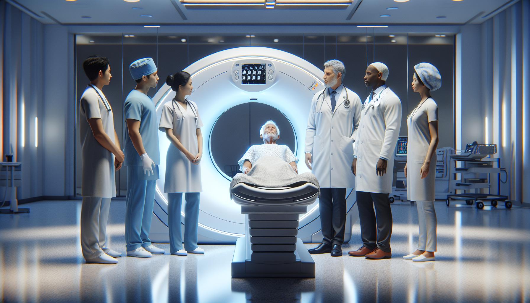
Understanding the Role of CT Scans in Stroke Diagnosis
In the race against time during a stroke, the importance of rapid and accurate diagnosis cannot be overstated. CT scans serve as a crucial tool in the diagnosis of strokes, effectively capturing detailed images of the brain that allow healthcare professionals to identify critical abnormalities. This imaging modality rapidly highlights areas affected by ischemic strokes (where blood supply is blocked) and hemorrhagic strokes (where bleeding occurs). For instance, while ischemic strokes may present as dark areas indicating insufficient blood flow, hemorrhagic strokes are typically shown as bright patches where blood has accumulated. Such clear visual distinctions are vital for determining the appropriate treatment pathway.
Speed and Efficiency
One of the standout benefits of CT scans is their speed. In emergency settings, a CT scan can be performed quickly, often within minutes, making it an invaluable asset in situations where every second can determine the outcome of the treatment. Patients lie on a table that moves through a ring-shaped machine, allowing the CT scanner to capture multiple cross-sectional images of the brain. This efficiency means that healthcare providers can swiftly decide on the next steps-whether it’s administering a clot-busting medication for ischemic strokes or preparing for surgical intervention in the case of a hemorrhagic event.
Patient Comfort and Safety
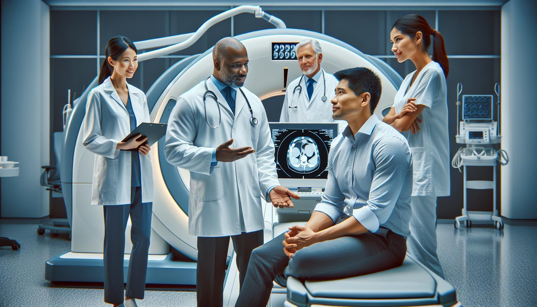
Understanding the procedure can significantly alleviate patients’ concerns. Generally, undergoing a CT scan does not require any special preparation or invasive techniques, which can make the experience less intimidating. Patients are asked to remain still during the scanning process, which typically lasts only a few minutes. The radiology team is highly trained to ensure that patients feel comfortable and well-informed throughout the entire experience. Additionally, CT scans are considered safe, utilizing a relatively low dose of radiation, and the benefits often far outweigh any potential risks.
In the context of stroke diagnosis, a CT scan is not only valuable for identifying the type of stroke but also plays a role in guiding treatment decisions. For anyone experiencing symptoms like sudden weakness, confusion, or difficulty speaking, immediate medical attention is essential. Trusting in the capabilities of modern imaging technology, such as CT scans, can empower patients and their families during a stressful time, offering hope and clarity in an otherwise uncertain situation.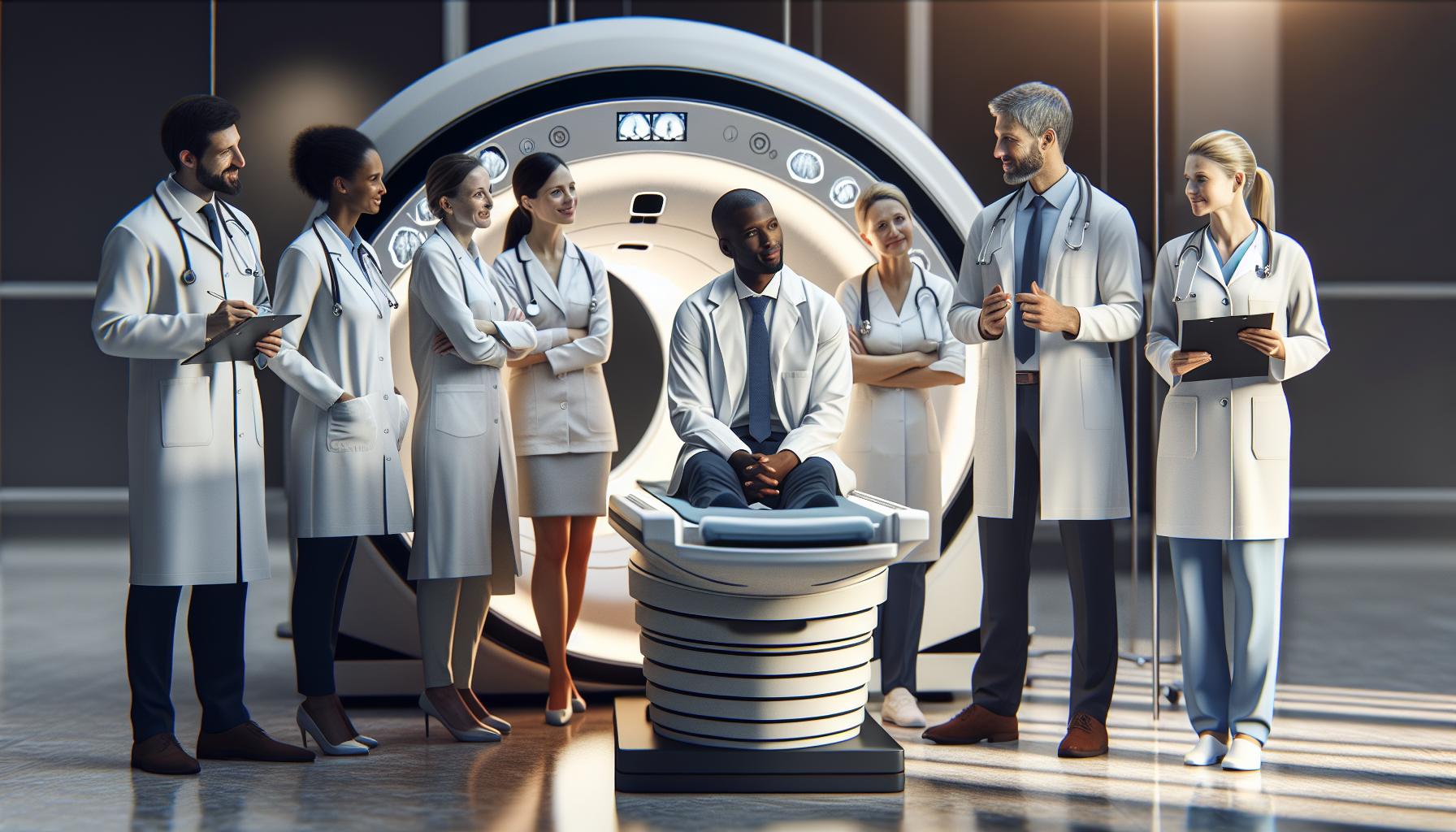
Types of Strokes: Is a CT Scan Effective?
When faced with the critical question of whether a CT scan can effectively identify different types of strokes, the answer lies in its remarkable imaging capabilities. CT scans are a frontline tool in emergency medicine, renowned for their speed and efficiency in diagnosing strokes. They can quickly distinguish between ischemic strokes, caused by blocked blood flow, and hemorrhagic strokes, characterized by bleeding in the brain. Each type leaves distinct imaging signatures: ischemic strokes often present as darker areas on the scan due to lack of blood, while hemorrhagic strokes typically appear as bright patches where blood has accumulated.
Understanding Stroke Types
To gain a better insight into the effectiveness of CT scans, it’s essential to recognize the two primary types of strokes they can diagnose:
- Ischemic Stroke: This occurs when a blood vessel supplying blood to the brain is obstructed, often due to a blood clot. In a CT scan, these areas may appear darker due to the absence of adequate blood flow.
- Hemorrhagic Stroke: This type is caused by bleeding within the brain, either from a ruptured blood vessel or an aneurysm. On a CT scan, this appears as bright areas, helping radiologists quickly determine the need for urgent interventions.
These clear visual distinctions are crucial for emergency responders and neurologists in determining treatment pathways. For instance, identifying an ischemic stroke allows for the rapid administration of clot-busting medications, which can significantly improve outcomes if given within the window of time. Conversely, recognizing hemorrhagic strokes can lead to immediate surgical options, potentially addressing life-threatening bleeding.
The Impact of Timeliness
In situations where every second counts, having access to a CT scan within the emergency room can make a profound difference. For individuals showing signs of a stroke-such as sudden weakness, confusion, or speech difficulties-the prompt use of a CT scan helps ensure timely intervention. Knowing the type of stroke not only accelerates care but enhances the chances of recovery and minimizes the risk of long-term complications.
Ultimately, while no diagnostic tool is without limitations, CT scans remain a cornerstone in the accurate and swift identification of stroke types, providing critical information that guides life-saving decisions. For anyone experiencing stroke symptoms, trusting in the advanced imaging capabilities of CT technology can bring clarity and relief during a moment of distress.
How Does a CT Scan Detect Stroke?
When considering how a CT scan detects stroke, it’s crucial to understand the underlying technology that powers this life-saving procedure. CT scans, or computed tomography scans, utilize a series of X-ray images taken from different angles around the body, which are then processed by a computer to create cross-sectional images, or “slices,” of the brain. This advanced imaging technique is particularly adept at revealing structural abnormalities and areas of concern in a matter of minutes, making it an essential tool in emergency medicine.
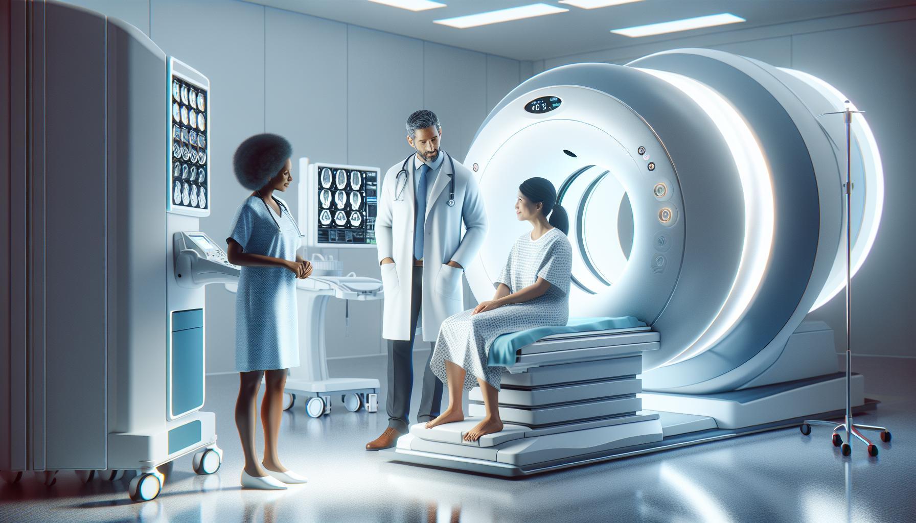
During a CT scan, the patient lies on a table that moves through a large, donut-shaped machine. As the machine rotates, it takes multiple X-ray images that help build a detailed 3D representation of the brain’s internal structures. In cases of stroke, the scan can reveal the type of stroke-ischemic or hemorrhagic-by showing the presence of blood clots or bleeding within the brain. For instance, an ischemic stroke appears as darker regions where blood flow is absent, while the presence of bright spots indicates bleeding, crucial for determining immediate treatment strategies.
It’s important to note that while a CT scan is an invaluable diagnostic tool, it does have its limitations. It may not always detect very small strokes or those that occur in specific areas of the brain. This is why medical professionals often use CT scans in conjunction with other evaluations or imaging techniques if they suspect a stroke or if initial results are inconclusive.
Understanding the CT scan process can alleviate anxiety for patients and their families. Knowing that this quick and non-invasive test provides critical information to healthcare providers empowers individuals to seek help promptly when stroke symptoms arise. In emergency scenarios, every minute counts; timely access to a CT scan can significantly influence treatment options and improve outcomes, reinforcing the importance of acting swiftly when stroke signs are evident.
The CT Scan Procedure: What to Expect
To understand what lies ahead during a CT scan, it’s reassuring to know that this examination is designed to be a quick and non-invasive procedure that delivers crucial information to healthcare providers. Most importantly, the entire process typically takes only a few minutes, allowing for prompt diagnosis in emergency situations like strokes. Patients can expect to lie down on a comfortable table that moves through a large, cylindrical machine-commonly referred to as a CT scanner.
The Procedure: Step-by-Step
- Preparation: When you arrive at the imaging center, you may be asked to change into a hospital gown and remove any metal objects, such as jewelry or glasses, which can interfere with the imaging process. Inform the staff of any medical conditions, especially if you have allergies, prior kidney issues, or if you’re pregnant, as these may influence your examination or may require additional precautions.
- Positioning: Once you are ready, you will lie on the table, which will steadily slide into the center of the CT scanner. It’s essential to remain still during the scan to ensure clear images; any movement can cause blurring. You may be asked to hold your breath for short periods while images are taken.
- Scanning Process: As the machine rotates around you, it captures multiple X-ray images from various angles. These images are then processed by a computer to create detailed cross-sectional “slices” of the brain. You will hear a whirring sound as the machine operates, which is perfectly normal. The technologist may monitor the procedure from an adjacent room and will be in contact with you throughout the process.
- Completion: After the scan is complete, you’ll be able to get up and resume your normal activities right away. In many cases, the physician will receive the scan results shortly thereafter, enabling timely decision-making for your care.
What to Expect After the Scan
Once the CT scan is completed, your doctor will explain the findings, which may take time based on the complexity of the images. While waiting for results can feel anxious, understanding that the procedure aids in assessing critical conditions-such as stroke-can provide some reassurance. Any significant findings will be addressed, and necessary steps or treatments will be discussed on an individualized basis.
In summary, knowing what to expect from a CT scan can ease apprehension, making the experience smoother for everyone involved. Should you have any concerns or questions during the process, don’t hesitate to direct them to the healthcare professionals present, as they are there to help guide you and ensure your comfort. Always remember, timely access to imaging can dramatically improve outcomes when it comes to conditions like strokes.
Preparing for a CT Scan: Step-by-Step Guide
Preparing for a CT scan can feel daunting, especially when it’s being done for something as serious as a suspected stroke. However, knowing what to expect can reduce anxiety and help you feel more at ease. First, it’s essential to understand that a CT scan is a non-invasive procedure designed to quickly provide detailed images of your brain, making it a critical tool in emergency situations.
When you arrive for your scan, you will typically be asked to change into a hospital gown to ensure that any clothing does not interfere with the imaging process. Additionally, you should remove all metal objects, including jewelry and eyeglasses, as they can obstruct the X-ray images. It’s important to communicate openly with the medical staff about any medical conditions you have, especially allergies or kidney issues, as these may influence the type of contrast material used during the procedure. For instance, if you are pregnant, this should be disclosed, as special precautions may be necessary.
Once you’re prepared, you’ll lie down on the scanning table, and the technician will position you carefully. Remaining still during the scan is paramount; even slight movements can affect the clarity of the images. You may be instructed to hold your breath for a few seconds while the machine captures the necessary images. As the scanner moves around you, it will emit various sounds, which is completely normal. The entire process typically lasts only a few minutes.
Following the scan, you will be able to resume your normal activities almost immediately. The results should be available shortly thereafter, allowing your healthcare provider to assess the situation quickly and make timely treatment decisions. Remember, while the prospect of a CT scan might be unsettling, it plays a vital role in diagnosing and managing stroke effectively. Always feel empowered to ask questions and express any concerns before, during, and after the procedure to ensure a supportive and transparent experience.
Interpreting CT Scan Results for Stroke
Interpreting the results of a CT scan after a suspected stroke can be pivotal in determining the appropriate treatment and intervention. CT scans are designed to provide quick and detailed images of the brain, which can reveal critical information about the presence and type of stroke. For patients and their families, understanding these results is essential for navigating the next steps in care.
When examining CT scan results, healthcare professionals typically look for signs of an ischemic stroke, caused by a blockage of blood flow, or a hemorrhagic stroke, resulting from bleeding in the brain. In cases of ischemic stroke, the damaged brain tissue may appear darker due to a lack of blood flow, which can often be identified within the first few hours after symptoms onset. Conversely, a hemorrhagic stroke may present as bright white areas on the scan where blood has leaked into surrounding tissues. It’s not uncommon for initial scans to appear normal within the first few hours following a stroke, so follow-up imaging may be necessary to detect changes over time.
It’s important for patients and their loved ones to communicate openly with their healthcare team about the results. Understanding whether the scan indicates a stroke can ease anxiety and help guide effective treatment decisions. Asking questions about the imaging findings, the interpretation of results, and the next steps is encouraged. For instance, if the scan reveals a stroke, discussing potential treatments, rehabilitation options, and lifestyle changes is vital to recovery.
While interpreting CT scan results can be complex, being an informed participant in the process can empower patients and their families. Knowledge about what the scans reveal helps demystify the medical process. Ultimately, timely and accurate interpretation of CT scans can make a significant difference in outcomes for stroke patients, highlighting the critical role they play in emergency diagnosis and treatment.
CT Scan vs. Other Imaging Techniques in Stroke Detection
When a stroke occurs, every second counts, making rapid and accurate diagnosis critical for effective treatment. CT scans, or computed tomography scans, have become a cornerstone in emergency stroke detection due to their ability to provide quick and detailed images of the brain. However, they are not the only imaging technique available, and understanding the differences can empower patients and their families in making informed decisions about care.
Why Choose CT Scans?
CT scans are particularly advantageous in emergency settings because they are fast and widely available. They help distinguish between types of strokes-ischemic (caused by a blockage) and hemorrhagic (caused by bleeding). In an acute setting, a CT scan can typically be completed in just a few minutes, allowing healthcare providers to quickly assess whether the brain has been affected by a stroke and determine the appropriate intervention. Their ability to visualize bleeding immediately after a stroke is crucial, as this can significantly influence treatment choices.
However, other imaging techniques also play important roles. Magnetic Resonance Imaging (MRI), for example, offers a more detailed view of the brain and can better detect the presence of ischemic strokes, especially in the early hours. MRIs use powerful magnets and radio waves to create images and may show brain tissue changes that a CT scan might miss, particularly in cases where blood flow reduction is subtle. Conversely, an MRI takes longer to perform and is less practical in acute situations where time is of the essence.
Comparing Imaging Techniques
In comparing CT scans to other modalities like MRI and ultrasound, each has its strengths and weaknesses:
| Imaging Technique | Advantages | Disadvantages |
|---|---|---|
| CT Scan | Quick; readily available; excellent for assessing bleeding. | Less detailed than MRI; may miss early ischemic changes. |
| MRI | More sensitive to ischemic stroke; detailed images. | Longer scan time; less available in some emergency settings. |
| Ultrasound | Non-invasive; useful for assessing blood flow in arteries. | Limited in visualizing brain structure; operator-dependent. |
While CT scans are invaluable in emergencies, the choice of imaging often depends on clinical factors and availability. In some cases, physicians may recommend follow-up MRI scans to provide a clearer picture of the brain’s condition once the immediate threat is addressed.
Ultimately, each imaging modality has its unique role in the stroke diagnostic process, and your healthcare team will determine which option is best suited for your specific situation. Understanding these options can alleviate some anxiety about medical procedures and empower you to engage in informed discussions with your healthcare providers about your treatment and care plans.
Safety Concerns: Are CT Scans Risky?
While CT scans are vital tools in the swift diagnosis of strokes, it’s natural to have concerns about their safety. On the one hand, the process involves exposure to ionizing radiation, which leads to questions about long-term health risks. However, it’s essential to put these risks into perspective. The radiation dose in a single CT scan is relatively low and is carefully managed to minimize exposure while ensuring that the diagnostic benefits far outweigh any potential harm. Medical professionals are trained to use imaging judiciously, ordering these scans only when clinically necessary.
Before undergoing a CT scan, patients often wonder about the preparation and procedure itself. Generally, there are no specific preparations required for a stroke CT scan. You may be asked to remove any metal jewelry or accessories that could interfere with the imaging process. In some cases, especially if a contrast agent is used, you might need to provide information about allergies, particularly to iodine, as well as your medical history regarding kidney function. This information is crucial as it helps the healthcare team provide safe and effective care.
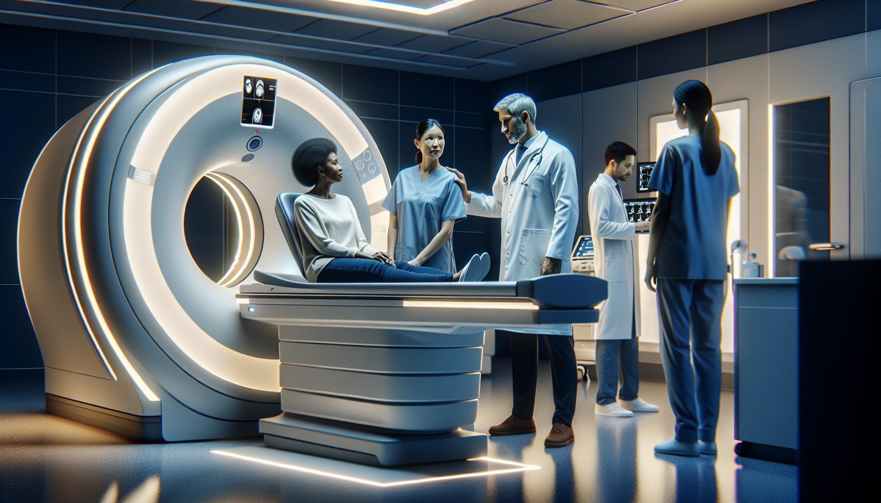
During the scan, the procedure is quick, usually lasting only a few minutes. You will lie still on a moving table as the scanner rotates around you, capturing images of your brain. Some patients might experience anxiety or claustrophobia; open communication with the imaging technician can help ease these feelings. It’s important to remember that CT scans are optimal for emergency scenarios because of their speed, allowing for immediate diagnosis and treatment-the very essence of preserving brain health in a stroke situation.
Lastly, while safety is a valid concern, it’s reassuring to know that healthcare professionals continuously strive to adhere to the principle of ALARA (As Low As Reasonably Achievable) when it comes to radiation exposure. If you have any lingering worries, discussing these with your healthcare provider can provide clarity and peace of mind, ensuring you feel comfortable and informed about the procedure and its necessity. Always prioritize open dialogue with your medical team regarding any personal health concerns or preferences you may have, empowering yourself in the decision-making process.
Costs of CT Scans: What You Should Know
When facing a medical emergency such as a stroke, understanding the financial implications of necessary diagnostic procedures like CT scans can add another layer of concern. It’s crucial to be aware of the potential costs associated with these scans, which play a vital role in swiftly diagnosing strokes. Typically, the cost of a CT scan can range from $300 to $3,000, depending on factors such as the facility’s location, whether the scan is part of an emergency visit, and if advanced imaging techniques are required.
Factors Influencing CT Scan Costs
The cost of a CT scan can be influenced by several factors:
- Type of Facility: Prices may vary significantly between hospitals, outpatient imaging centers, and emergency departments.
- Insurance Coverage: Patients with health insurance may only pay a portion of the cost, depending on their specific plan and the deductible status.
- Contrast Agents: If a contrast dye is used during the scan, additional fees may be incurred, contributing to higher overall expenses.
- Geographical Location: Costs can fluctuate based on local healthcare market dynamics, with urban areas typically being more expensive than rural ones.
Patient Considerations
It’s important to discuss with your healthcare provider about the reasons for the scan and its urgency. Many patients find comfort in understanding that, despite the costs, the potential benefits of an early diagnosis can outweigh the financial burden. If cost is a concern, inquire about options such as payment plans, sliding scale fees based on income, or financial assistance programs that some facilities may offer.
Being proactive by checking your insurance coverage prior to the scan can also reduce surprise expenses. Have your insurance card handy and request clarification on what will be covered related to your CT scan for stroke evaluation. Knowledge is empowering, and understanding the financial aspects can help you focus on what truly matters: receiving appropriate care promptly.
Real-Life Impact: Stories of Timely Stroke Detection
Recognizing the warning signs of a stroke can mean the difference between life and death, as well as the potential for major recovery versus long-term disability. For instance, a man named John experienced sudden weakness in his face, slurred speech, and confusion while watching television. His partner quickly recognized these as classic stroke symptoms and immediately called emergency services. Within minutes, he was at the hospital, where a CT scan was performed. The imaging revealed a blockage in a critical artery, allowing doctors to act swiftly to restore blood flow to his brain. John’s timely diagnosis and treatment meant he could return to his regular activities within weeks, a fate that might have looked very different had he delayed seeking help.
Another poignant example involves a woman named Maria, who was at work when she suddenly felt dizzy and struggled to coordinate her movements. Concerned colleagues urged her to get checked out immediately. At the hospital, her doctor ordered a CT scan, which showed signs of a small stroke. Thanks to the quick intervention facilitated by the CT imaging, Maria began treatment with medication aimed at preventing further complications. Witnessing her journey from a critical health scare to a full recovery underscored not only the effectiveness of CT scans but also the importance of acting quickly when stroke symptoms appear.
Timely stroke detection through CT scans can significantly alter patient outcomes. Advances in imaging technology allow healthcare providers not just to diagnose but also to tailor interventions quickly and effectively. Patients and their families should understand that these rapid diagnostics are critical in emergencies; knowing what to do when symptoms arise can lead to lifesaving care. It’s crucial to have conversations with healthcare providers about the importance of recognizing stroke symptoms and seeking immediate help, as the ability to evaluate and act on a CT scan result in real time is vital.
In any health crisis, empowering yourself through knowledge is essential. Patients are encouraged to familiarize themselves with the FAST acronym (Face drooping, Arm weakness, Speech difficulties, Time to call emergency services) to identify stroke symptoms promptly. Understanding these warning signs can facilitate faster responses that lead to the effective use of advanced imaging techniques like CT scans, making a significant impact on recovery and long-term health outcomes.
Expert Insights: When to Get a CT Scan for Stroke
When faced with potential stroke symptoms, timing is critical, and understanding when to get a CT scan can be lifesaving. CT scans are a frontline tool in emergency medicine for diagnosing strokes, often used to quickly determine whether a patient is experiencing an ischemic stroke (caused by a blockage) or a hemorrhagic stroke (caused by bleeding). If you or someone you know shows any signs of a stroke-such as sudden weakness on one side of the body, difficulty speaking, or severe headaches-it’s imperative to seek medical attention immediately.
A CT scan is typically ordered by a healthcare professional in emergency situations to quickly visualize the brain and assess the incidence of a stroke. It can provide immediate insights within minutes, allowing doctors to discern the type of stroke and decide on appropriate treatment plans. Here are a few scenarios that emphasize the importance of timely CT scanning:
- Sudden Onset Symptoms: If someone suddenly exhibits symptoms such as facial drooping, arm weakness, or difficulty speaking, this warrants an immediate trip to the emergency room where a CT scan will likely be part of the diagnostic process.
- History of Stroke: Individuals with a previous stroke history or risk factors such as hypertension and diabetes should be vigilant. If they experience any new symptoms, they should prompt immediate evaluation and a CT scan may be indicated even with mild symptoms.
While CT scans are highly effective in diagnosing strokes, it’s vital to remember that the scan is one part of a broader assessment. Medical professionals will consider the patient’s history, physical exams, and other tests before making a treatment decision. Being prepared can make a considerable difference in how efficiently healthcare providers can intervene. Always consult with healthcare professionals to ensure personalized advice and intervention based on specific circumstances. Empowering yourself with knowledge about stroke symptoms can significantly enhance outcomes and underscore the importance of rapid medical response.
Faq
Q: How quickly can a CT scan detect a stroke?
A: A CT scan can quickly detect a stroke, often within minutes of a patient arriving at the hospital. This rapid assessment is crucial for prompt treatment decisions, particularly in cases of ischemic stroke where time is critical for restoring blood flow. For more information, see our section on “Understanding the Role of CT Scans in Stroke Diagnosis.”
Q: What type of stroke is best identified by a CT scan?
A: CT scans are particularly effective in identifying hemorrhagic strokes, which are caused by bleeding in the brain. They are less effective at detecting early ischemic strokes, but can show signs of damage after several hours. Refer to “Types of Strokes: Is a CT Scan Effective?” for a deeper dive.
Q: Can a CT scan distinguish between a stroke and a migraine?
A: A CT scan can help differentiate between a stroke and a migraine by revealing abnormal brain activity and structural changes associated with a stroke. However, migraines may sometimes present similarly, requiring a comprehensive clinical evaluation to make an accurate diagnosis.
Q: What symptoms indicate the need for a CT scan for stroke?
A: Symptoms like sudden numbness or weakness, particularly on one side of the body, confusion, difficulty speaking or understanding speech, and sudden vision problems warrant immediate medical attention and may lead to a CT scan to rule out stroke.
Q: Are there any risks associated with having a CT scan for stroke detection?
A: While CT scans are generally safe, they involve exposure to low doses of radiation. It’s essential for patients to discuss potential risks, especially pregnant individuals or those needing multiple scans. See “Safety Concerns: Are CT Scans Risky?” for more details.
Q: How do doctors interpret CT scan results for a stroke?
A: Doctors analyze CT scan images for signs of bleeding, ischemia, or swelling in the brain. They look for specific patterns and changes that indicate different types of strokes, helping to determine the appropriate treatment. For a better understanding, check our section on “Interpreting CT Scan Results for Stroke.”
Q: When should a CT scan be prioritized in stroke cases?
A: A CT scan should be prioritized when a patient shows signs of an acute stroke, such as sudden neurological deficits. Rapid scanning within the first few hours of symptoms can significantly influence treatment outcomes, especially for thrombolysis.
Q: What alternatives exist to CT scans in stroke diagnosis?
A: Alternatives to CT scans include MRI and ultrasound imaging. MRI offers greater detail without radiation and is more sensitive for detecting ischemic strokes. However, CT scans remain the first choice due to their speed and effectiveness in emergency settings. For insights on imaging techniques, see “CT Scan vs. Other Imaging Techniques in Stroke Detection.”
The Conclusion
Understanding whether a CT scan can reveal a stroke is crucial for timely emergency intervention. Remember, swift diagnosis can significantly improve treatment outcomes, so if you or someone you know is showing signs of a stroke, don’t hesitate to seek medical attention right away. For more insights, explore our related articles on “CT Scans Explained: What to Expect” and “Understanding Stroke Symptoms” to enhance your knowledge.
Stay informed and empowered about your health-consider signing up for our newsletter for the latest updates in medical imaging and health tips. Every moment counts, and being proactive can make a difference. If you have questions or need personalized assistance, our team is here to support you. Let’s navigate your healthcare journey together, ensuring you have all the resources you need at your fingertips. Share this information with loved ones to spread awareness, and visit us for more resources on CT scans and emergency care.

