Hernias are a common medical concern that can lead to discomfort and potential complications if left untreated. Understanding whether a CT scan can effectively detect hernias is crucial for patients experiencing abdominal pain or unusual bulges. With advancements in imaging technology, many people might wonder about the visibility and detection rates of hernias on these scans.
When faced with persistent abdominal issues, the uncertainty can be daunting. A CT scan is often a go-to diagnostic tool, capable of revealing the presence of a hernia, which may sometimes evade physical examination, especially in their early stages. This insight not only helps in accurate diagnosis but also guides the appropriate course of treatment.
Exploring the nuances of how CT scans contribute to hernia detection can empower patients with knowledge, alleviating anxiety about their symptoms and the diagnostic process. Join us as we delve into the visibility of hernias on CT scans and what to expect during this essential imaging procedure.
Understanding Hernias: Types and Symptoms
Hernias are common conditions where an organ protrudes through an abnormal opening in surrounding tissue, often causing discomfort and pain. Understanding the types and symptoms can empower individuals to seek timely medical attention, which is crucial in preventing complications. The most common types of hernias include inguinal hernias, which occur in the groin; femoral hernias, found in the upper thigh; umbilical hernias, seen near the belly button; and incisional hernias, which can develop at the site of surgical scars.
Symptoms may vary depending on the type and severity of the hernia. Patients often report a noticeable bulge or swelling at the site, especially when standing or straining. Other common symptoms include a feeling of heaviness, pain when lifting, and uncomfortable pressure in the abdomen or groin area. In some cases, a hernia may not exhibit any noticeable symptoms, which can lead to dangerous complications if left untreated. Recognizing these early signs and understanding the various types can provide individuals with vital information to discuss with their healthcare provider.
If you’re experiencing symptoms suggestive of a hernia, it’s important not to delay seeking medical evaluation. Hernias can become incarcerated or strangulated, leading to serious health risks. A thorough assessment by a healthcare professional, including imaging tests like CT scans or ultrasounds, plays a key role in diagnosis and treatment planning. Understanding hernias is the first step toward effective management and recovery, ensuring that individuals can maintain their overall health and quality of life.
How CT Scans Work for Hernia Detection
One of the most effective tools for diagnosing hernias is the CT scan, a non-invasive imaging technique that allows for detailed visualization of the internal structures. A CT, or computed tomography scan, uses a series of X-ray images taken from different angles to create cross-sectional images of the body. These images can reveal the presence of a hernia, including its size and location, which is essential for determining the appropriate treatment plan. This advanced imaging technique is particularly beneficial for detecting complex cases, such as those involving internal hernias or when distinguishing a hernia from other abdominal masses, like tumors or hematomas [3[3].
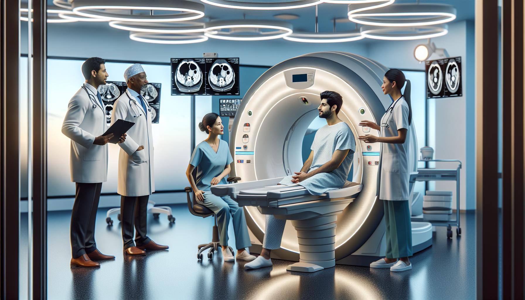
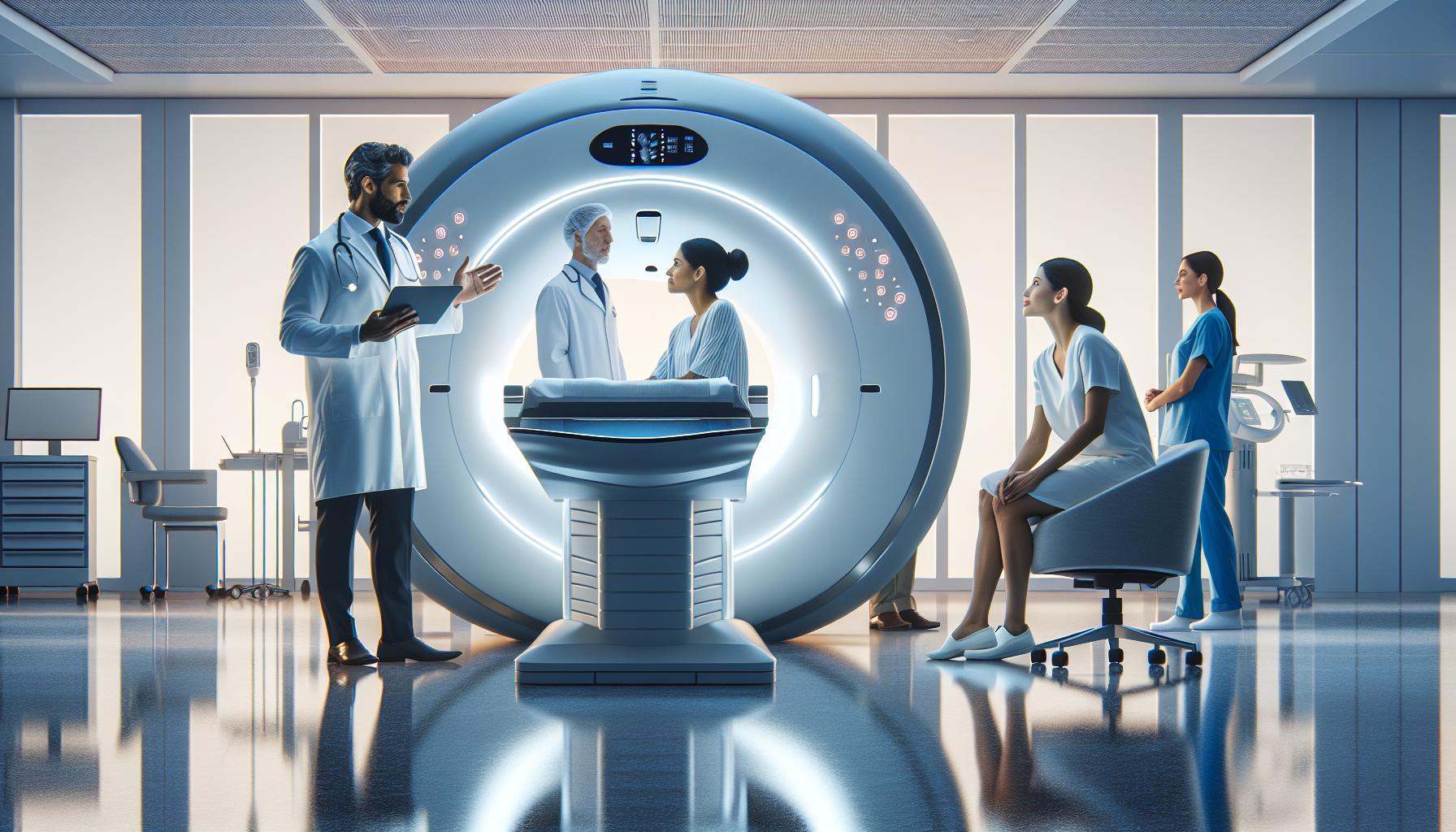
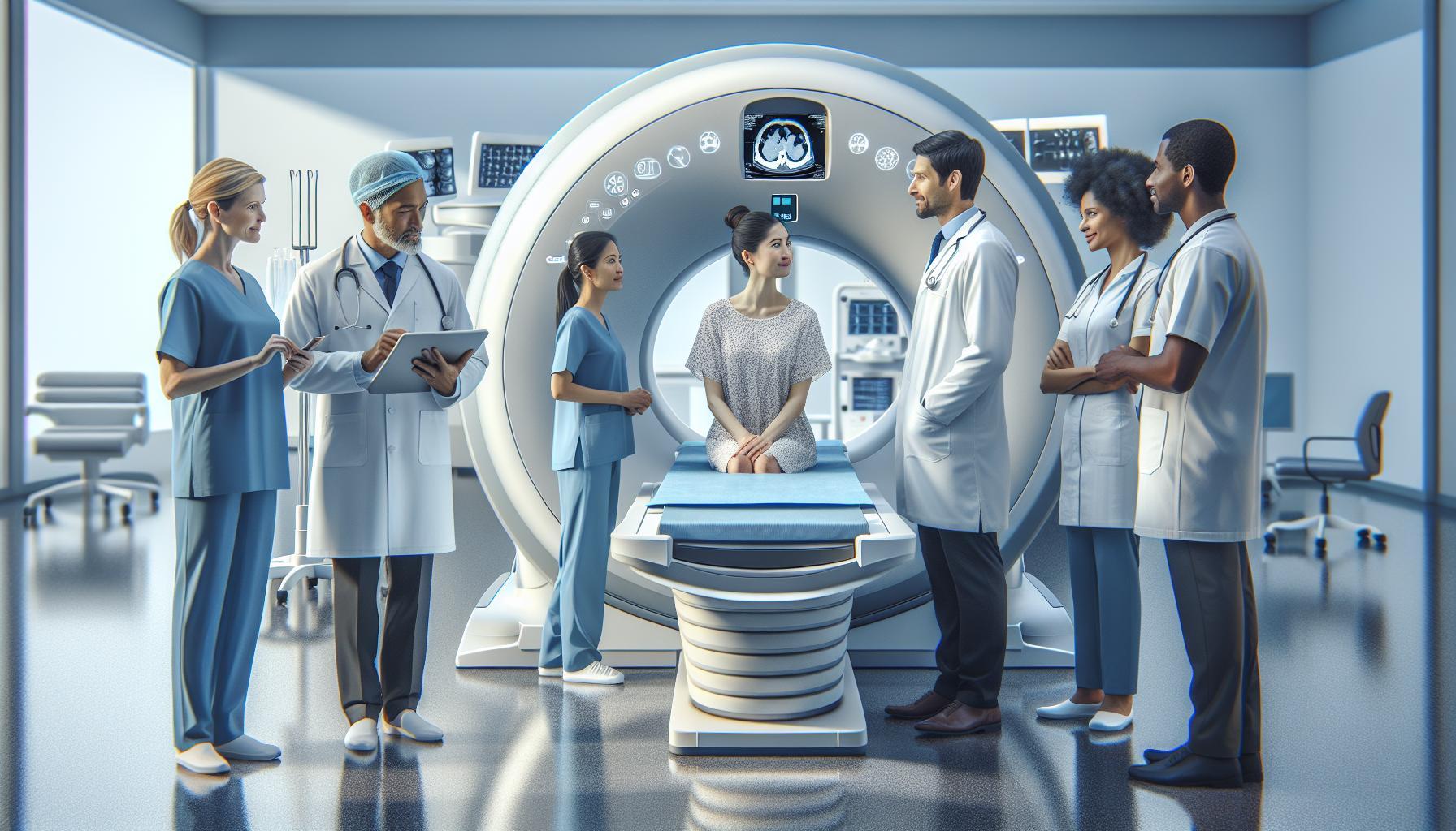
Before undergoing a CT scan for suspected hernia, it’s important for patients to be informed about the procedure. Patients will typically lie on an examination table that moves through a large, doughnut-shaped machine. During the scan, the machine will produce a series of images while the patient remains still. Depending on the specific circumstances, contrast dye may be used to enhance visibility, providing clearer images of the abdominal structures. It’s crucial to communicate any pre-existing health conditions or allergies to your healthcare provider prior to the test, as this information can influence the use of contrast materials [2[2].
Understanding how CT scans work can help ease the anxiety that often accompanies medical procedures. The entire scanning process generally takes only a few minutes, yet it can yield valuable insights into the presence and characteristics of hernias. Most critically, the results will guide healthcare professionals in determining whether surgical intervention is necessary, thus facilitating timely treatment and minimizing the risk of complications. Always remember to discuss your results with your doctor to understand their implications and to outline the next steps in your care.
Visibility of Hernias on CT Scans: What to Expect
CT scans are a remarkable technology that can reveal the presence of hernias with impressive accuracy, helping to alleviate some of the uncertainty and worry that often accompanies abdominal discomfort. When undergoing a CT scan, patients can anticipate receiving detailed images of their internal structures, which can highlight not just the location of a hernia, but also its size and any potential complications. This clarity is crucial in differentiating between a hernia and other possible abdominal masses, such as tumors or inflammatory conditions, which might present similarly but require different management approaches.
During the scan, the machine captures multiple slice images from various angles, providing a three-dimensional view of the abdomen. This allows radiologists to detect even small hernias that may not be visible through physical examinations alone. In particular, CT imaging has proven effective in identifying internal hernias-where portions of the intestines protrude into other areas of the abdomen-which can be notoriously difficult to diagnose. Patients should be prepared for the possibility of a contrast dye being used, which can improve the visibility of the surrounding tissues and vascular structures.
It’s also important to manage expectations regarding visibility. Although many hernias are detectable on a CT scan, certain factors can influence the effectiveness of the imaging. The patient’s body habitus, the size of the hernia, and the presence of abdominal contents can all play a role in how clearly the hernia is visualized. Those undergoing a CT scan should keep in mind that the results can be detailed, but sometimes further evaluation may still be required based on findings. If the scan does reveal a hernia, the next steps will typically focus on discussing treatment options that align with the severity and characteristics of the hernia detected. Remember, maintaining open communication with healthcare professionals about all findings is key to navigating the journey towards recovery effectively.
Detection Rates of Hernias by CT Scan Type
When it comes to the accuracy of detecting hernias, the type of CT scan performed plays a crucial role. Studies have shown that different imaging techniques yield varying detection rates, impacting both diagnosis and subsequent treatment plans. Generally, CT scans are highly effective in illustrating hernias, especially in complex cases involving internal structures. For instance, multi-phasic CT scans that involve both contrast and non-contrast phases can significantly enhance the visibility of subtle hernias, particularly in patients with a history of prior abdominal surgery where scar tissue complicates the imaging.
Hernia detection rates are often categorized based on the region of the body being examined. For abdominal wall hernias, including umbilical and incisional types, a standard CT scan typically demonstrates high sensitivity, often exceeding 90% in experienced hands. Groin hernias, such as inguinal and femoral types, can vary more widely; while CT scans still provide reliable evaluations, factors like the patient’s body type and the size of the hernia may influence results. Additionally, the presence of bowel loops or fluid can obscure readings, potentially lowering detection rates.
Patients preparing for their CT scans can support the imaging process by communicating openly with their healthcare providers. This includes discussing any previous surgeries, current symptoms, and concerns regarding the examination. Such dialogue ensures that radiologists can tailor their approach, possibly opting for advanced imaging techniques when necessary.
Ultimately, while CT scans present a powerful tool for diagnosing hernias, understanding the specifics of detection rates based on scan type is essential. Awareness of the limitations, combined with thorough preparation and clear communication with medical professionals, can enhance the diagnostic process, allowing for timely and effective treatment plans tailored to individual needs.
Factors Affecting Hernia Visibility on CT
The visibility of hernias on CT scans can be influenced by a variety of factors that vary from patient to patient. Understanding these factors can enhance one’s expectations and improve the overall diagnostic process. One major consideration is the type and location of the hernia. For instance, abdominal wall hernias like umbilical or incisional hernias are generally more visible during a CT scan due to their position and the nature of surrounding tissues. Conversely, groin hernias, such as inguinal or femoral hernias, may present challenges in detection due to their anatomical complexity.
Another crucial factor is patient characteristics, including body habitus and existing medical conditions. For example, obesity can complicate imaging results as increased adipose tissue may obscure views of the hernia itself. Additionally, a patient’s prior surgical history adds another layer of complexity, as scarring can interfere with the visibility of hernias. The presence of bowel loops or fluid collections in the abdominal cavity can also obscure hernias and reduce detection rates.
To facilitate accurate imaging, preparation is key. Patients are encouraged to communicate any prior surgeries or symptoms to their healthcare providers. This dialogue might influence decisions regarding the imaging technique used, such as considering a multi-phasic CT that combines both contrast and non-contrast phases. Such approaches can significantly enhance visibility and accuracy in diagnosing subtle hernias, especially in complex cases.
In summary, while CT scans are an effective tool for detecting hernias, various factors can influence visibility. By understanding these elements, patients can better prepare for their scans and engage meaningfully with their healthcare team to ensure the best possible outcomes.
Preparing for a CT Scan: What Patients Should Know
Before a CT scan, it’s essential to focus on preparation to ensure the process goes smoothly while maximizing the quality of the images taken. One of the most important things to remember is to follow any dietary instructions provided by your healthcare provider. Often, you might be advised to refrain from eating or drinking for a specified period before the scan. This helps reduce the presence of gas in the intestines, which can interfere with the results by obscuring the view of any hernias and other structures.
Moreover, inform your healthcare team about any medications you are taking or medical conditions you have, as this information can influence the type of contrast material that may be used during the scan. If you’re likely to require IV contrast, your doctor may advise blood tests before the procedure to check kidney function, as this is crucial for preventing potential complications.
Getting comfortable with the scan environment can also alleviate anxiety. A CT scan typically takes about 10 to 30 minutes. During this time, you’ll lie on a table that moves through a large, donut-shaped machine. It’s important to remain as still as possible to allow for clear images. You might be asked to hold your breath briefly during certain parts of the scan-this is normal and will be clearly communicated by the technologist.
Understanding that CT scans are generally painless can help ease fears. If you have concerns about radiation exposure, discuss them with your doctor, who can explain the necessity of the scan and why the benefits typically outweigh the risks. Finally, reassure yourself that you’re taking a proactive step in managing your health. Preparing adequately for the scan is beneficial not just for the quality of the imaging but also for your own peace of mind. Always consult your healthcare provider if you have any questions or need further clarification before the procedure.
Interpreting CT Scan Results for Hernias
Interpreting the results of a CT scan is a pivotal step in understanding the presence and nature of a hernia. CT scans provide detailed images that can help radiologists differentiate between a true hernia and other abdominal wall conditions such as tumors or abscesses. When reviewing CT images, radiologists look for specific features that indicate not only the presence of a hernia but also its size, type, and any potential complications, such as strangulation or incarceration.
Typically, the assessment begins by examining the adhesions, the contents of the hernia sac, and the surrounding tissues. Key indicators include the location of the hernia, whether it’s located at previous surgical sites (incisional hernias) or in typical areas such as the inguinal, umbilical, and femoral regions. Additionally, certain criteria are assessed, such as the presence of intestinal loops within the hernia sac, which can signify the type and urgency of treatment required.
Common Findings:
- Size and Content: The size of the hernia can influence treatment options. A larger hernia may present more risk for complications.
- Strangulation: Observing if the hernia contents lack a blood supply is critical for determining if emergency surgery is needed.
- Associated Complications: Radiologists will also look for any signs of bowel obstruction, which can arise from a hernia.
Once the radiologist has interpreted the CT scan, they will generate a report detailing their findings. This report is crucial as it guides your healthcare team in determining the appropriate course of action. It’s advisable to discuss these results with your physician, who can provide clarity on the implications and potential next steps, directly addressing any concerns you may have. Remember, interpreting CT scan results is not about diagnosing you on the spot; it’s a starting point for a thorough discussion about your health and the best way forward.
Alternatives to CT Scans for Hernia Diagnosis
While CT scans are a valuable tool for diagnosing hernias, they are not the only option available. For patients concerned about exposure to radiation or those who have contraindications to CT imaging, other imaging modalities may be more suitable. Understanding these alternatives can empower patients to have informed discussions with their healthcare providers about the best approach for their individual situation.
Ultrasound is often the first-line imaging test for hernias, especially in cases involving children or pregnant individuals due to its non-invasive nature and absence of radiation. It provides real-time imaging and is especially effective for identifying superficial hernias, such as inguinal or umbilical hernias. By applying the Valsalva maneuver, where the patient may be instructed to hold their breath and bear down, ultrasound can enhance visibility, making it easier to detect the presence of a hernia even when it may not be obvious at rest.
Magnetic Resonance Imaging (MRI)
Another non-radiative option is MRI, which offers high-resolution images and is particularly useful for complex cases or when soft tissue differentiation is required. MRI can provide detailed information on the contents of a hernia sac and can be helpful in diagnosing rare types of hernias or complications. Although it is usually more costly and less accessible than ultrasound or CT, it can be invaluable in certain cases where precise anatomical details are necessary, such as in patients with a history of previous surgeries or complex clinical presentations.
- Advantages of Ultrasound: No radiation exposure, real-time imaging, cost-effective.
- Advantages of MRI: Excellent soft tissue contrast, no radiation, useful for complex cases.
Ultimately, the choice of imaging modality should be guided by the specific clinical situation, the patient’s overall health, and the expertise of the healthcare provider. Open discussions about these alternatives can not only alleviate anxiety but also provide comfort in knowing that safe, effective diagnostic options exist to identify and evaluate hernias. Always consult with a healthcare professional to determine the best imaging strategy tailored to your needs.
When to Seek Further Evaluation After a CT Scan
After undergoing a CT scan to investigate a potential hernia, it’s natural to feel a mix of relief and anticipation about the results. However, there are specific scenarios where seeking further evaluation becomes essential. If your CT scan indicates the presence of a hernia but does not provide sufficient information regarding its size, contents, or complications, it’s crucial to have an open dialogue with your healthcare provider. For instance, if your provider identifies the need for additional imaging to clarify the nature of the hernia, they may recommend an ultrasound or MRI. These alternatives can help assess the hernia’s characteristics more precisely, which is particularly important if surgery might be considered or if complications such as strangulation could be a risk.
In some cases, the CT scan might reveal suspicious findings that are not definitively identified as a hernia, such as masses or other abnormalities in the abdominal wall. If there are concerns regarding these unclear results, further evaluations, like additional imaging or a referral to a specialist, would be prudent. Maintaining a proactive stance in your healthcare journey is vital, so don’t hesitate to ask questions or express any concerns about the scan results. This partnership with your provider can ensure a comprehensive understanding of your condition.
Moreover, immediate follow-up is recommended if you experience worsening symptoms, such as increased pain, swelling, nausea, or changes in bowel habits, even after a CT scan has been performed. These symptoms may indicate potential complications that require urgent attention. Regular communication with your healthcare team will help navigate these challenges effectively, ensuring that any necessary interventions are timely and appropriate. Remember that your comfort and safety are paramount, and staying actively engaged in your health decisions enhances the overall experience.
Cost Considerations for Hernia CT Scans
The cost of a CT scan can often be a significant concern for patients seeking diagnosis for potential hernias. This imaging technique, while highly effective for visualizing abnormalities in the abdomen, can come with varying price tags depending on several factors. Understanding these elements can empower patients to make informed decisions regarding their healthcare.
One of the primary factors influencing the cost is the facility where the CT scan is performed. Prices can fluctuate widely between hospitals, outpatient imaging centers, and academic medical institutions. On average, patients can expect to pay anywhere from $300 to over $3,000 for a CT scan, depending on the location and whether additional imaging (like contrast dye) is utilized. Factors such as insurance coverage, co-pays, and deductibles will also greatly affect out-of-pocket expenses. For those without insurance, it’s advisable to inquire about cash pricing or payment plans that many facilities offer.
Another aspect to consider is the necessity for follow-up imaging or additional procedures that may arise based on CT scan findings. If a hernia is detected, subsequent evaluations, such as ultrasounds or MRIs, might be required to determine the best course of action, thereby adding to overall costs. It’s crucial for patients to have open discussions with their healthcare providers about the financial aspects of their imaging services, particularly when further evaluation may be warranted.
Ultimately, it’s important to prioritize health and well-being, but being proactive about costs can also alleviate some financial stress. When scheduling a CT scan, patients should inquire about expected costs, consult with their insurance providers, and explore potential financial assistance options available through healthcare facilities. Taking these steps can help turn an often-worrisome experience into a more manageable one.
The Role of Radiologists in Hernia Diagnosis
The expertise of radiologists is paramount in the accurate diagnosis of hernias through CT scans. Radiologists are specialized medical doctors trained to interpret medical images, and their role is crucial in identifying conditions that may not be evident during a physical examination. As the first readers of CT scans, they have the skills needed to differentiate hernias from other abdominal conditions, such as tumors or infections, ensuring that patients receive appropriate care tailored to their unique situations.
When a CT scan of the abdomen is performed, the resulting images produce detailed views of internal structures, including any hernia present. Radiologists employ their training and advanced imaging technology to assess the images thoroughly. During a typical evaluation, they will not only look for the presence of hernias but also analyze various factors such as the size, shape, and content of the hernia. This thoroughness can help in distinguishing between different types, like inguinal, femoral, umbilical, or incisional hernias, which may require different surgical approaches if treatment is needed.
The communication skills of radiologists also play a critical role in patient care. After analyzing the CT scan results, they provide detailed reports to the referring physician, outlining findings and suggesting next steps. This collaboration ensures that the right course of action, whether that’s further imaging, observation, or surgical intervention, is taken. Patients can take comfort in knowing that the interpretation of their results involves a comprehensive process aimed at their health and safety.
Ultimately, the involvement of radiologists in hernia diagnosis epitomizes a team-based approach to healthcare. Their astute observations and recommendations not only support accurate diagnosis but also enhance overall patient outcomes. For anyone undergoing a CT scan, understanding the radiologist’s role can help to alleviate any anxiety, knowing that a dedicated professional is working diligently to assess their condition accurately. Remember, your healthcare team is always available for any questions or clarifications you may have about your results and the next steps in your care journey.
Patient Experiences: Real Stories About CT Scans and Hernias
The journey through the diagnosis of a hernia can feel daunting, but many patients find reassurance and clarity through CT scans. These imaging tests often reveal the precise nature and extent of the hernia, which in turn aids in determining the appropriate course of action. For instance, Emma, a 45-year-old woman, experienced persistent abdominal pain and swelling, leading her to seek medical advice. Her physician recommended a CT scan, which effectively illuminated an umbilical hernia, previously unnoticed during physical examinations. Thanks to the CT scan’s comprehensive imagery, Emma was able to undergo timely treatment and regain her quality of life.
Another patient, John, shared his encounter with a groin hernia. He had been living with discomfort for months, attributing it to exercise-related strain. After an initial examination, his doctor suggested a CT scan to rule out any underlying issues. The scan provided detailed views of his abdominal wall, which not only confirmed the hernia but also revealed the size and position affecting his daily activities. Knowing his condition allowed John to make informed decisions regarding his treatment plan, leading to a successful surgical intervention.
For many, the prospect of a CT scan can elicit anxiety about what to expect. However, the process is straightforward: patients lie down on a table that moves through a large, donut-shaped machine. Keeping still is essential, as the images produced must be clear for accurate interpretation. Most report that the experience is quick, lasting under 30 minutes, with some even feeling reassured as they realize the preparation is less invasive than anticipated.
To ease concerns further, preparation for a CT scan may include dietary restrictions, such as fasting for a few hours before the appointment. Understanding these requirements in advance helps smooth the process. Overall, patient narratives reveal a common thread of relief: knowing that CT scans provide a clearer picture enables individuals to navigate their hernia diagnosis with confidence and understanding, fostering effective communication with their healthcare providers about the next steps in their treatment journey.
Faq
Q: Can CT scans detect all types of hernias?
A: CT scans are effective for diagnosing many types of hernias, including inguinal and femoral hernias. However, they may have limitations in detecting small or complicated hernias, particularly in obese patients or those with overlapping structures. For certain hernias, additional imaging may be required.
Q: How reliable are CT scans for hernia detection?
A: CT scans are considered highly reliable for hernia detection, often boasting detection rates above 90%. However, factors such as the patient’s body composition and the experience of the radiologist can affect outcomes. It’s essential to discuss individual circumstances with healthcare providers for tailored guidance.
Q: What other imaging techniques can complement CT scans for hernia diagnosis?
A: Ultrasound and MRI are alternative imaging techniques that can complement CT scans for hernia diagnosis. Ultrasound is particularly useful for evaluating superficial hernias in real-time, while MRI may be considered for complex cases or when radiation exposure is a concern.
Q: Do you need to prepare for a CT scan for hernia evaluation?
A: Yes, preparation for a CT scan may include fasting for several hours before the procedure and discussing any medications with your healthcare provider. Drinking contrast materials may also be necessary to enhance image clarity. Always follow specific instructions given by your medical team.
Q: What are the risks associated with CT scans for hernia diagnosis?
A: The main risk associated with CT scans is radiation exposure, which can contribute to long-term health risks. However, modern CT technology minimizes exposure, and the benefits of accurate diagnosis typically outweigh the risks. Discuss concerns with your doctor to understand your personal risk factors.
Q: How long does a CT scan take for hernia diagnosis?
A: A CT scan for hernia diagnosis usually takes about 30 minutes to an hour, including preparation and imaging time. The actual scanning process is relatively quick, often lasting only 10-15 minutes, depending on the complexity of the case.
Q: What information do doctors look for in CT scans for hernias?
A: Doctors look for the size, location, and characteristics of the hernia, as well as any complications like strangulation or obstruction. This information is crucial for determining the appropriate treatment strategy. Consult your doctor for specific insights based on your CT scan results.
Q: How can I discuss CT scan results with my doctor effectively?
A: To discuss CT scan results effectively, prepare questions in advance, ask for clarifications on any confusing terms, and understand the next steps in your treatment plan. Request to see the images, if possible, to visualize the findings. Open communication is key to comprehending your health situation.
In Conclusion
Understanding the visibility and detection rates of hernias on CT scans is crucial for anyone concerned about their health. As we’ve explored, while CT scans can be effective for diagnosing hernias, factors such as the size and type of hernia can influence visibility. If you still have questions or concerns, it’s essential to consult with a healthcare professional who can provide personalized guidance based on your unique circumstances.
Don’t hesitate to explore more about related topics; check out our articles on diagnosing hernias and the latest in hernia treatment options. If you found this information helpful, consider signing up for our newsletter for continued insights into health topics that matter to you. Your health is important, and staying informed is the first step in taking charge of it. Feel free to leave a comment below or share this with someone who might benefit from it. Your journey toward better health starts with knowledge!


