Heart disease remains a leading cause of mortality worldwide, making early detection of potential blockages crucial. A chest CT scan, often praised for its ability to provide detailed images of the heart and lungs, can help identify areas where arteries may be narrowed or blocked. This non-invasive imaging technique serves as a vital tool in assessing your cardiovascular health, allowing physicians to gauge the severity of blockages without requiring invasive procedures.
Many patients often wonder how such scans impact their health outcomes. Understanding whether a chest CT scan can reveal heart blockages can empower you to take informed steps toward managing your heart health. As you explore this article, you’ll uncover essential information regarding the capabilities of chest CT imaging, the underlying technology, and how these insights can lead to proactive measures in your healthcare journey. Keep reading to discover how this vital tool can play a role in safeguarding your heart.
Understanding Chest CT Scans and Heart Health
A chest CT scan can reveal critical information about your heart health, including the presence of blockages that may not be visible through traditional X-rays. This advanced imaging technique uses a series of X-ray images taken from different angles to create cross-sectional views of the chest, allowing for a detailed assessment of the heart’s structures and surrounding tissues. It is particularly useful in diagnosing various conditions such as pulmonary nodules, lung diseases, and even heart abnormalities that could lead to serious issues like heart attacks.
When patients undergo a chest CT scan, it is typically ordered based on symptoms such as chest pain, shortness of breath, or abnormal results from other tests. The scan can help healthcare providers evaluate the anatomy of the heart and blood vessels, identifying potential obstructions caused by plaque buildup or other vascular issues. However, it’s important to understand that while a chest CT scan can indicate the presence of blockages, it may not provide the complete picture. Certain blockages may go undetected if they are too small or in locations that are not well visualized by a CT scan, leading to the possibility of additional tests such as coronary angiography, which provides a more detailed view of the coronary arteries.
Preparation for a chest CT scan involves several straightforward steps. Patients are usually instructed to avoid eating or drinking for a few hours before the procedure, particularly if contrast dye is to be used. It’s also vital to inform your healthcare provider about any allergies, especially to iodine, as this could impact the safety of the contrast material used during the scan. During the scan, which typically lasts only a few minutes, patients are asked to lie still on a table that moves through the scanner. The process is quick and painless, although the rapid rotation of the X-ray device may create a whirring noise.
Once the images are captured, they will be analyzed by a radiologist, who will provide a report to your doctor. Understanding the results is crucial for your health. If any abnormalities are noted, your doctor will discuss the implications and outline the necessary follow-up steps. This could involve further testing or immediate treatment options if serious blockages are found. Open communication with your healthcare provider ensures that any questions or concerns regarding the CT scan and its findings can be addressed, providing you with the necessary reassurance and guidance for your heart health journey.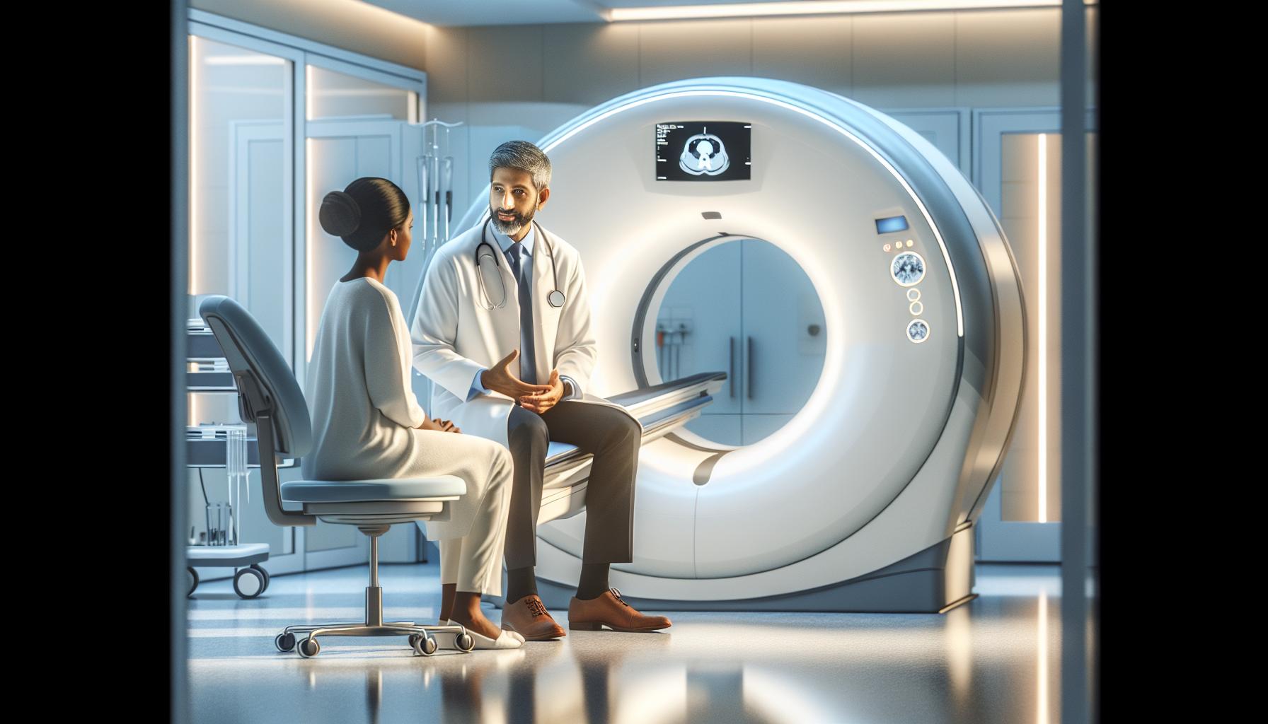
How Chest CT Scans Work: A Detailed Overview
A chest CT scan utilizes sophisticated imaging technology that can be crucial for assessing heart health and identifying potential blockages. This non-invasive procedure employs a series of X-ray images taken from multiple angles. The information is then processed by a computer to produce detailed cross-sectional images, known as slices, of the chest area. These images provide a comprehensive view of the heart, lungs, and surrounding structures, allowing for more accurate diagnoses than standard X-rays.
The advanced nature of CT scans allows healthcare providers to visualize the heart’s anatomy effectively, including the coronary arteries where blockages may form. The process often involves the use of contrast material, which enhances the visibility of the blood vessels. This dye is usually administered through an IV line, helping to distinguish blood flow and highlight areas where blockages might exist. Although CT scans are powerful tools for detecting issues such as coronary artery disease, it’s critical to recognize their limitations. For instance, small blockages or those in less accessible areas may be missed, necessitating further testing like coronary angiography for a more thorough investigation.
When preparing for a chest CT scan, patients should follow several key steps to ensure the best possible results. It’s typically advised to refrain from eating or drinking for a few hours before the scan, particularly if contrast dye is used. Informing your healthcare provider of any allergies, especially to iodine, is essential, as it may influence the use of contrast material. Understanding the entire process, from preparation to what happens during the scan and how results are interpreted, can alleviate anxiety and promote a positive experience during the procedure.
In addition to the technical aspects of the procedure, fostering open communication with healthcare professionals is vital. Discussing any concerns about the scan or its implications helps ensure clarity and support throughout the examination and follow-up stages. Remember, having a well-rounded understanding of how chest CT scans work can equip patients with the knowledge they need to make informed decisions about their heart health and further evaluations if necessary.
Common Reasons for Chest CT Scans
Chest CT scans serve a vital role in accurately diagnosing various cardiac and pulmonary conditions. Not only do they provide a detailed look at heart structures, but they can also assist in evaluating critical health issues that may arise. A chest CT scan is often recommended for patients exhibiting symptoms that may indicate heart problems or other complications. Common reasons for undergoing this imaging procedure include:
- Suspected Coronary Artery Disease: When patients present with chest pain, shortness of breath, or abnormal stress test results, a chest CT scan can help visualize the coronary arteries to assess for blockages or narrowing.
- Assessment of Heart Structures: Conditions such as cardiomyopathy or congenital heart defects may necessitate a chest CT scan to evaluate the heart’s anatomy and function more thoroughly than standard imaging might allow.
- Pulmonary Issues: Symptoms like persistent cough or unexplained weight loss could prompt a chest CT scan to look for lung diseases, which can indirectly affect heart health by impacting oxygen supply and overall cardiovascular function.
- Follow-up on Previous Diagnoses: For patients with existing cardiovascular problems or prior interventions, a CT scan can provide crucial follow-up information to ensure that treatments are effective or to adjust management plans accordingly.
Additionally, understanding why a chest CT scan is recommended can help demystify the procedure for patients. For instance, individuals with risk factors for heart disease, such as a family history, smoking, or high cholesterol, may benefit from early imaging to catch potential issues before they escalate. Ultimately, discussing any concerns or questions with a healthcare provider can enhance patient understanding and comfort with the process. Medical professionals can tailor recommendations based on individual health profiles and provide insights on how imaging might influence future management or treatment strategies.
Limitations of Chest CT Scans in Detecting Blockages
While chest CT scans are invaluable tools in detecting various conditions, they do have notable limitations when it comes to identifying heart blockages. The precision of chest CT imaging is influenced by several factors, and understanding these can help patients make informed decisions regarding their heart health.
One significant limitation of chest CT scans is motion artifacts. As patients breathe or experience anxiety during the scan, these movements can distort the images, making it challenging to obtain a clear view of the coronary arteries. For individuals who have irregular heart rhythms or difficulty remaining still, this can impact the scan’s effectiveness. Furthermore, calcified plaque in the arteries can complicate interpretation. While chest CT scans can identify calcium deposits, which suggest coronary artery disease, they are less effective in detecting non-calcified plaque that could lead to blockages.
The diagnostic accuracy of chest CT scans can also be affected by factors such as patient body habitus and the quality of the imaging equipment. Obesity can obstruct the clarity of imaging, while older CT machines might not provide the level of detail needed to detect all blockages accurately. Additionally, the presence of overlapping structures, like lung tissue, can further obscure the view of the heart and vessels.
When assessing heart blockages, it’s essential to consider that other imaging modalities, such as coronary angiography, may provide complementary or more definitive information. This method involves a direct visualization of the coronary arteries and is often referred to as the gold standard for diagnosing blockages. Engaging with healthcare providers about the most appropriate imaging strategy based on individual health circumstances can lead to better management of heart health.
Ultimately, while chest CT scans are powerful tools, understanding their limitations empowers patients to actively participate in discussions about their health and to seek additional insights from professionals when necessary. Always follow up with your healthcare team to discuss the results of any imaging and to explore the best next steps for your cardiovascular health.
Alternatives to Chest CT Scans for Heart Assessment
When it comes to assessing heart health, particularly in the context of potential blockages, there are several effective alternatives to chest CT scans. Each option offers unique benefits and can provide valuable insights depending on the individual’s circumstances. Here are some notable alternatives:
Coronary Angiography
One of the most definitive methods for visualizing coronary arteries is coronary angiography. This procedure involves injecting a contrast dye into the blood vessels to illuminate blockages or narrowing on X-ray images. It is often considered the gold standard for diagnosing coronary artery disease. If you have symptoms such as chest pain or shortness of breath, your doctor may recommend this test for a clear view of your heart’s blood flow.
Stress Testing
Stress testing is another alternative that can help assess heart function under physical stress. These tests can be performed with or without imaging, known as echocardiographic stress tests or nuclear stress tests. During the procedure, patients engage in exercise or are administered medication to simulate exercise effects on the heart. If you’re concerned about your heart health but want to avoid radiation exposure from imaging, discussing a stress test with your doctor could be beneficial.
Echocardiography
Echocardiograms use sound waves to create images of the heart and can assess its structure and function. This non-invasive test allows doctors to observe how well your heart is pumping and whether the valves are functioning properly. It is particularly useful for identifying issues related to heart walls, valves, and overall cardiac function without exposing the patient to radiation.
Cardiac MRI
Cardiac MRI (Magnetic Resonance Imaging) is another alternative that provides detailed images of the heart’s structures and blood vessels. It is particularly helpful in assessing areas of ischemia, which occurs when blood flow to the heart muscle is reduced due to blockages. This imaging method doesn’t involve radiation and can give comprehensive information about the heart’s condition, making it a valuable tool for evaluating heart disease.
While each alternative has its own advantages, the choice of the best imaging method should be made in consultation with healthcare professionals. They will consider your medical history, symptoms, and overall health to recommend the most appropriate assessment tool. Engaging in open conversations with your healthcare team can alleviate concerns and guide you to the right testing avenues for your heart health.
Understanding Heart Blockages: Symptoms and Risks
Heart blockages are a critical concern for many individuals, potentially leading to serious health issues such as heart attacks. It’s essential to recognize the symptoms and understand the associated risks. Symptoms of heart blockages can vary significantly; they may be subtle or even mistaken for less serious ailments. Common indicators include chest pain or discomfort (often described as pressure or aching), shortness of breath, fatigue during physical activity, and episodes of dizziness or light-headedness. Individuals might also experience heart palpitations or a sense of light-headedness, especially with exertion, which could signal that the heart is not receiving adequate blood flow due to a blockage.
It’s important to note that some individuals may have blockages without any symptoms, making regular check-ups essential, particularly for those with risk factors such as high blood pressure, diabetes, high cholesterol, or a family history of heart disease. The risk associated with blockages is significant; over time, untreated blockages can lead to ischemia, where oxygen-rich blood cannot reach the heart muscle, potentially resulting in tissue damage or a heart attack. If you’re experiencing symptoms, or if you fall into a higher risk category, consult with a healthcare professional promptly.
While symptoms serve as alert signs, understanding the underlying risks can empower individuals to take proactive steps. Leading a heart-healthy lifestyle, which includes regular exercise, a balanced diet, and managing stress levels, can significantly reduce the likelihood of developing heart blockages. Additionally, lifestyle changes can complement diagnostic imaging tests, such as chest CT scans, which are effective tools for identifying these blockages early. A timely diagnosis, paired with appropriate lifestyle adjustments or medical interventions, can help mitigate the risks associated with heart blockages and improve overall cardiovascular health.
Comparison of chest CT and coronary angiography
When evaluating heart health, two common imaging techniques emerge at the forefront: chest CT scans and coronary angiography. Each has its unique strengths and applications, which can significantly influence diagnosis and treatment. It’s crucial to understand how these methods work and when one may be preferred over the other.
Chest CT (computed tomography) scans create detailed cross-sectional images of the chest, providing a comprehensive view of the lungs, heart, and surrounding structures. These scans are particularly effective for identifying blockages in large coronary arteries and assessing the overall condition of the heart. A notable advantage of chest CT is its non-invasive nature; the procedure is swift and typically discomfort-free, allowing for rapid imaging. However, it is essential to note that while chest CT can reveal abnormalities, it may not always conclusively diagnose the extent of a blockage.
In contrast, coronary angiography, often considered the gold standard for evaluating coronary artery disease, involves threading a catheter through the blood vessels to inject a contrast dye directly into the coronary arteries. This method provides real-time feedback and detailed images of the blood flow within the heart’s arteries. It allows cardiologists to visualize the precise location and severity of any blockages. While this technique is highly accurate, it is more invasive, requiring a hospital stay and a longer recovery time due to the associated risks, such as bleeding and infection.
Both imaging techniques play critical roles in assessing heart conditions. For individuals exhibiting symptoms of heart disease, coronary angiography might be recommended for a definitive diagnosis and potential treatment plan. Meanwhile, healthcare providers might utilize chest CT scans as an initial investigation for those at risk or with less obvious symptoms to evaluate the need for further invasive procedures. Ultimately, the choice between these imaging modalities depends on individual patient circumstances, including risk factors, symptoms, and the specific information that needs to be gathered. Discussing options with a healthcare provider is essential for ensuring the most appropriate and effective approach to heart health.
Factors Affecting the Accuracy of Chest CT Scans
When it comes to understanding the effectiveness of chest CT scans, it’s vital to recognize several factors that can influence their accuracy in detecting heart blockages. Despite being a powerful imaging tool, various aspects can affect the reliability of the results, ultimately shaping the diagnostic journey.
Technical Parameters
One of the primary factors affecting accuracy is the quality of the imaging equipment. Modern CT scanners utilize advanced technology that can significantly enhance image clarity and detail. For instance, scanners with higher slice counts and improved algorithms can produce sharper images, aiding in the detection of minute abnormalities. Additionally, the timing of image acquisition is crucial; scans performed during optimal phases of the cardiac cycle can offer clearer views of blood flow and potential blockages.
Patient Factors
Patient-specific characteristics are equally significant. Factors such as body mass index (BMI), age, and heart rate can impact image quality. Obesity might require higher radiation doses for clear images, while rapid heart rates can blur images, making it challenging to identify blockages. Preparing for the scan by following pre-exam instructions-like fasting or avoiding certain medications-can help control these variables, ensuring the best possible outcomes.
Interpretative Skills
Moreover, the expertise of the radiologist interpreting the CT images plays a critical role. A skilled radiologist can differentiate between significant and insignificant findings, minimizing the chances of misinterpretation. Engaging with a healthcare provider who has experience in cardiac imaging can ensure that the most accurate assessments are made, underpinning the importance of communication in the diagnostic process.
In light of these factors, it’s clear that while chest CT scans are valuable in detecting heart blockages, several conditions must be optimized for accurate results. If concerns arise regarding heart health, consulting with a healthcare professional is essential for personalized advice and to determine the most appropriate imaging strategy tailored to individual health needs.
Preparing for a Chest CT Scan: What Patients Should Know
Preparing for a chest CT scan can feel daunting, but understanding what to expect and how to prepare can significantly ease any anxiety. This non-invasive imaging technique plays a crucial role in assessing heart health, particularly in detecting potential blockages. Knowing a few key facts can help you navigate the process with confidence, ensuring that you are well-prepared and informed.
Before your scan, you may receive specific instructions from your healthcare provider. It’s essential to follow these carefully to achieve the most accurate results. Typically, you might be advised to refrain from eating or drinking for several hours before the procedure. This fasting helps ensure that the images are as clear as possible and minimizes the risk of any complications. Additionally, if you’re on certain medications, your doctor may recommend temporarily discontinuing them. Always feel free to seek clarification on these guidelines, as your provider is there to ensure your safety and comfort.
On the day of your appointment, wearing comfortable clothing is advisable. You may need to change into a hospital gown, but avoiding clothing with metal fasteners-like zippers or hooks-will expedite the process and reduce any discomfort during scanning. It’s also a good idea to inform the staff about any allergies or existing medical conditions, including kidney issues, as these factors can impact the use of contrast materials, which are sometimes utilized to enhance imaging results.
Lastly, consider bringing a supportive companion with you. Having someone you trust can provide emotional comfort and assistance with any questions that may arise both before and after the scan. This support can be invaluable, particularly as you navigate any follow-up discussions regarding your results. Remember, your healthcare team is your best source of information, so don’t hesitate to engage with them about any concerns or inquiries you have; their empathy and expertise are your allies throughout this experience.
What to Expect During Your Chest CT Scan
During a chest CT scan, you’ll find that the experience is quite streamlined and designed to minimize any discomfort while maximizing diagnostic accuracy. These scans are swift, often taking just a few minutes, and understanding what happens during the procedure can alleviate pre-scan anxiety.
Once you arrive at the imaging center, a technician will greet you and ask a few questions regarding your medical history and current medications. This is a crucial step as it ensures the safety and effectiveness of your scan, particularly if contrast dye is used. The technician will guide you to the scanning room, where you’ll be asked to change into a gown that’s free from metal fasteners to avoid interference with the imaging process.
As you lie down on the scanning table, the CT machine will position itself around you. It’s important to remain still during the imaging to ensure clear results. You might need to hold your breath briefly-this common instruction helps capture clearer images of your heart and lungs. The scanner will make a series of whirring sounds as it takes pictures from various angles. You may feel a slight warmth if contrast dye is administered, but these sensations are temporary and typically harmless.
After the scan is complete, you can resume your normal activities almost immediately. The results will be evaluated by a radiologist, who will share the findings with your healthcare provider. This collaborative approach enables you to understand your heart health and any potential blockages effectively, paving the way for personalized follow-up discussions.
By familiarizing yourself with this process, you can approach your chest CT scan with confidence, knowing that each step is designed to support your health and well-being. Always remember to discuss any lingering questions or concerns with your healthcare team-they’re dedicated to guiding you through your medical journey.
Interpreting Chest CT Scan Results: Key Takeaways
Understanding the results of a chest CT scan can be key to managing your heart health. When you receive your reports, they will typically detail the presence of any abnormalities, including potential blockages. It’s essential to know that while a CT scan can offer valuable insights, not every blockage will be visible. This is particularly true for smaller blockages or those located in very small blood vessels, which may require further imaging techniques for confirmation.
When reviewing your results, look for terms such as “stenosis,” which indicates a narrowing of the arteries. This can be a sign of a blockage that might restrict blood flow. The report may also include descriptions of the size, shape, and location of any identified issues, helping your healthcare provider formulate an appropriate plan for further testing or treatment. It’s also helpful to remember that findings listed may not always point to a direct cause for symptoms; some individuals may have significant blockages yet experience few or no symptoms, a phenomenon known as silent ischemia.
If you have questions about your results, don’t hesitate to engage with your healthcare provider. They can explain how the findings relate to your overall heart health and discuss any additional tests or treatments that may be recommended based on your specific condition. Some key points to discuss might include:
- Next Steps: What follow-up tests might be needed?
- Treatment Options: Are medication or lifestyle changes needed to address any identified risks?
- Coping Strategies: What should you be mindful of in your daily life following these results?
Always keep in mind that your health journey is a collaborative process. Just as the imaging process aims to provide clarity about your heart health, understanding your results thoroughly allows you to take an active role in discussions about your care and decisions moving forward.
Consulting Your Doctor: Next Steps After CT Scan Results
Receiving your chest CT scan results can be a pivotal moment in understanding your heart health. If abnormalities are detected, the next steps are crucial for ensuring timely and appropriate care. Engaging openly with your healthcare provider can significantly impact how you manage any concerns that arise from your scan results.
It’s essential to prepare for your consultation by noting any specific questions or symptoms you’ve been experiencing. For instance, if the scan indicates potential blockages, ask your doctor about the nature and significance of these findings. They can clarify whether follow-up tests, such as echocardiograms or additional imaging studies, are necessary to get a clearer picture of your cardiovascular status.
When discussing treatment options, inquire about lifestyle changes or medications that may help mitigate risks associated with identified blockages. For example, ask about dietary adjustments, exercise recommendations, and whether specific medications may be beneficial. Understanding these aspects can empower you to take an active role in your health management.
Remember that care isn’t just about addressing immediate findings; it’s also about understanding your long-term health trajectory. Consider discussing coping strategies for maintaining heart health and managing stress. Your healthcare provider can guide you on how to stay informed and engaged with your treatment plan, ensuring you feel supported throughout your health journey.
FAQ
Q: Can a chest CT scan detect heart disease?
A: A chest CT scan can provide insights into heart health, but it is primarily designed for assessing lung conditions. While it may show signs of heart disease, such as calcified coronary arteries, dedicated cardiac imaging tests like coronary angiography are more effective for directly evaluating heart blockages.
Q: How accurate are chest CT scans for identifying blockages in coronary arteries?
A: Chest CT scans can be useful for detecting coronary artery disease but have limitations in accuracy. They might miss some blockages, especially if there is significant artery narrowing. For precise evaluation, consult your healthcare provider about more specialized tests like coronary angiography.
Q: Are there risks associated with a chest CT scan for heart assessment?
A: Yes, while chest CT scans are generally safe, they involve exposure to radiation, which can increase cancer risk over time. It’s advisable to discuss the potential risks and benefits with your doctor, especially if multiple scans are needed for continuous assessment.
Q: What preparation is needed before undergoing a chest CT scan?
A: Preparation for a chest CT scan typically includes avoiding food or drink for 4-6 hours before the procedure, depending on whether contrast material will be used. Always follow specific instructions from your healthcare provider to ensure accurate results.
Q: Can I see my heart’s condition on a chest CT scan report?
A: Yes, a chest CT scan report may include information on the condition of your heart, such as chamber sizes and heart artery status. However, for detailed analysis, it’s best to discuss findings with your healthcare provider who can explain the implications of the report.
Q: What should I expect after a chest CT scan regarding results?
A: After a chest CT scan, results are typically available within a few days. Your healthcare provider will review the images and discuss any findings, including potential heart health implications, and outline next steps based on your condition.
Q: How does a chest CT scan compare to other cardiac imaging tests?
A: A chest CT scan provides a quick overview of heart and lung structures, whereas tests like echocardiograms or angiograms offer detailed insights into heart function and blockages. Discuss with your provider which test is best suited for your specific health needs.
Q: Can a chest CT scan be used to monitor known heart conditions?
A: Yes, a chest CT scan can be used to monitor existing heart conditions by tracking changes over time. However, other imaging modalities may provide more detailed information about heart function and blood flow, so consult with your doctor on the best monitoring approach.
In Summary
Understanding whether a chest CT scan can reveal heart blockages is crucial for your health journey. While this imaging technique primarily focuses on the lungs and surrounding structures, it can also provide indirect insight into the heart, especially if there are associated complications. If you’re concerned about heart health, don’t wait-consult with your healthcare provider about scheduling a cardiac imaging evaluation tailored to your needs.
For further information, explore our detailed guides on CT scans and their applications in diagnosing lung issues, or learn about preparation steps for your upcoming scan. Stay informed about your health journey-sign up for our newsletter to receive updates on the latest in medical imaging and health tips. Remember, empowered patients make the best health decisions. If you have questions or need personalized advice, don’t hesitate to reach out to our team. Your path to clarity starts now!

