Understanding how to read a CT scan of the brain is crucial for patients eager to grasp their health conditions. With approximately 70 million CT scans performed annually in the U.S., this imaging technique is invaluable for diagnosing various neurological issues.
Navigating the intricacies of a CT scan can feel overwhelming, but being informed can empower you during your healthcare journey. Whether you’re anticipating a scan, awaiting results, or trying to comprehend a diagnosis, knowing how to interpret these images can significantly alleviate anxiety and grant you greater insight into your health.
This guide equips you with the knowledge to decode your CT scan results with confidence. Discover the key components of brain scans and learn what the images reveal about your condition. Understanding these critical details helps put you in control and fosters a deeper partnership with your medical team.
Understanding CT Scans: A Patient’s Overview
Understanding the technology behind CT scans can empower patients as they navigate their medical journeys. A CT scan, or computed tomography scan, is a powerful imaging tool that provides detailed images of the body, including the brain. Utilizing a series of X-ray images taken from different angles, a CT scan compiles these into cross-sectional views, allowing doctors to assess conditions that may not be visible through standard X-rays. This capability is particularly vital in diagnosing and evaluating brain injuries, tumors, and other neurological disorders.
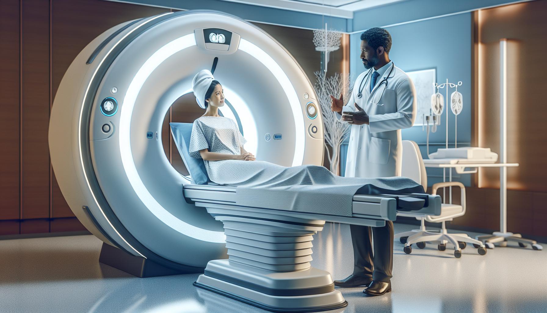
When preparing for a CT scan, it’s important to understand aspects of the procedure that may impact your experience. The process begins with the patient lying on a movable table that slides into a donut-shaped machine. The scan itself is swift, usually lasting only a few minutes, but it may require patients to remain still to ensure clear images. Understanding what occurs during this brief period can alleviate anxiety. For those with claustrophobia or difficulty staying still, discussing options with a healthcare provider can offer reassurance and strategies to cope.
Patients often wonder about the safety and risks associated with CT scans. While they are generally safe, concerns about ionizing radiation exposure are common. It’s crucial to discuss with your healthcare provider whether the benefits of the scan outweigh the risks. Furthermore, if contrast dye is required, understanding its role-enhancing the visibility of blood vessels and organs-can demystify part of the process and make the experience feel more manageable.
Empowerment through knowledge is key; hence, if questions arise about the results or subsequent steps, patients should not hesitate to ask their healthcare provider for clarity. Engaging actively in your healthcare can promote better understanding and ease apprehensions surrounding your CT scan experience.
How a CT Scan Works: Step-by-Step Explanation
During a CT scan, advanced imaging technology provides a detailed look at the body, especially the brain. The process begins with the patient positioned on a movable examination table, which is gently guided into a large, donut-shaped scanner. This machine emits X-rays, which are invisible beams of radiation. Unlike traditional X-rays that provide only two-dimensional images, a CT scanner captures multiple images from various angles, allowing it to create three-dimensional cross-sectional views of the brain’s structures.
The imaging process typically lasts only a few minutes, but it’s crucial for patients to remain still during this time to ensure the clarity and accuracy of the images. Prior to the scan, patients may be asked to remove any metal objects, such as jewelry or glasses, that could interfere with the imaging. They may also receive specific instructions related to diet or medications, especially if a contrast dye is to be used to enhance image quality. This contrast dye, which helps delineate blood vessels and tissues, is administered either orally or through an intravenous line, contributing to more precise examination results.
Throughout the procedure, the technician will monitor the patient from a separate room using a video screen. Patients can communicate with the technician via an intercom. This supportive setup is designed to keep the patient informed and comfortable, addressing any concerns about the scanning process.
As the scan completes, images are processed and interpreted by a radiologist, who will analyze the resulting data for any abnormalities or concerns. Understanding how a CT scan operates-from the patient’s experience lying on the table to the advanced imaging technology at play-can help alleviate anxiety and empower patients to engage in their healthcare discussions with their providers more confidently.
Preparing for Your CT Scan: Essential Tips
Undergoing a CT scan can feel daunting, especially when it involves the intricacies of your brain. Being well-prepared is essential not only for the efficacy of the scan but also for your peace of mind. A bit of foreknowledge can turn a potentially stressful experience into a smoother, more manageable one. Here are some key tips to facilitate a seamless preparation process.
Follow Instructions Carefully
One of the most critical aspects of preparing for your CT scan is adhering to the specific instructions provided by your healthcare provider. These can include guidelines on fasting before the procedure, particularly if contrast dye is to be used. If instructed to avoid food or drink, ensure you follow these recommendations closely, as they can significantly affect the quality of your images.
Dress Comfortably
Wear loose, comfortable clothing without metal fasteners. It’s advisable to opt for something simple like a T-shirt and sweatpants. Avoiding clothes with zippers, belts, or buttons is crucial since these metal items could interfere with the imaging. You may need to change into a hospital gown, but starting with suitable attire can make the transition easier.
Discuss Medication and Health Conditions
Before your scan, have a candid conversation with your healthcare provider about any medications you are taking, allergies, or pre-existing conditions. These factors may influence your scan procedure or necessitate special preparations, especially concerning the use of contrast dyes. For example, patients with renal issues might require a different approach when using certain types of contrast agents.
Plan for Comfort and Support
It’s perfectly normal to feel anxious about medical procedures. Consider bringing a family member or friend along for emotional support. Additionally, familiarize yourself with the scanning process to alleviate fear of the unknown. Don’t hesitate to ask your healthcare team about what to expect, and remember, they are there to help guide you every step of the way.
By taking these proactive steps to prepare for your CT scan, you can enhance your comfort and contribute to the overall success of the procedure. Understanding the significance of proper preparation will empower you to approach your scan with greater confidence and assurance. Always feel free to reach out to your healthcare provider with any questions or concerns you may have before the appointment.
What to Expect During a CT Brain Scan
Undergoing a CT brain scan can be a pivotal step in diagnosing various neurological conditions, but knowing what to expect can alleviate many concerns. Once you arrive at the imaging center, a technologist will guide you through the preparation for your scan. The process is typically smooth, with the entire procedure taking about 15 to 30 minutes, depending on the specifics of your examination.
You will lie on a padded table that slides into a large, doughnut-shaped machine. It’s important to remain as still as possible during the scan, as any movement can obscure the images. You may hear a whirring sound and feel slight vibrations during the scan, which are perfectly normal. If a contrast dye is needed to enhance the images, it will be administered either via an intravenous line or through an injection. This may cause a warm sensation or a metallic taste in your mouth, but these effects are temporary and generally harmless.
After the procedure, you can usually resume your normal activities unless your doctor advises otherwise, especially if contrast dye was used. The images obtained will be reviewed by a radiologist, who will provide a report to your healthcare provider. They will discuss the results with you to interpret findings and recommend any next steps if necessary. Always remember that you can ask questions throughout the process to enhance your understanding and comfort.
Interpreting CT Scan Images: A Patient Guide
Interpreting CT scan images can initially feel daunting, yet understanding the basics can empower you to engage more actively in your healthcare. A CT scan, or computed tomography scan, produces cross-sectional images of the brain, allowing for a detailed view of structures and potential abnormalities. These images are generated using a combination of X-rays and computer technology, creating precise and intricate pictures of the brain that can detect conditions ranging from tumors to brain injuries.
Once the scans are completed, a radiologist analyzes the images, looking for any unusual signs. Typically, normal brain tissue will appear lighter in color than denser structures like bones or fluid-filled spaces. Areas of concern might show up as darker regions, indicating potential issues such as swelling or mass. It’s important to remember that while the radiologist is trained to identify these conditions, they will provide their findings to your physician, who will then communicate the results to you in a way that is clear and understandable.
To better understand your CT images, consider the following:
- Know the Appearance of Normal Structures: Familiarize yourself with what healthy brain tissue should look like, which can help you feel more comfortable discussing your results.
- Ask Questions: Don’t hesitate to ask your doctor to explain what the images show. They can give context and meaning to areas of concern, as well as address any uncertainties.
- Utilize Visual Resources: Many healthcare facilities offer resources such as diagrams or educational videos explaining CT scans and typical findings, which can enhance your understanding.
Your participation in understanding your CT scan results not only demystifies the process but also helps ease anxiety about potential diagnoses. Each individual’s case is unique, so working closely with your healthcare team allows for personalized discussions about what the images indicate and the next steps based on the findings.
Common Findings in Brain CT Scans
When looking at CT scans of the brain, it’s important to understand some of the most common findings that radiologists may identify. Recognizing these can help demystify the process and allow you to engage more meaningfully with your healthcare team. A CT scan employs advanced imaging techniques to reveal a variety of conditions, potentially affecting your brain’s structure and function.
One of the most frequently encountered findings is acute hemorrhage, often resulting from head trauma, stroke, or aneurysm rupture. This appears as areas of high density on the images, which can signify bleeding inside the brain or surrounding spaces. In contrast, ischemic strokes, which occur when there’s a blockage restricting blood flow, can present as darker regions indicating areas where brain tissue has been deprived of oxygen.
Another significant category of findings includes tumors, which may manifest as well-defined masses that disrupt the normal architecture of the brain. These lesions can vary in appearance based on their type-benign tumors might have smooth edges, while malignant ones may show irregular borders and surrounding edema. Also common in assessments are ventricular enlargement and cerebral atrophy, often indicative of conditions like hydrocephalus or dementia, respectively. These findings may appear as larger fluid-filled spaces or a general shrinkage of brain tissue, prompting further evaluation and potential intervention.
Recognizing the Importance of Unusual Findings
It’s also essential to be aware that findings such as cysts, calcifications, or chronic changes resulting from aging can often be benign but warrant monitoring. Radiologists use established criteria for evaluating these conditions; however, the interpretation can sometimes vary based on individual cases. Patients should feel empowered to ask their doctor for clarity on any findings, ensuring they understand what these mean for their health. Creating a space for open dialogue can alleviate anxiety and foster a collaborative environment in managing potential health issues.
Using this knowledge, you can approach your CT scan results with a more informed perspective, helping you engage effectively with your medical team while reducing any anxiety that might be associated with the findings.
Potential Risks and Safety Measures of CT Scans
While CT scans are invaluable tools that help diagnose a range of medical conditions, it’s essential to be aware of the potential risks and safety measures associated with them. One of the primary concerns with CT scans is exposure to radiation. Although the amount of radiation used in a typical brain CT scan is generally considered low, repeated exposure over time can increase the risk of developing cancer. The key is to weigh the benefits of obtaining critical diagnostic information against any potential risks. If you have had multiple scans or if a scan is necessary for children, discuss these concerns with your doctor to determine the appropriate need for the procedure.
Preparation can also play a crucial role in minimizing risks. Patients are typically advised to inform their healthcare provider about any pre-existing conditions, allergies, or medications they are taking, particularly those that may affect kidney function or interact with contrast dyes if they will be used during the scan. In certain cases, healthcare professionals may opt out of using contrast dye or may suggest alternatives if there are significant concerns. Fasting for a specified period before the procedure can also be necessary, especially if contrast will be used, to reduce the risk of complications.
Another important consideration is the psychological aspect of undergoing a CT scan. Many patients experience anxiety about the procedure itself or about the potential findings. It is vital to communicate any fears or concerns with your healthcare provider, who can provide reassurance and address specific questions. Knowing what to expect can greatly alleviate anxiety; for instance, the procedure is generally quick, and you will be lying still for only a few minutes while the scans are taken.
Finally, following the scan, your physician will discuss the results with you, which can help clarify any uncertainties and reduce stress regarding the outcomes. Remember, effective communication with your healthcare team is not just beneficial for your understanding but also for tailoring your care plan according to your unique health needs.
Understanding Contrast Dye in CT Scans
During a CT scan, contrast dye often plays a crucial role in enhancing the clarity of the images taken, providing your doctor with better visuals to diagnose conditions accurately. This specialized dye, which may be administered via injection or through oral consumption, helps in highlighting specific areas inside the body-especially the blood vessels, organs, and soft tissues-making it easier to identify abnormalities such as tumors or inflammation.
When preparing for a CT scan involving contrast dye, it’s essential to disclose any allergies, particularly to iodine, as many contrast agents contain this element. If you have a known allergy, your healthcare provider may recommend alternative imaging routes or take precautionary measures. Additionally, informing your doctor about any existing health issues-especially kidney problems-is important because contrast materials can sometimes affect kidney function, particularly in those who are already at risk.
How Contrast Dye Works
The contrast dye enhances the visibility of certain systems in the body through the following mechanisms:
- Absorption: The dye absorbs X-rays differently than surrounding tissues, allowing for improved contrast in the images. This differentiation can reveal structures and conditions that would otherwise remain hidden.
- Distribution: When injected, the dye circulates through the bloodstream, providing a real-time view of how blood flows through different areas, which can help in assessing vascular conditions.
What to Expect
Before the scan, you’ll typically receive instructions, including whether to fast or stay hydrated, depending on the type of contrast used. Post-procedure, it’s common to experience mild side effects, such as a warming sensation or metallic taste, which are generally not a cause for concern. However, if you experience severe reactions, such as trouble breathing or swelling, it’s essential to seek immediate medical attention.
In summary, while the prospect of receiving contrast dye may feel daunting, understanding its purpose and following preparation guidelines can significantly enhance the accuracy of your CT scan and ultimately support your healthcare provider in ensuring your best health outcomes. Always feel free to discuss any concerns or questions about contrast dye with your medical team; they can provide personalized advice tailored to your situation.
Decoding Your CT Scan Results: What They Mean
Understanding your CT scan results can often feel overwhelming, but it’s a crucial step in taking charge of your health. CT scans of the brain provide detailed images that can reveal various conditions, from tumors to hemorrhages, and understanding these images helps you engage meaningfully with your healthcare provider. Each scan is unique, and radiologists will assess multiple aspects, including the structure and density of the brain tissue, to interpret the findings accurately.
The report generated by your CT scan will highlight key observations that your doctor will discuss with you, often categorizing findings as normal, abnormal, or requiring further investigation. Here are some common terms and what they may indicate:
- Hyperdense Areas: These may suggest the presence of blood, such as in a hemorrhage, or calcifications related to aging or certain conditions.
- Hypodense Areas: These darker regions could indicate swelling, edema, or lesions that require further examination.
- Midline Shift: This refers to a displacement of brain structures, often indicative of increased intracranial pressure, which is a serious condition.
While your report may include professional terminology, remember that your healthcare provider is there to help translate these findings into actionable insights. It’s perfectly normal to have questions about what specific findings mean for your health and what the next steps may be. Knowledge is empowering-asking for clarification on terms or abnormal findings can foster a more collaborative relationship with your medical team.
Engaging in this dialogue is essential, as many conditions discovered through CT scans can be managed effectively with the right interventions. Depending on the findings, follow-up imaging, medication, or referrals to specialists may be recommended. Always advocate for yourself by seeking answers about your health; understanding your CT scan results is a vital part of your healthcare journey.
When to Follow Up: Next Steps After Your Scan
Receiving your CT scan results can feel like a momentous occasion, filled with both anticipation and uncertainty. After all the imaging and evaluations, understanding the next steps can help ease your mind and allow you to plan for what lies ahead. Following up after a CT scan typically hinges on two primary factors: the findings reported and your healthcare provider’s recommendations.
If the scan reveals normal results or findings that do not require immediate action, your doctor might suggest continuing with routine healthcare practices, including regular check-ups. However, if any abnormal findings are detected, you may be advised to schedule follow-up appointments to discuss these results in detail. This discussion is crucial in determining whether additional imaging, such as an MRI or another CT scan, is necessary, or if other types of evaluations like blood tests or referrals to specialists are warranted.
Key Points to Consider for Follow-Up
- Ask Questions: Don’t hesitate to reach out to your healthcare provider for clarification about any findings or terminology you don’t understand. It’s your health, and seeking to understand it better empowers you.
- Schedule Appointments Promptly: If your doctor recommends further tests or observational visits, try to schedule these as soon as possible to maintain continuity in your healthcare and monitoring your condition.
- Keep a Record: Maintain a folder with copies of your reports and test results. This will help you and your healthcare team make informed decisions as you move forward.
- Monitor Symptoms: Be vigilant about any new or worsening symptoms following your CT scan. If you notice anything concerning, contact your healthcare provider without delay.
Following these guidelines will not only assist you in navigational next steps but also ensure that your journey toward understanding and managing your health remains smooth and supportive. You hold an integral role in your healthcare journey, and open communication with your medical team is the foundation for effective care. Remember, the path to clarity often involves more discussions and decisions, and taking these proactive steps contributes to achieving the best possible health outcomes.
Cost of CT Scans: What to Know Beforehand
The financial aspect of undergoing a CT scan can be a significant concern for many patients, but understanding the costs involved can help alleviate some anxiety surrounding the procedure. On average, a CT scan can cost between $300 to $3,000, depending on various factors such as the location of the scan, the facility’s pricing policies, and whether contrast dye is used during the procedure. It’s essential to clarify the expected costs with your healthcare provider or the imaging center ahead of time, so there are no unexpected bills later.
Insurance coverage plays a crucial role in determining out-of-pocket expenses for a CT scan. Many insurance plans cover part, if not all, of the cost, especially when the scan is deemed medically necessary. It’s advisable to verify your specific coverage details beforehand. In some cases, facilities may offer discounted rates or payment plans for patients without insurance or those facing high deductibles, which can ease the financial burden.
Key Considerations Regarding CT Scan Costs
- Discuss Costs Upfront: Before scheduling your scan, inquire about the estimated cost, and ask if the facility offers any payment options.
- Insurance Verification: Contact your insurer to confirm coverage for the scan, and understand any co-pays or deductibles you might incur.
- Inquire About Additional Fees: Be aware that costs can improve if follow-up procedures, such as additional imaging or consultations, are required.
- Stay Informed: Research local imaging centers, as prices can vary significantly from one facility to another.
Planning for the financial aspects of your CT scan doesn’t have to be overwhelming. With clear communication with your healthcare providers and insurance company, you can navigate the costs confidently, allowing you to focus on your health and the outcomes of your scan. Remember, your well-being is the priority, and understanding the financial commitment helps in achieving that peace of mind.
Frequently Asked Questions About Brain CT Scans
Patients often have a multitude of questions when it comes to brain CT scans, especially when facing potential health concerns. One of the most common queries revolves around the purpose of the scan. Simply put, a brain CT scan is a diagnostic tool that uses X-ray technology to create detailed images of your brain. This imaging helps doctors identify conditions such as tumors, bleeding, or abnormalities that could be causing symptoms like headaches, confusion, or weakness.
Another frequent concern is about the safety and side effects associated with CT scans. While CT scans do involve exposure to radiation, the amount is considered low and is often outweighed by the diagnostic benefits. Health professionals take necessary precautions to minimize risks, ensuring that scans are performed at the lowest effective dose. It’s important to discuss any potential allergies or previous reactions to contrast dye with your technician prior to the scan, as this can influence the procedure.
Patients also inquire about the scan duration and discomfort level. A typical brain CT scan is quick, usually lasting about 10 to 30 minutes, and is painless. Most patients find the experience to be quite manageable; they simply need to lie still on the scanning table, and the machine does the rest. If you’re anxious about being in the enclosed space of the scanner, let the staff know; they can provide reassurance and support.
After your CT scan, understanding the results can be another source of confusion for patients. Your healthcare provider will interpret the images and explain what they mean. It’s crucial not to jump to conclusions based on the images alone; always consult your doctor for a comprehensive understanding of your results and any necessary next steps. This dialogue is vital in making informed decisions about your health.
Keep in mind, it’s natural to feel apprehensive about medical procedures. By gathering information and having open conversations with your healthcare team, you can navigate the process more comfortably and confidently.
Q&A
Q: How can I prepare for my brain CT scan?
A: To prepare for your brain CT scan, follow your doctor’s instructions regarding fasting, especially if contrast dye will be used. Discuss any medications you’re taking, and inform the technician if you’re pregnant or have kidney issues. Comfort is key, so wear loose clothing and avoid accessories.
Q: What are the typical findings on a brain CT scan?
A: Typical findings on a brain CT scan include hemorrhages, tumors, cysts, and signs of stroke. The scan can also detect abnormalities in structure and swelling. For detailed information about common findings, check the “Common Findings in Brain CT Scans” section of our guide.
Q: How long does it take to get CT scan results?
A: CT scan results are usually available within a few days to a week, depending on facility protocols and how busy the radiologist is. For immediate interpretation, some facilities may provide preliminary results within hours. Always consult your healthcare provider for final results.
Q: What should I do if I feel anxious about my upcoming CT scan?
A: If you’re feeling anxious about your CT scan, consider discussing your concerns with your healthcare provider who can provide reassurance and might suggest relaxation techniques. Bringing a friend or family member for support can also help ease anxiety.
Q: Are there any risks associated with brain CT scans?
A: Yes, risks include exposure to radiation and allergic reactions to contrast dye. However, the risk is generally low. Inform your medical team about any allergies or previous reactions to contrast materials. Review the “Potential Risks and Safety Measures of CT Scans” section for more details.
Q: What does it mean if my CT scan includes contrast dye?
A: Contrast dye enhances the visibility of certain areas in the brain, helping to highlight abnormalities. It allows radiologists to differentiate structures more clearly. For more information about its use, visit “Understanding Contrast Dye in CT Scans” in the patient guide.
Q: Can I drive after having a CT scan?
A: Yes, you can typically drive after a CT scan unless sedative medication was used. If contrast dye is administered, you might feel slightly disoriented; it’s best to have someone accompany you. Always follow your doctor’s specific advice regarding post-scan activities.
Q: How is a CT scan different from an MRI for brain imaging?
A: A CT scan uses X-rays to create images and is faster, making it ideal for emergencies. An MRI, on the other hand, uses magnetic fields and provides more detailed images of soft tissue but takes longer. Both have unique benefits depending on the clinical situation. For a deeper comparison, refer to the sections on imaging in our guide.
In Conclusion
Understanding how to read a CT scan of the brain can significantly enhance your confidence in discussing your health with healthcare professionals. By grasping the fundamental aspects of brain imaging, you empower yourself with knowledge that aids in making informed decisions regarding your health care. Don’t hesitate to explore our detailed resources on CT scan safety and preparation tips for scans to ensure you are well-equipped for your next appointment.
For continuous support and information, consider signing up for our newsletter, where you’ll get the latest updates on medical imaging and health tips. If you have questions or need personalized guidance, please consult your healthcare provider-they’re your best resource. Remember, your health journey is important, and we’re here to help you navigate it. Share your thoughts in the comments below and stay engaged for more insights!


