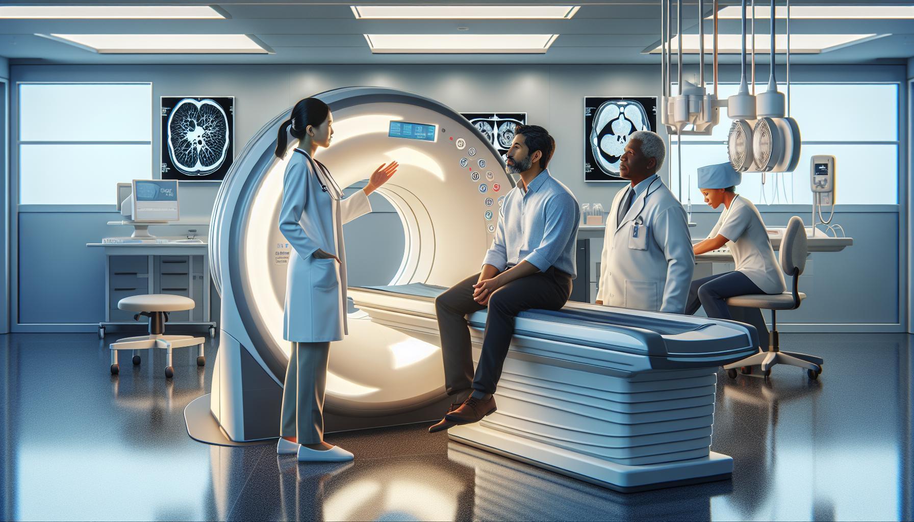
Understanding how to read a CT thorax scan can empower patients and improve outcomes in diagnosing and managing respiratory conditions. A CT scan provides detailed images of the chest, offering crucial insights into lung health, heart conditions, and potential abnormalities. With the increasing reliance on advanced imaging in today’s healthcare landscape, familiarizing yourself with the basics of these scans is both beneficial and necessary.
Many patients feel anxious about interpreting their medical imaging results, unsure of what the images reveal. By demystifying the process and highlighting key features to look for within a CT thorax scan, this guide will not only aid in your understanding but also alleviate concerns about what your healthcare provider might find. Engaging with this knowledge is the first step in taking charge of your health, and we invite you to explore further to uncover the essential details that can lead to more informed discussions with your medical team.
Understanding the Basics of CT Thorax Scans
A CT thorax scan, or chest CT scan, is a powerful diagnostic tool that allows medical professionals to visualize the internal structures of your chest in great detail. This imaging technique uses computer-processed combinations of many x-ray measurements taken from different angles to produce cross-sectional images of specific areas within the body. The images created resemble a series of slices, providing a comprehensive view of the lungs, heart, blood vessels, airways, and other chest components.
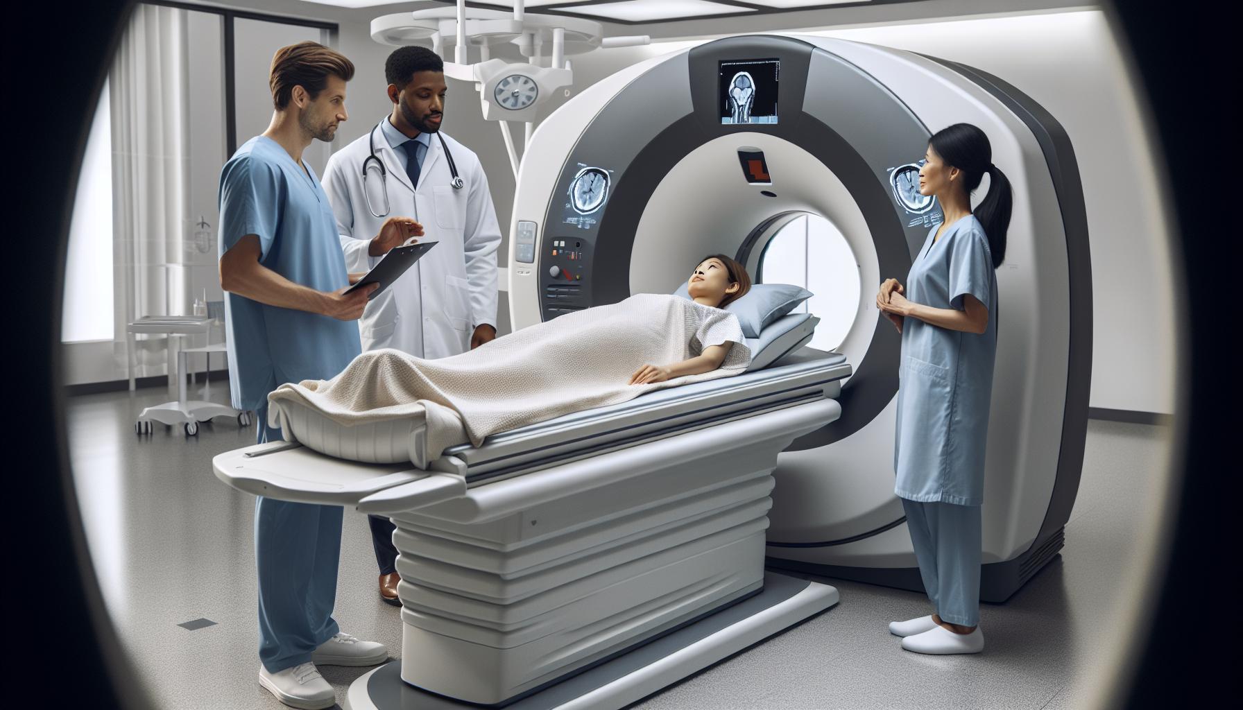
Understanding how a CT thorax scan works can significantly alleviate pre-scan anxiety. During the procedure, you will lie on a table that slides into a large, donut-shaped machine. The machine rotates around you, capturing images from multiple angles. A contrast dye may be administered through an intravenous line to enhance the visibility of certain areas. This process enables the detection of abnormalities such as tumors, cysts, or signs of diseases like pneumonia or pulmonary embolism.
In addition to its diagnostic capabilities, a CT scan is distinguished from traditional x-rays by its ability to generate three-dimensional images, allowing for a more thorough examination of complex structures. It’s important to remember that seeing images from a CT scan is only part of the equation; interpreting these images requires professional expertise. As you navigate this process, engaging with your healthcare provider can clarify what the images reveal about your health and ensure you receive the most appropriate treatment based on the findings.
Key Anatomy: What to Look for in a Chest Scan
A CT thorax scan can reveal critical insights into your chest anatomy, making it a vital tool for diagnosing various health conditions. Understanding what to look for in these scans can be empowering and alleviate some anxiety surrounding the procedure. During a scan, radiologists focus on several key anatomical structures to identify potential issues.
One of the primary areas of interest in a chest scan is the lungs. The technician assesses the lung tissue for signs of infections, nodules, or tumors. It’s crucial to note the distribution of any abnormalities and the presence of fluid, which may indicate conditions like pneumonia or pleural effusion.
Another vital structure is the heart. Scans often evaluate the size and shape of the heart chambers, identifying any enlargement or signs of heart disease. The coronary arteries, which supply blood to the heart, may also be visible, helping in the assessment for blockages or other coronary artery conditions.
When looking at the blood vessels, radiologists will examine the aorta and pulmonary arteries. They assess for abnormalities such as aneurysms or embolisms, which can be life-threatening if not detected promptly. Additionally, health professionals will analyze the mediastinum, the area between the lungs that contains the heart, major blood vessels, and other vital structures, ensuring no enlarged lymph nodes or masses are present.
In summary, recognizing these key anatomical features during a CT thorax scan can help you better understand your health. If you have specific concerns or questions regarding the scan results, discussing them with your healthcare provider will offer the most accurate insights and guidance tailored to your unique situation.
Common Conditions Diagnosed by CT Thorax Imaging
A CT thorax scan is instrumental in diagnosing a variety of conditions that affect the chest area, providing detailed images that allow healthcare professionals to see inside the body with remarkable clarity. These scans are particularly effective in identifying issues related to the lungs, heart, blood vessels, and other structures in the chest. Understanding the common conditions diagnosed through CT imaging can empower patients and enhance their engagement in the diagnostic process.
One of the most common conditions detected is lung cancer, where the scan reveals abnormal masses or nodules in the lung tissues. Early detection through imaging can lead to timely intervention, which is crucial for improving outcomes. Additionally, pneumonia can be identified as areas of consolidation or fluid accumulation in the lungs, giving physicians insight into the severity of the infection and guiding treatment decisions.
Another significant finding in a CT thorax scan may be pulmonary embolism, a potentially life-threatening condition where blood clots block the pulmonary arteries. The scan can provide clear images of these clots, allowing for immediate and effective treatment. Similarly, the scan can reveal signs of chronic obstructive pulmonary disease (COPD), including emphysema and chronic bronchitis, by showing structural changes in the lungs and airways.
Alongside these conditions, the scan also facilitates the detection of heart-related issues, such as enlargement of the heart chambers or signs of coronary artery disease. Aortic aneurysms, which can rupture and pose serious risks, can also be spotted through careful examination of the aorta.
By understanding what a CT thorax scan can detect, patients can better appreciate the importance of the procedure and its role in diagnosing significant health issues. Always consult with healthcare providers for tailored advice and to address any concerns regarding the scan’s findings or your overall health.
Step-by-Step: Preparing for Your CT Thorax Scan
Before undergoing a CT thorax scan, preparation plays a crucial role in ensuring that the procedure is effective and that the resulting images are of high quality. A well-prepared patient can help facilitate a smoother process and accompany a more accurate diagnosis. Here are some essential steps and tips to consider as you prepare for your CT thorax scan.
First and foremost, it’s important to follow any specific instructions provided by your healthcare provider or the imaging center. Generally, you may be advised to avoid eating or drinking for a few hours prior to the scan, especially if a contrast dye is going to be used. This helps minimize any potential for nausea and ensures that the contrast material can work effectively. If you’re prescribed medications, confirm with your doctor whether you should take them as normal.
When it comes to clothing, wear comfortable, loose-fitting garments and avoid wearing any metal accessories such as jewelry, hairpins, or belts, as these can interfere with the imaging process. You may be asked to change into a hospital gown to ensure that the scanning area is clear of obstruction.
If you have a history of allergies, particularly to iodine or contrast dyes, inform the radiology team prior to the scan. This will allow them to take appropriate precautions or consider alternative imaging methods if necessary. It’s also beneficial to create a list of your current medications and any existing health conditions to share with the radiologist, as this information can aid in interpreting your results accurately.
Lastly, consider bringing a family member or friend along for support. The presence of a loved one can help alleviate anxiety about the procedure, making the experience more comfortable for you. By being well-prepared and informed, you can approach your CT thorax scan with greater confidence and peace of mind. Always remember that your healthcare team is there to answer any questions or address concerns you may have before the procedure.
What to Expect During a CT Thorax Procedure
During a CT thorax procedure, patient comfort and clarity are paramount. When you arrive at the imaging center, you’ll be greeted by a team that will guide you through the entire process. One of the first steps is usually a brief consultation where you can discuss any concerns, and the radiologic technologist will ensure you’re aware of what to expect.
Once ready, you’ll be asked to change into a hospital gown to allow for a clear scan without any obstruction. You’ll then lie down on the CT scanner bed, which is equipped with a cushion to maximize your comfort. As the bed slides into the CT machine, you might hear a soft whirring noise as the scanner rotates around you, capturing high-resolution images of your thoracic region. It’s important to remain still during the scanning process for the best image quality, but the radiologic staff will provide clear instructions and reassurance throughout.
Before the scan begins, you might be asked to hold your breath for a few seconds at certain points. This is a standard procedure that helps eliminate any movement during the imaging, ensuring clearer, more defined results. Depending on the purpose of the scan, a contrast dye may be administered either orally or through an IV. While some patients may feel a slight warmth or metallic taste, this is typically temporary.
The entire CT thorax scan usually takes around 10 to 30 minutes, depending on the complexity and any additional imaging required. After the procedure, you’ll be directed to wait briefly while the technologist checks that the images are satisfactory. Once completed, you can usually resume normal activities unless otherwise advised. Knowing what to anticipate can ease anxiety and enhance your overall experience during this essential diagnostic procedure.
Decoding CT Thorax Images: A Guide for Patients
Interpreting a CT thorax scan can initially seem daunting, but understanding some fundamentals can greatly enhance your confidence and ease your concerns about the process. These scans produce detailed cross-sectional images of your chest, capturing the anatomy of structures such as the lungs, heart, and blood vessels. This high level of detail allows healthcare professionals to spot various conditions and make informed decisions about your health.
When viewing a CT thorax image, it’s important to understand what to look for. Radiologists analyze different densities and contrasts within the images, as various tissues absorb x-rays differently. For example, healthy lung tissue appears darker compared to any abnormal fluid accumulation or solid masses, which would appear brighter. Here are some key elements to consider:
- Lungs: Look for symmetric lung fields. The absence of nodules, masses, or areas of consolidation is ideal.
- Heart: The heart should appear in the center of the chest, with normal size and shape, without significant enlargement or unusual contours.
- Blood Vessels: The aorta and other vessels should have clear outlines, with no signs of blockages or abnormalities.
- Mediastinum: This central compartment of the thoracic cavity should show no unusual swelling or masses.
While patients may be curious about their scans, it’s essential to remember that only a qualified radiologist can accurately interpret these images. They will consider your medical history, symptoms, and the reason for the scan alongside the imaging results to provide a comprehensive analysis. During your follow-up consultation, if any findings are observed, your healthcare provider will discuss what they mean in your specific context and recommend any necessary next steps.
Overall, while understanding the basics of CT thorax images can empower you, always seek professional guidance for a precise and personalized interpretation of your results. This collaboration with your healthcare team can help calm any fears and facilitate informed decision-making regarding your health journey.
Interpreting Results: What CT Findings Mean
Interpreting a CT thorax scan is crucial for understanding your respiratory health and overall chest anatomy. The findings from a CT scan can reveal a lot about the structures it captures – from the lungs and heart to the blood vessels and surrounding tissues. Knowing how to interpret these findings can empower you, providing a greater sense of involvement in your healthcare journey.
When reviewing your CT results, radiologists focus on several key aspects. For instance, they look at the density of lung tissue. Healthy lung areas appear darker on the scan, signifying normal air-filled spaces. Any unusual bright spots might indicate the presence of fluid, infections, or tumors. Additionally, the appearance of the heart is carefully assessed; it should have a normal size and be centrally located. Enlarged or irregularly shaped hearts may signal underlying issues such as cardiomyopathy or heart disease.
It’s also essential to examine the blood vessels. Any blockages or irregularities in the aorta or pulmonary arteries can significantly impact blood flow and overall health. The mediastinum, the space between your lungs, should be clear of any abnormal masses or swellings. Radiologists look for signs that could suggest conditions such as lymphadenopathy, which can indicate infections or malignancies. Keep in mind, however, that while understanding these factors is beneficial, the interpretation of your results must be left to a qualified radiologist who will integrate your medical history and symptoms into a personalized assessment.
Ultimately, your healthcare provider will discuss any significant findings during a follow-up appointment, addressing your concerns and outlining potential next steps. This collaborative approach ensures that you receive informed care tailored to your specific situation, transforming potential anxiety into actionable plans for better health.
Comparative Techniques: CT vs. Other Imaging Methods
Modern medical imaging offers various techniques, each with unique advantages and limitations in diagnosing thoracic conditions. While CT scans have become a cornerstone in evaluating chest-related issues, it’s beneficial to understand how they compare with other imaging modalities, such as X-rays, MRIs, and ultrasound.
CT scans provide detailed cross-sectional images of the body, particularly the lungs, heart, and surrounding structures. They excel in revealing intricate details that standard X-rays may miss, making them particularly useful for detecting conditions like pulmonary embolism, tumors, or complex vascular anomalies. In contrast, traditional X-rays remain valuable for initial assessments, often serving as a first-line tool for identifying gross abnormalities, such as fractures or significant lung infiltrates. However, they do not offer the same depth of detail as CT imaging.
Another imaging method to consider is magnetic resonance imaging (MRI), which uses powerful magnets and radio waves to create images of internal structures. While MRIs are exceptional for soft tissue contrast and assessing heart conditions, their application in chest imaging is limited compared to CT scans. This limitation stems from longer scan times and the inability of MRI to capture rapid changes like those that occur in breathing. Therefore, CT is often preferred for thoracic imaging, particularly in emergencies.
Ultrasound, on the other hand, uses sound waves to visualize soft tissues and fluid collections in the thoracic cavity. This method is especially beneficial for guiding procedures or evaluating conditions like pleural effusions. While it provides real-time imaging, it is less effective in visualizing fine details within the lung parenchyma or the structures behind the rib cage when compared to CT.
In conclusion, each imaging modality plays a crucial role in patient care. CT scans often stand out for their comprehensive views and depth of detail, making them indispensable for definitive thoracic evaluations. However, patients should always consult their healthcare professionals to determine the most appropriate imaging technique based on their specific symptoms and medical history, ensuring tailored and effective treatment plans.
Safety Measures: Risks and Precautions in CT Scans
Undergoing a CT scan can feel daunting, but understanding the safety measures, risks, and precautions involved can help alleviate anxiety and empower patients. CT scans are a powerful diagnostic tool, providing detailed images that aid in understanding various chest conditions. However, like any medical procedure, they come with specific considerations to ensure patient safety and health.
One of the primary concerns with CT scans is exposure to ionizing radiation, which can increase the risk of cancer over time. The amount of radiation from a single CT scan is generally equivalent to several years’ worth of background radiation an average person would encounter in daily life. While the benefits of clear and accurate scans typically outweigh these risks, healthcare providers usually employ strategies to minimize exposure. If possible, doctors might recommend alternative imaging methods like ultrasound or MRI, especially for populations more sensitive to radiation, such as children.
Another important precaution relates to the use of contrast material, often administered intravenously to enhance the images. Although this contrast is generally safe, some patients may experience allergic reactions or kidney issues. Before a CT scan, it’s vital to inform medical staff about any allergies, particularly to iodine or shellfish, and pre-existing renal conditions. Healthcare teams will monitor for any adverse reactions and are equipped to manage them promptly.
Preparing for the scan can also impact safety. Patients are usually advised to remove any metal objects, such as jewelry, which can interfere with imaging. Following your healthcare provider’s instructions carefully-such as fasting or hydration guidelines-can further enhance the safety and effectiveness of the procedure. Always feel free to ask your radiologic technologist any questions or express concerns, as they are there to ensure you are comfortable and informed.
In summary, while CT scans are invaluable for diagnosing thoracic conditions, understanding the associated risks and precautions can make the process more reassuring. Always consult with your healthcare provider for personalized recommendations and discuss any concerns you may have to ensure a safe and positive experience during your CT thorax scan.
Cost Considerations: Understanding Your CT Scan Expenses
Understanding the financial aspect of a CT scan can help alleviate some of the anxiety surrounding the procedure. The costs associated with a CT thorax scan can vary significantly depending on factors such as the location of the facility, whether you have health insurance, and the specific procedures involved. On average, a CT scan can cost anywhere from $300 to $3,000, depending on these variables. Patients should always inquire about costs beforehand, as facilities often provide estimates based on individual insurance plans and financial situations.
When considering the expense, it’s essential to keep in mind that many insurance plans will cover a significant portion of the cost for medically necessary CT scans. It’s advisable to check with your insurance provider to understand your coverage limits, co-pays, and deductibles. This can help gauge your out-of-pocket expenses. If you are uninsured or if the procedure is not covered, some imaging centers offer payment plans or discounts for upfront payments, making it easier to manage costs.
Furthermore, discussing potential cost concerns with your healthcare provider can open up opportunities for more personalized care. They might suggest cost-effective imaging facilities or alternative diagnostic methods if appropriate. It’s also worth exploring whether a facility participates in any community programs that aim to reduce scanning costs for patients who face financial difficulties.
Lastly, remember that while cost is a significant factor, the most important aspect is obtaining an accurate diagnosis. Clear communication about financial concerns with healthcare providers and scanning facilities can lead to a more manageable experience, ensuring you receive the necessary care without added financial strain. Being proactive in understanding the cost can empower you to make informed decisions regarding your health and financial well-being.
Follow-Up Care: Next Steps After Your CT Thorax Scan
After completing a CT thorax scan, you might feel a mix of relief and curiosity about what comes next. Understanding the follow-up process can greatly ease any lingering anxiety and help you prepare effectively for the next steps in your healthcare journey.
First and foremost, your healthcare provider will review the images produced during the scan. The results are typically available within a few days, and your doctor will reach out to discuss the findings. This communication is important; it allows you to understand what the images show and how they relate to your health concerns. Be sure to prepare any questions you might have for this discussion, such as what the results mean in the context of your symptoms, any further tests that may be needed, or changes to your treatment plan.
In the days following the scan, it’s also beneficial to keep track of any ongoing symptoms or new issues you might experience. This information can be crucial for your healthcare provider in determining your next steps. Don’t hesitate to reach out to your healthcare team with any concerns. They can provide guidance on managing symptoms or suggest ways to maintain your overall health.
Another aspect to consider is the importance of follow-up appointments. Your doctor may recommend scheduling a visit after the imaging results are discussed, especially if further evaluation or treatment is necessary. Staying engaged with your healthcare process is vital; it ensures that all your questions are answered and that your care is adjusted as needed based on the scan outcomes.
Finally, remember that understanding your health is a partnership between you and your healthcare provider. If any treatment options are discussed, feel empowered to ask for clarification on how they will affect your health. The path forward might seem daunting, but with the right information and support, you can navigate it with confidence. Regularly checking in with your medical team and being proactive about your health will help ensure you receive the best care moving forward.
Frequently Asked Questions About CT Thorax Imaging
Understanding the nuances and details of CT thorax imaging is vital for patients, as it helps alleviate concerns and enhances comprehension of the process. Many individuals have questions regarding preparation, procedure, safety, costs, and what to expect after their scan. By addressing these common inquiries, you can empower yourself with knowledge and feel more prepared for your upcoming imaging appointment.
What should I do before my CT thorax scan?
Before your scan, it’s essential to follow specific preparation guidelines to ensure the best results. Generally, you may be advised to avoid eating or drinking for several hours beforehand, especially if contrast dye is to be used. Inform your technician about any medications you are taking and any allergies, particularly to iodine or shellfish, as they may affect your ability to receive contrast material. Additionally, wear comfortable clothing without metal fastenings, as metal can interfere with the imaging process.
What happens during the scan?
During the CT thorax scan, you will lie on a movable examination table that slides into the CT scanner. It’s common to hear whirring sounds as the machine takes images of your chest from various angles. While you may need to hold your breath briefly at times, the entire procedure usually lasts around 10 to 30 minutes. Your comfort is a priority, so communicate any concerns with the technician before and during the scan.
Are there risks associated with CT scans?
CT scans use X-rays, which means exposure to radiation is a consideration. However, the risk of potential adverse effects from radiation is generally low, especially when weighed against the benefits of accurate diagnosis. Your healthcare team should discuss any risks relevant to your specific condition. Always feel encouraged to ask about safety measures or express any concerns regarding radiation exposure during your appointment.
How and when will I receive my results?
After the scan, a radiologist will analyze the images and prepare a report that your healthcare provider will review. Typically, results are available within a few days, and your doctor will contact you to discuss the findings and any next steps. It’s a good practice to write down any questions you have about your results or what they mean for your health, enabling you to have a thorough discussion during your follow-up appointment.
In summary, being informed about the CT thorax imaging process can significantly reduce anxiety and enhance your understanding. Don’t hesitate to reach out to your healthcare provider for personalized advice tailored to your unique situation, and remember that preparation and communication are key to a smooth imaging experience.
Q&A
Q: What is a CT thorax scan and what does it reveal?
A: A CT thorax scan is a detailed imaging test that uses X-rays to create cross-sectional images of the chest. It reveals structures like the lungs, heart, blood vessels, and bones, aiding in diagnosing various conditions such as tumors, infections, or structural abnormalities.
Q: How can I prepare for a CT thorax scan?
A: To prepare for a CT thorax scan, follow your doctor’s instructions. Typically, you’ll need to refrain from eating for a few hours before the scan and may need to remove jewelry or clothing covering the chest. Discuss any medications or allergies with your healthcare provider.
Q: What are the common conditions diagnosed by CT thorax imaging?
A: Common conditions diagnosed by CT thorax imaging include lung cancer, pneumonia, pulmonary embolism, and chronic obstructive pulmonary disease (COPD). The scan can also identify abnormalities in the heart and blood vessels.
Q: How are CT thorax images interpreted?
A: CT thorax images are interpreted by radiologists who look for abnormalities such as masses, fluid accumulation, or changes in lung tissue. They compare images to normal anatomy and evaluate the size, shape, and position of any findings.
Q: What are the risks associated with a CT thorax scan?
A: Risks of a CT thorax scan include exposure to radiation and potential allergic reactions to contrast materials used during the procedure. However, the benefits of accurate diagnosis usually outweigh these risks. Always consult your doctor about specific concerns.
Q: Is a CT thorax scan better than a chest X-ray?
A: Yes, a CT thorax scan provides more detailed images than a chest X-ray, allowing for better visualization of structures within the chest. This enhanced detail can help in diagnosing conditions that might not be visible on a standard X-ray.
Q: How long does it take to receive results from a CT thorax scan?
A: Typically, results from a CT thorax scan are available within a few hours to a couple of days, depending on the facility and radiologist’s workload. Your doctor will discuss the findings with you during a follow-up appointment.
Q: Can I eat or drink before a CT thorax scan?
A: In most cases, you should avoid eating or drinking for a few hours before a CT thorax scan. However, follow your healthcare provider’s specific instructions regarding dietary restrictions to ensure accurate imaging results.
In Conclusion
As we wrap up our exploration of CT thorax readings, remember that understanding your chest scans is crucial to your health journey. We’ve highlighted essential techniques and knowledge that can empower you to engage with your medical team more effectively and make informed decisions. Don’t hesitate-the sooner you apply this knowledge, the better prepared you’ll be for your next appointment.
If you found this content valuable, check out our related articles on “Understanding CT Scan Results” and “Preparing for Your First CT Scan” to deepen your understanding further. Consider subscribing to our newsletter for ongoing insights into medical imaging and health tips, ensuring you’re always informed and ready to take action. Your health matters-take the next step in your education by exploring these resources now!
Remember, while this guide provides essential information, always consult with a healthcare professional for personalized advice. Your journey toward clarity in medical imaging starts here; let’s navigate it together! Share your thoughts or questions in the comments below-your engagement is invaluable to us!

