Did you know that brain tumors can sometimes go undetected even during imaging exams? Understanding whether a CT scan can reveal the presence of a brain tumor is crucial for anyone experiencing concerning symptoms or seeking answers about their health. While CT scans are commonly used for initial assessments, their effectiveness can vary depending on the type of tumor and its location.
For patients and their families, the uncertainty surrounding brain health can be daunting. Questions about detection accuracy and the next steps in diagnosis are common. This article delves into how CT scans work in identifying brain tumors, their strengths and limitations, and what you can expect during the diagnostic process. Understanding these elements can empower you with knowledge and help ease the anxiety associated with medical evaluations. Keep reading to uncover valuable insights that could shape your path toward a clearer understanding of your health.
Understanding CT Scans for Brain Tumor Detection
When it comes to detecting brain tumors, a CT scan serves as a valuable tool for medical professionals. This imaging technique utilizes a combination of X-rays and computer technology to produce detailed cross-sectional images of the body, including the brain. While CT scans are indeed helpful, their effectiveness can vary based on several factors, including the type and size of the tumor. Certain brain tumors may be more difficult to identify with a CT scan, particularly smaller lesions or tumors located deep within the brain.
Getting a CT scan involves a few essential considerations and steps that can make the experience more manageable. Patients are typically advised to discuss any concerns or previous medical history with their healthcare provider. Staying calm and following the preparation guidelines can significantly reduce anxieties regarding the procedure. Patients may need to refrain from consuming food or drink for a few hours prior to the scan, especially if a contrast dye will be used. This dye helps enhance the imaging contrast, providing clearer pictures of the brain and making it easier for doctors to identify any abnormalities.
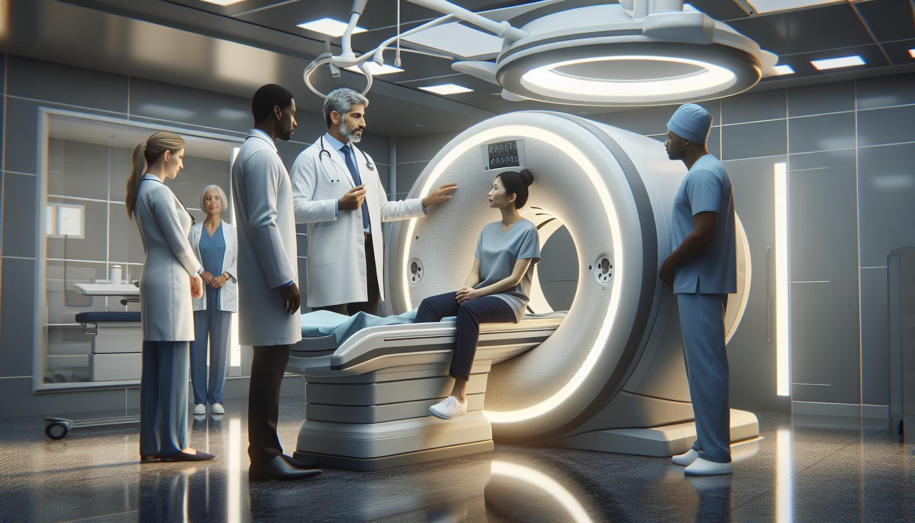
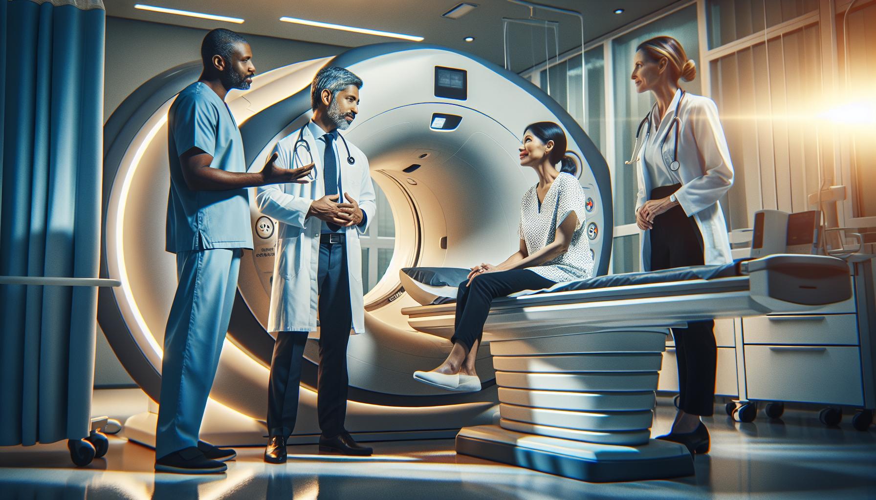
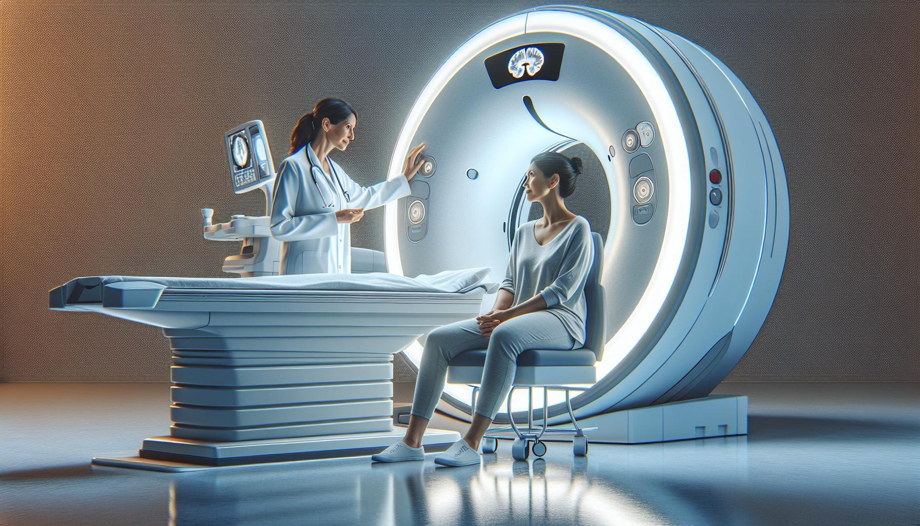
During the CT scan, patients will lie down on a narrow table that slides into the CT scanner-a large, doughnut-shaped machine. It’s crucial to remain still and follow any instructions given by the technician, as movement can blur the images and lead to less accurate results. The procedure typically lasts about 10 to 30 minutes. After the scan, patients can usually resume normal activities right away, but they may need to wait for the physician to review the images before discussing the results.
While CT scans are beneficial, it’s important to understand their limitations compared to other imaging techniques, particularly MRI, which tends to provide clearer images of soft tissues like the brain. Patients should engage in conversations with their healthcare provider about the most suitable imaging options based on individual symptoms and clinical history. Remember, understanding the purpose and procedure of a CT scan can help alleviate concerns and empower patients to take an active role in their health care journey.
How Accurate Are CT Scans for Tumor Detection?
When considering the efficacy of CT scans for tumor detection, particularly in the brain, it’s vital to recognize their capabilities and limitations. CT scans offer detailed, cross-sectional images that can help in identifying larger masses or tumors. However, the accuracy of these scans can be influenced by various factors, such as the tumor’s size, location, and type. For instance, smaller lesions or tumors situated deep within the brain may evade detection, leading to potential false negatives. In some studies, inaccuracies in tumor evaluation via CT scans have been noted, reinforcing the importance of complementary imaging modalities when necessary.
CT scans excel in rapidly assessing acute situations, making them a valuable first-line tool in emergency settings. Yet, their accuracy varies significantly based on tumor characteristics. According to recent findings, certain types of brain tumors, especially those presenting as smaller lesions, pose a greater challenge for CT imaging. For example, these scans may not adequately visualize tumors with indistinct borders or those that do not substantially alter the brain’s structural appearance. Thus, healthcare providers often adopt a nuanced approach, sometimes recommending an MRI for more detailed soft tissue evaluation when there is a suspicion of a brain tumor.
Understanding the inherent limitations of CT scans is essential for patients. Engaging in open discussions with healthcare professionals about the appropriateness of a CT scan in the context of specific symptoms and medical history is crucial. It helps to clarify expectations and make informed decisions regarding further imaging or alternative diagnostic methods. Empowering oneself with knowledge about these nuances not only alleviates anxiety but also encourages proactive involvement in one’s healthcare journey. Always remember, timely detection of any abnormalities significantly improves treatment outcomes, making the role of accurate imaging indispensable.
Common Symptoms That May Prompt a CT Scan
Certain symptoms can often serve as red flags that may prompt healthcare providers to recommend a CT scan for evaluating potential brain tumors. Recognizing these signs is crucial for timely assessment and intervention. Symptoms can vary widely, but they often relate to changes in neurological function, mood, or overall health.
Common Symptoms to Watch For
- Persistent Headaches: Unexplained, chronic headaches that differ from typical patterns can be significant. Many patients report their worst headaches ever experienced, or headaches that worsen over time.
- Cognitive Changes: Difficulty with concentration, memory problems, or confusion can be indicative of brain involvement. If these changes appear suddenly or progress rapidly, they warrant further investigation.
- Nausea and Vomiting: Frequent nausea or unexpected vomiting, especially if accompanied by other symptoms, may suggest increased intracranial pressure, potentially related to a mass in the brain.
- Visual Disturbances: Sudden changes in vision, including blurriness, double vision, or peripheral vision loss, can point towards a problem in the brain that needs further examination.
- Seizures: The onset of seizures, particularly in individuals without a prior history of epilepsy, is a critical symptom that should prompt immediate medical evaluation.
- Personality or Behavioral Changes: Subtle alterations in behavior, personality, or mood such as increased irritability or depression can sometimes be linked to brain abnormalities.
These symptoms alone do not equate to a diagnosis of a brain tumor, but they highlight the importance of seeking medical advice. If you’re experiencing any combination of these signs, it’s essential to consult your healthcare provider. They can evaluate your symptoms thoroughly and determine whether a CT scan or other diagnostic imaging is necessary. Early detection can significantly influence treatment options and outcomes, providing peace of mind during a potentially stressful time. Always prioritize open communication with your medical team to address any concerns you may have regarding your health.
Preparing for a CT Scan: What Patients Need to Know
Preparing for a CT scan can feel daunting, but understanding the process and what to expect can greatly alleviate any anxiety. A CT scan, or computed tomography scan, is essential for detecting abnormalities in the brain, including potential tumors. Taking a few preparatory steps ensures the process is smooth and efficient, helping you feel more comfortable during your visit.
To prepare, it’s vital to inform your healthcare provider of any health conditions you may have, including allergies or if you’re pregnant. You may also need to adjust your medication regime beforehand. Often, patients are advised to refrain from eating or drinking for a few hours prior to the scan, particularly if contrast dye will be used. This dye enhances the images and provides clearer insight into possible abnormalities. If you’re unsure about dietary restrictions, consult your medical team ahead of time.
Arriving at the facility a bit early can help you settle in and complete any required paperwork. Depending on the center, you might change into a gown to ensure images are not obstructed by clothing. During the scan, you will lie on a table that moves through the CT scanner, and it’s crucial to stay very still to ensure high-quality images. The procedure itself usually takes about 30 minutes. While the machine might produce loud noises, it’s harmless and indicates that the imaging process is ongoing.
After the scan, the results are typically reviewed by a radiologist, and your healthcare provider will discuss them with you in follow-up visits. If contrast dye was administered, staying hydrated post-scan is beneficial to help flush it from your system. Remember, if any concerns or questions arise, you should feel empowered to reach out to your healthcare team for clarity and support throughout the process. This proactive step in your health management can make a significant difference in understanding your condition and any necessary next steps.
The CT Scan Process: Step-by-Step Overview
A CT scan can be a crucial tool in detecting brain tumors, providing detailed images that help healthcare providers assess any abnormalities. Understanding the step-by-step process of undergoing a CT scan can alleviate concerns and prepare patients for what to expect during their visit to the imaging facility.
Upon arrival, you’ll check in at the reception, where you’ll provide any necessary information about your health history and the reason for the scan. It’s a good idea to arrive early to complete any paperwork without feeling rushed. If you are required to use contrast dye, the healthcare provider will explain this process, ensuring you are informed about its purpose in enhancing the clarity of the images.
Next, a medical professional will guide you to the imaging room, where you may need to change into a gown. As you lie on the CT scanner table, it’s essential to remain as still as possible during the imaging process. The machine will move around you, capturing multiple angles of your brain. While it’s natural to be anxious about the scanner’s noises – which can be quite loud – they are merely part of the imaging process and not harmful.
After the scan is completed, which usually takes about 10 to 30 minutes, you’ll be instructed to wait for a brief period while the radiologist reviews the images for quality. If you received contrast dye, drinking plenty of water post-scan will help flush it from your system. You’ll then schedule a follow-up appointment with your healthcare provider to discuss the results, allowing you to understand the findings and the next steps in your care. This awareness and involvement can significantly reduce anxiety and facilitate a clearer understanding of your health status.
What Happens After the CT Scan?
After undergoing a CT scan, patients often experience a mix of anticipation and anxiety as they await the results. To help alleviate concerns, it’s crucial to understand what happens after the imaging is completed. Once the scan finishes, which typically takes between 10 to 30 minutes, the radiology team will review the images for quality and clarity. This preliminary check ensures that no critical information is missed, and if any issues arise, such as needing a repeat scan, they can address those immediately.
While you’re waiting, it’s recommended to stay hydrated, especially if a contrast dye was used during the procedure. Drinking water helps to clear the dye from your system, which not only promotes your comfort but also supports overall health. Most patients can resume normal activities after the scan, but it’s wise to follow any specific instructions given by your healthcare provider.
Once the images are deemed satisfactory, a radiologist will analyze them in detail and prepare a report. This report is typically shared with your primary healthcare provider, who will discuss the findings with you during a follow-up appointment. It is important to prioritize this follow-up, as it provides the opportunity to understand the implications of the scan results, whether they are positive or suggest further diagnostic steps might be needed. Engaging in this discussion helps you take an active role in your healthcare decisions and can reduce the anxiety that often accompanies medical testing.
In the meantime, remain proactive by jotting down any questions or concerns you might have for your healthcare provider. Open and clear communication can lead to a better understanding of your health situation and empower you to make informed choices as you navigate any potential next steps in your care.
Comparing CT Scans with Other Imaging Techniques
When seeking to diagnose brain tumors, healthcare providers often choose between several imaging techniques, each with advantages and limitations. While a CT scan is a widely used and effective tool for quickly detecting certain abnormalities within the brain, it may not be the best choice for every situation. For instance, a magnetic resonance imaging (MRI) scan is frequently preferred when a high-resolution image is necessary to differentiate complex structures and assess the full extent of a tumor. MRIs provide detailed images of soft tissues, making them particularly useful for identifying brain tumors and their relationship to surrounding structures.
CT Scans vs. MRI
CT scans are especially valuable in emergency situations due to their speed and ability to visualize bleeding, fracture, or substantial mass effects in the brain. However, their effectiveness can diminish for smaller, more subtle lesions since they have lower soft tissue contrast compared to MRIs. Studies have shown that while CT scans can detect larger tumors, MRIs excel in revealing tumors that may not be readily visible on a CT scan. As a result, providers may often use both modalities. A CT scan might be employed first, particularly if urgent issues like a hemorrhage are suspected, followed by an MRI for a more detailed evaluation.
Other Imaging Techniques
In addition to CT and MRI, other imaging techniques can play a role in brain tumor diagnosis. For example, positron emission tomography (PET) scans are useful in assessing the metabolic activity of tumors, helping to differentiate between benign and malignant lesions. Functional MRI (fMRI) can also provide insights into brain activity and its relationship to tumor location, which is invaluable when planning surgical interventions.
While choosing the most appropriate imaging technique, healthcare providers consider various factors, including the patient’s symptoms, medical history, and specific concerns about the tumor. It’s essential for patients to openly discuss their options with healthcare professionals, as they can provide tailored recommendations based on individual circumstances. Understanding the differences among imaging modalities can empower patients, reduce anxiety during the diagnostic process, and contribute to informed decision-making regarding their healthcare journey.
Factors That Influence CT Scan Accuracy
Certain factors can significantly influence the accuracy of CT scans in detecting brain tumors, making it essential for patients to understand these elements as they prepare for this diagnostic tool. One of the most critical aspects is the specific type and size of the tumor. Larger tumors with distinct shapes and clear borders are generally easier to detect on CT scans compared to smaller lesions or those that blend in with surrounding brain tissue. Subtle lesions may be missed due to the limited soft tissue contrast that CT imaging provides, highlighting the importance of follow-up imaging, such as MRI, which can reveal abnormalities that CT scans might overlook.
The location of the tumor also plays a vital role in detection accuracy. Tumors situated in complex areas of the brain or near critical structures may present challenges for CT detection, as overlapping anatomical features can obscure clear imaging. Additionally, the use of contrast agents can enhance the visibility of certain types of tumors, helping radiologists distinguish between healthy and diseased tissue more effectively. However, this tactic depends on proper clinical judgment regarding when to use contrast material and understanding each patient’s health status.
Patient-related factors, such as body size and the presence of motion during the scan, can affect the quality of the CT images. For instance, patients who are unable to remain still may inadvertently introduce motion artifacts, leading to less clear images. Taking appropriate measures, such as having patients hold their breath when instructed, can minimize these issues. Furthermore, discussing any concerns with healthcare providers beforehand can help ensure the scan is conducted smoothly and effectively.
Lastly, advancements in CT technology, including higher-resolution machines and improved scanning techniques, have dramatically enhanced the ability to detect brain tumors. Staying informed about these developments can empower patients to advocate for the most effective imaging strategies suited to their specific needs. As such, it is crucial for patients to maintain open communication with their healthcare providers to ensure a comprehensive understanding of the factors affecting their CT scan results and to receive personalized advice tailored to their diagnostic journey.
The Role of Advanced CT Technology in Detection
Advancements in CT technology have significantly transformed the landscape of brain tumor detection, offering patients and healthcare providers enhanced tools for diagnosing and monitoring these serious conditions. Modern CT scanners now utilize higher resolution capabilities, which provide clearer and more detailed images, aiding radiologists in identifying small tumors and subtle abnormalities that previous generations of scanners may have missed. These improvements are particularly important for patients where early detection can lead to more effective treatment options.
Moreover, the integration of multi-slice CT scanning has further enhanced diagnostic accuracy. This technology allows simultaneous imaging of multiple slices of the brain, resulting in a faster and more comprehensive examination. As a result, patients spend less time on the scanner while still achieving high-quality imaging. This swift process can help alleviate anxiety by reducing the duration of the procedure. Conducting the scan in a comfortable and efficient manner ensures that patients feel cared for and supported throughout their experience.
In addition to these imaging enhancements, advanced CT technology often incorporates artificial intelligence (AI) algorithms that assist radiologists in interpreting scans more effectively. AI can analyze patterns and detect anomalies that might be overlooked by the human eye, thus streamlining the diagnosis process. Patients can feel reassured knowing that cutting-edge technology is actively being used to enhance the accuracy of their examinations.
While these advancements undeniably improve tumor detection, it remains essential for patients to communicate openly with their healthcare providers about any concerns they may have. Discussing personalized circumstances, such as individual risk factors and imaging needs, will help in determining the best course of action related to diagnostic imaging. Ultimately, the role of advanced CT technology not only fortifies medical practices but also fosters a sense of empowerment and partnership between patients and caregivers in the journey towards health and recovery.
Understanding Results: Positive and Negative Outcomes
Understanding the results of a CT scan is a critical step for patients navigating potential brain tumor issues. The outcomes of these scans can yield both positive and negative news, and knowing how to interpret these results can greatly alleviate anxiety. A positive result typically indicates the presence of a tumor or abnormality, while a negative result suggests that no significant issues were identified. However, it’s essential to understand that the absence of findings does not always rule out the possibility of a brain tumor, as smaller or less aggressive tumors may not be easily detectable on a CT scan.
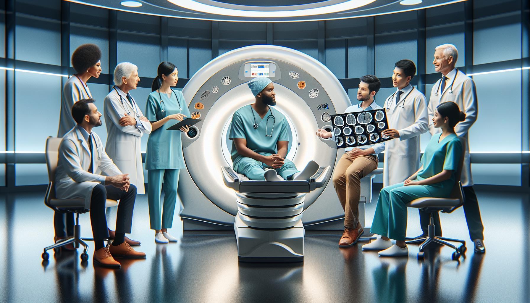
When receiving the results, patients might feel overwhelmed. Here, it’s beneficial to consult with your healthcare provider to discuss the findings in detail. They can explain technical terms and clarify the implications of the results. For instance, if a CT scan shows a mass, the doctor may recommend further testing with an MRI, as MRI imaging provides more detailed images of the brain tissue, which can help in assessing the characteristics of the tumor more effectively.
It’s also important to know that while CT scans are a valuable diagnostic tool, they do have limitations. False negatives can occur, especially in less aggressive tumors that may not be readily identifiable on the scan. To address this, a healthcare provider may recommend follow-up scans or different imaging methods based on symptoms and medical history. Consistent communication with the care team allows for personalized management of the situation, ensuring that patients feel supported through each step of the process.
Ultimately, whether the results are positive or negative, understanding them in the context of one’s health is key. Engaging in open discussions with healthcare professionals, seeking clarity on any uncertainties, and asking about subsequent steps helps empower patients in their healthcare journey. Knowing that your care team is there to assist can significantly reduce stress and foster a sense of partnership in managing your health.
Financial Considerations: Cost of a CT Scan
The cost of a CT scan can be a significant concern for patients; understanding the financial implications is crucial for informed decision-making. Typically, without insurance, a CT scan can range from $300 to over $3,000, depending on the facility, the type of scan, and geographical location. It’s vital to be aware that prices can vary dramatically, so acquiring a precise estimate from the healthcare provider beforehand can help avoid unexpected expenses.
Many insurance plans cover CT scans, particularly when they are deemed medically necessary. Patients should check with their insurer to understand any required copays, deductibles, or prior authorization processes. For individuals without insurance, some hospitals and imaging centers offer payment plans or financial assistance programs, ensuring that necessary imaging is accessible even for those facing financial hardships. It can also be beneficial to inquire about self-pay rates, which may offer discounted prices for upfront payments.
Tips for Managing Costs
- Confirm Coverage: Always confirm that the imaging center is in-network if you have insurance. This can significantly reduce out-of-pocket expenses.
- Ask for a Quote: Request a detailed quote from the imaging center before scheduling the scan. This ensures transparency regarding all possible fees.
- Consider Alternatives: If cost is an issue, discussing alternative imaging options, such as MRI or ultrasound scans, with your healthcare provider may be beneficial, as costs can vary.
- Financial Aid Inquiry: Don’t hesitate to ask about financial assistance programs if you’re encountering difficulties with costs.
Patients encouraged to proactively engage with their healthcare providers about financial matters related to their CT scan can alleviate stress and foster a supportive partnership. While the financial aspect of undergoing such diagnostic tests can be daunting, ensuring open communication will empower individuals to make choices that are best for both their health and their financial well-being.
Consulting Your Healthcare Provider: Next Steps
Following a CT scan to investigate potential brain tumors, it’s essential to consult your healthcare provider to understand the next steps in the diagnostic process. Having an open dialogue will provide clarity and reassurance during a potentially stressful time. Your healthcare provider can explain the results from the CT scan, whether they indicate the presence of a tumor or provide insights into other concerns that might have been identified.
When discussing findings, don’t hesitate to ask questions. Inquire about the implications of the results, any further tests that might be necessary, and the timeframe for next steps. For example, if a tumor is detected, your doctor might recommend additional imaging tests, such as an MRI, which can provide more detailed information about the tumor’s size and location. They may also discuss a referral to a specialist, such as a neurosurgeon or oncologist, to explore treatment options.
It’s equally important to discuss any symptoms you may be experiencing that prompted the CT scan, as these details can guide your provider in tailoring a care plan that’s best suited to your needs. Keeping a list of your symptoms, their duration, and any changes can be helpful during the appointment. Your healthcare provider will appreciate this information, as it aids in understanding the overall clinical picture.
Before concluding your appointment, ensure you understand the next steps and the rationale behind them. Ask for clarification on any medical jargon or procedures that seem complex. This will empower you to take an active role in your health care decisions and alleviate some of the anxiety associated with awaiting further treatment or monitoring. Lastly, don’t forget to discuss follow-up appointments and any other necessary future tests to maintain comprehensive care. Remember, you are not alone; your healthcare team is there to support and guide you through this journey.
Q&A
Q: Can a CT scan detect all types of brain tumors?
A: No, while CT scans can identify many brain tumors, they may not detect all types, especially smaller or less aggressive ones. MRI scans are often more effective for detailed imaging and better characterization of brain tumors. Consider discussing imaging options with your healthcare provider for comprehensive evaluation.
Q: How effective are CT scans compared to MRIs in detecting brain tumors?
A: CT scans are effective for identifying significant abnormalities in the brain, but MRIs provide more detailed images and can detect smaller tumors more accurately. Combining both imaging techniques can enhance detection capabilities. For a thorough assessment, refer to the section that compares imaging techniques in your article.
Q: What symptoms might indicate a need for a CT scan to check for a brain tumor?
A: Symptoms that may prompt a CT scan include persistent headaches, seizures, vision changes, difficulty with speech, and unexplained nausea. If you experience these symptoms, consult your healthcare provider who may suggest a CT scan or other imaging tests based on your condition.
Q: How can I prepare for a CT scan if brain tumor concerns are present?
A: Preparation for a CT scan generally includes wearing comfortable clothing without metal and informing your provider of any medications or allergies. A pre-scan consultation will help alleviate anxiety and clarify the process. Refer to your article’s preparation section for detailed steps.
Q: What can a patient expect during the CT scan process for brain tumor detection?
A: During a CT scan, you will lie on a table as it moves through the scanner, which takes X-ray images of your brain. The procedure is quick-typically 10 to 30 minutes-and painless. Ensure to remain still to enhance image quality; follow your article’s step-by-step overview for more details.
Q: How long does it take to receive CT scan results for brain tumor detection?
A: CT scan results are usually available within a few hours to a couple of days, depending on the facility. Once available, your healthcare provider will discuss the findings and any next steps. Make sure to consult your article section on understanding results for more insights.
Q: What factors can affect the accuracy of a CT scan in detecting brain tumors?
A: Factors impacting CT scan accuracy include the tumor’s size, location, and type, as well as the quality of the scan and the patient’s body composition. Regularly updating imaging technology can also improve detection accuracy. Look into your article’s section on this topic for further details.
Q: What happens if a CT scan indicates a brain tumor?
A: If a CT scan suggests a brain tumor, your healthcare provider will likely recommend further evaluation, which may include an MRI or a biopsy for definitive diagnosis and treatment planning. It’s essential to discuss next steps outlined in your article to understand the process.
Key Takeaways
Understanding whether a CT scan can reveal a brain tumor is crucial for your peace of mind and health management. Remember, a CT scan uses advanced imaging technology to provide detailed insights, aiding in accurate diagnosis. If you’re still feeling uncertain about the process, consider discussing your concerns with your healthcare provider to clarify how this imaging can benefit you directly.
For further information, check out our resources on preparing for a CT scan and what results to expect post-procedure. Exploring how imaging works in detecting other conditions can also enhance your knowledge-be sure to read about the benefits of low-dose CT scans in preventative care.
Don’t hesitate-take charge of your health today! Sign up for our newsletter to stay informed, or schedule a consultation to discuss your options and ensure you have the information needed for your peace of mind. Your health journey is important, and we’re here to support you every step of the way.


